Protein Translocation Through Artificial Nanopores
←
→
Page content transcription
If your browser does not render page correctly, please read the page content below
Protein Translocation Through
                    Artificial Nanopores

                               Marc Creus
                            University of Basel

                                ERBM4 Liège

A WHOLE nano-world to be explored!

Institute of Microtechnology (Neuchâtel)   Institute of Chemistry (Neuchâtel)
          Dr Urs Staufer                             Dr Marc Creus
          Dr Anpan Han                               Prof Thomas Ward
          Prof Nico de Rooij
First patent application for Coulter Counter: 1949

      “You cannot patent a hole!”
US Patent granted: 1953, USPT 2656508

                                          Wallace H. Coulter (1913-1998)
                                    Engineer, Inventor, Entrepreneur, Visionary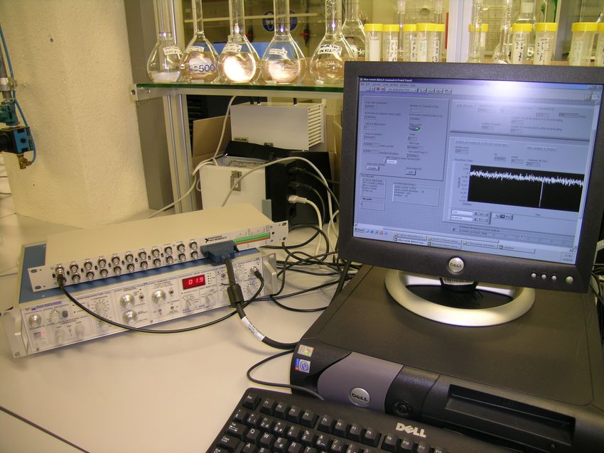
Application of Coulter Principle:
                     Blood-Cell Counter

The complete blood count or “CBC” is
one of the most commonly ordered
diagnostic tests worldwide.

Today, ninety-eight percent of CBCs
are performed on instruments using the
Coulter Principle.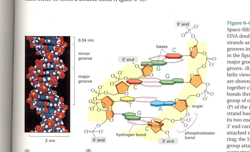
Micro vs Nano

             10µm

                 1nm
           (e.g. diametre of
           a small protein,
           which is
           10 000 x smaller
           than this small cell)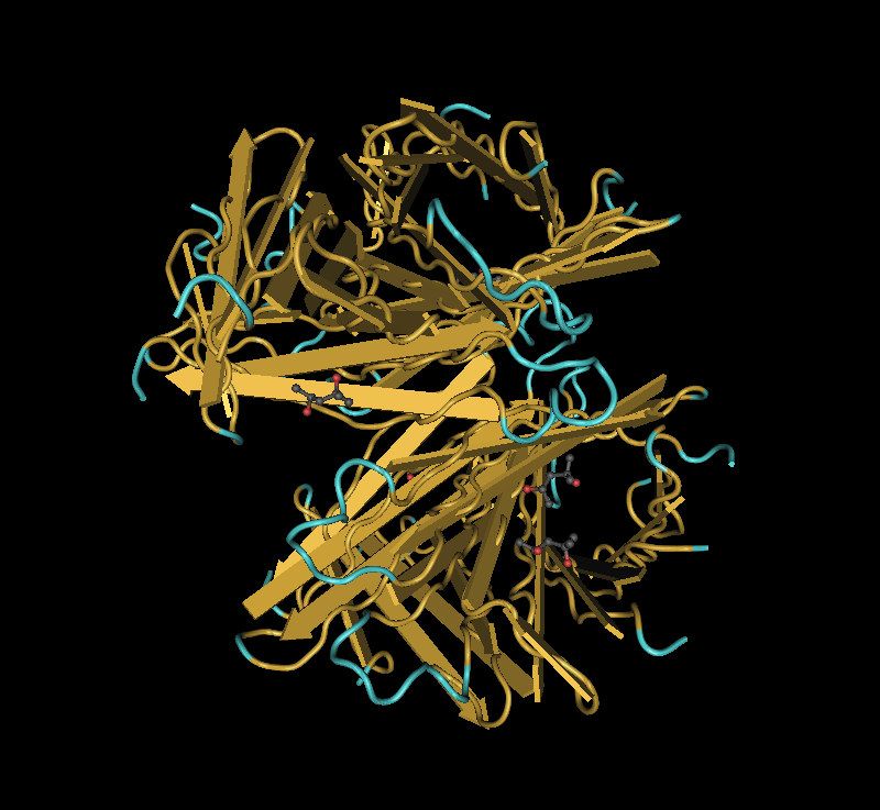
Since the nano-scale corresponds to the size of
   biological macromolecules, nanopores could be
   useful in biochemical analyses of proteins.

                                                                    ~5nm

“We have friends in other fields---in biology, for instance. We physicists often look at them and
say, (…) ``You should use more mathematics, like we do.'' They could answer us (…) ``What
you should do in order for us to make more rapid progress is to make the electron microscope
100 times better.''
                                                  Richard Feynman, December 29th 1959
                                                  “There’s plenty of room at the bottom”
                                                  http://www.zyvex.com/nanotech/feynman.html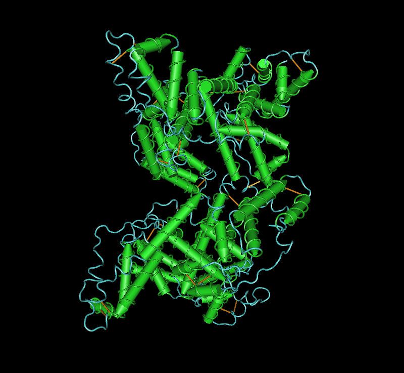
Structure of DNA
•   Genetic data
•   Primary structure
    – Polymer of A, T, C, G
•   Secondary structure
    - B helix
    – Diameter
      2 nm
•   Tertiary structure              Alberts et al.
Structure of Proteins
•   Primary structure
    – Polymer of 20 amino acids
•   Secondary structure
    – α-helix, β-strands, coils
•   Tertiary structure
•   Quaternary structure
    – Multi protein complex, filaments
•   Typical diameter: 1 - 20 nm                 Ovalbumin
                                         Deposition: Stein , Leslie,
                                            1990 PDB: 1OVA
Properties of macromolecules:

•      Surface charge: positive, neutral or negative

                      -DNA (an acid) is usually negatively
                      charged
    Acid (low) pH                                      Basic (high) pH

                    -Proteins can be basic or acidic
                    and have different charges
                    depending on the pH
                                                                         Alberts et al.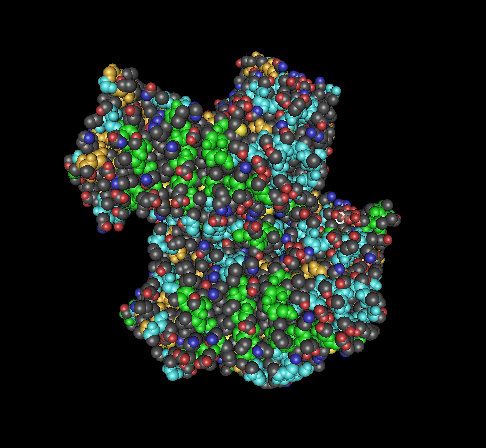
More properties of macromolecules:
               Specific interactions

•   Proteins are designed for recognition:
    antibodies, hormones, enzymes,
    structural proteins, toxins, etc…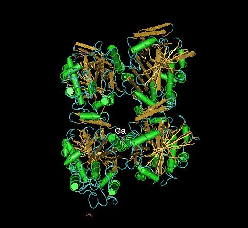
How can a biochemist make use of
         synthetic nanopores?
•   Measure size, charge, structural
    properties and interactions of
    proteins, in real-time and in solution?
Process flow chart nanopore fabrication
500 nm SiO2 AZ 1518 both sides KOH etching rinsing
20 nm Si3N4
spin PMMA backside alignment oxidation
optical lithography
20nm
e-beam litho. RIE, stripping chip level PDMS bonding
silicon Si3N4 SiO2
RIE, stripping
PMMA PDMS AZ 1518Wafer-level nanopore fabrication process
PDMS
lp= 20nm
25 nm
Si3N4
SiO2
Si
SiO2
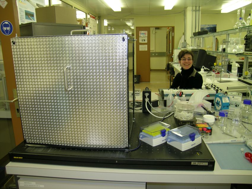
Si3N4Experiment setup
Measured parameters
•   Count the numbers of
    spikes per minute
    – Number of spikes
      proportional to
      concentration

•   Individual spikes
    – Duration: ∆t
    – Current change: ∆I
                              ∆I
                                     ∆t
Four different proteins, differing in size and
charge properties
Streptavidin Mut. S27a Human Serum Albumin Ovalbumin Avidin comp. Biotin.
Le Trong et al. 2002 S.Sugio, et al., 1998 Stein , Leslie, 1990 Livnah et al 1993,
PDB: 1N9Y PDB: 1BM0 PDB: 1OVA PDB: 2AVI
Notes Mass (kDa) rstoke (nm) pIisof pIef
Streptavidin (SAV) Recombinant 66 - 6.5
BSA >99% electrophoresis 66 3.5 5.3 4.25
Ovalbumin (OA) Grade VII (>98% elph.) 44 2.7 4.54 4.6
Avidin (AV) Heterogeneous 72/62 - 10.5Translocation by electrophoresis Electrode bias set at 50 mV (or -50mV) pH 6, citrate, 1M KCl, 1 µg BSA/mL Since pore is considerably larger than proteins, at a first approximation we can ignore protein-pore interactions
Protein charge explored by nanopores

Valleys (50mV)                              Peaks (-50mV)
Protein charge explored by nanopores

                                                                                                           BSA

BSA is reported to have pI 4.2 in
presence of KCl

(reports that pI is reduced from 5.3
due to binding of Cl-)

Suggests importance of counterions?

                  Han, Creus, Schürmann,Lindner, Ward, Staufer, Analytical Chemistry (2008) 89:4651-4658
Spikes: pH-dependence of shape and duration
BSA
Duration of blockage-events
varies with pH: longer (and
more complex) signals closer
to pI
Fewer, sharp spikes when pH
is distant from pI
Suggests time resolution is a
critical issue
Han, Creus, Schürmann,Lindner, Ward, Staufer, Analytical Chemistry (2008) 89:4651-4658Variety of spikes with complex fine-structure
BSA pH3 BSA pH6 AV pH6 SAV pH5
Han, Creus, Schürmann,Lindner, Ward, Staufer, Analytical Chemistry (2008) 89:4651-4658Time resolution
Our calculations suggest
that at pH8 BSA BSA
BSA
translocates the 20nm pore-
length in about 2µs
Even with 100kHz
bandwidth, practical time
resolution is only 40µs
Very fast translocations will
not be resolved
Can slow down by
measuring with pH close to
pI
Han, Creus, Schürmann,Lindner, Ward, Staufer, Analytical Chemistry (2008) 89:4651-4658Slowing down by pH
r
E I
pH close to pI
pH far from pI
tProtein translocation explored by nanopores
BSA
BSA is reported to have pI 4.2 in
presence of KCl
(reports that pI is reduced from 5.3
due to binding of Cl-)
Very few translocations of BSA at
pH4
Han, Creus, Schürmann,Lindner, Ward, Staufer, Analytical Chemistry (2008) 89:4651-4658Protein diameter measured by nanopores
Pore Protein I (nA) dp (nm) ΔI (nA) dm (nm)
A OA 10.7 21.9 0.21 7.1
B OA 10.8 22.0 0.25 7.5
pH 6, 100mV
OA, BSA, SAV C OA 14.0 26.3 0.23 7.3
D BSA 14.0 26.4 0.31 8.6
E BSA 14.5 26.5 0.27 8.7
F BSA 15.9 28.9 0.31 8.8
dOA = 7.3 nm ± 0.2 nm
dBSA = 8.7 nm ± 0.1 nm
1 nm = 10 hydrogen atoms (10 Å)Quantifying molecules by exploiting
specific interactions of proteins
IgG (hCG)= 4µg/ml
0 2 4 6 8 10 0 2 4 6 8 10
Time (s) Time (s)
0 2 4 6 8 10
Time (s)Interpretation
r
E I
tNanopore bioassays
• The principle of the assay is general & can be applied wherever
two molecules combined give a different signal from signals of
either molecules alone
A+B=C
•
Titrations can be employed for
quantification (e.g. measures of affinity)
Statistical calculations: 1000 counts (C.V. 3.2%)
Counting 1000 proteins in 1ml volumes is not “zeptoM sensitivity”,
due to limitations:
- Time: 500 counts/min (25nM antibody)
- Affinities (for biomolecular interactions)New methods bring surprising outcomes…
• SAV (calculated pI= 6.5) is
apparently very heterogeneous, SAV
with both positively and
negatively-charged tetramers at
any given pH
SDS-PAGE gelSAV apparently pure?
Mass Reconstruction of Streptavidin Wildtype.
Applied Biosystems/ Sciex QTrap Mass Spectrometer:
Electrospray Low Resolution, Positive Ion Mode
Acetonitrile/Water (1:1) + 1%HFo
Avi Sav
16430.0
Sav (theory)= 16423 Da
Sav (found) = 16430 Da
Sav + Ca2+= 16470 Da
Sav + 2x Ca 2+= 16510 Da
Sav + 3x Ca2+ = ~16552 Da
Isoelectric Focusing
Lutter et al. Electrophoresis 2001, 22: 2888-2897New methods bring surprising outcomes… • SAV (calculated pI= 6.5) is apparently very heterogeneous, SAV with both positively and negatively-charged tetramers at any given pH • Charge heterogeneity? • Binding to counterions?
Summary

•   Protein sensing using nanopores: label-free, in solution, in
    real time
    – Exquisitely sensitive: proteins analysed one-by-one
    – Diameter precisely determined with 0.2nm reproducibility
    – Charge-properties and interactions between proteins can be
      measured
    – Label-free immunoassays
    – Counting just 1000 molecules is required for accuracy, which
      could be found in tiny volumes
Questions and outlook
What are the effects of counterions?
What is the significance of the fine-structure of spikes?
Structural/ biophysical properties:
Explore orientation of translocation
Sequence proteins: beyond genomics?
Protein folding (time resolution)
Domain movements (time resolution)
Nanopore Assays:
Protein heterogeneity
Biomolecular interactions & affinities
Paradigm shift (beyond DNA):
Since nanopores are easy to use and informative, they
may become a useful analytical tool for the biochemistAcknowledgements
• Canton de Neuchâtel
• Swiss National Science Fund
• Danish Research Agency
for financial support
• The staff of COMLab & the joint clean-room facility of IMT and
CSEM for their technological support
• Prof. Urs Staufer (now at Delft Technical University)
• Dr Anpan Han (now in Copenhagen)Wafer-level nanopore fabrication process Si3N4 SiO2 Si SiO2 Si3N4
Wafer-level nanopore fabrication process

Resist
 Si3N4
  SiO2

   Si

  SiO2
 Si3N4
Wafer-level nanopore fabrication process
                 e-beam exposure

Resist
 Si3N4
  SiO2

    Si

   SiO2
  Si3N4
Resist

                optical lithography
You can also read