Rare paratesticular aggressive angiomyxoma mimicking an epididymal tumor in an 82-year-old man: Case report
←
→
Page content transcription
If your browser does not render page correctly, please read the page content below

Open Medicine 2021; 16: 973–977
Case Report
Chi-Fang Chen, Tao-Yeuan Wang, Marcelo Chen, Yung-Chieh Lin*
Rare paratesticular aggressive angiomyxoma
mimicking an epididymal tumor in an 82-year-old
man: Case report
https://doi.org/10.1515/med-2021-0317 involving the scrotum, pelvic region, perineum, and ingu-
received March 12, 2021; accepted June 7, 2021 inal area [3]. AAM is difficult to diagnose without patholo-
Abstract: Aggressive angiomyxoma (AAM) is a rare gical proof, and misdiagnosis as hydrocele, spermatocele,
mesenchymal myxoid tumor, and most cases occur in the testicular, and paratesticular neoplasia is common [2,4]. In
pelvic region or perineum of adult females. AAM is very rare the literature review, only four cases were identified. Herein,
in males. Most of these cases have been diagnosed in we report on an 82-year-old male patient who had parates-
patients aged 30–60 years, and the tumors involved the ticular AAM, and to the best of our knowledge is the oldest
pelvic cavity, scrotum, or spermatic cord. AAM can mimic case in the literature. Informed consent was obtained from
inguinal hernia, hydrocele, or paratesticular neoplasm. Four the patient.
male cases have been reported with paratesticular AAM
mimicking a testicular/epididymal tumor, and to the best
of our knowledge, this is the oldest patient in the literature.
Because of its rarity, making an exact diagnosis before sur- 2 Case report
gery is difficult. Herein, we present a case of AAM in an
82-year-old man and review the literature. An 82-year-old man recently diagnosed with prostate
cancer, which was under surveillance, presented to our
Keywords: AAM, deep angiomyxoma, mesenchymal tumor, outpatient department with a right palpable painless
paratesticular mass, older paratesticular mass. The physical examination revealed
a round paratesticular mass that was firm in consistency
without tenderness. It was fixed on the lower pole of the
right testis and mimicked an epididymal or testicular
1 Introduction neoplasm. Scrotal sonography showed a heterogenous
round mass over the tail of epididymis (Figure 1a and b).
Aggressive angiomyxoma (AAM) is a rare mesenchymal Rich blood supply was noted under Color Doppler sono-
tumor. In 1983, Steeper et al. [1] described nine cases of graphy (Figure 1c). The patient denied a history of cryptorchi-
female pelvis and perineum tumors, which they called dism or previous trauma. Further imaging, including whole
AAMs [2]. In men, less than 50 cases have been reported, abdominal computer tomography scan and bone scan,
showed no lymphadenopathy or distant tumor metastasis.
The tumor markers Lactate dehydrogenase, beta-Human
* Corresponding author: Yung-Chieh Lin, Department of Urology, Chorionic Gonadotropin, alpha-fetal protein, and CEA were
Hsinchu Branch, MacKay Memorial Hospital, No. 690, Sec. 2, at normal levels.
Guangfu Rd., East Dist., Hsinchu City, Taiwan (R.O.C.),
Epididymal tumor excision by a scrotal approach was
e-mail: cooldong.tw@yahoo.com.tw
Chi-Fang Chen: Department of Urology, MacKay Memorial Hospital, arranged. Because it presented as a non-testicular tumor,
Taipei City, Taiwan (R.O.C.), e-mail: s850292@gmail.com orchiectomy from the inguinal approach did not take into
Tao-Yeuan Wang: Department of Pathology, Tamsui Branch, MacKay consideration before surgery. However, the operation
Memorial Hospital, New Taipei City, Taiwan (R.O.C.), was shifted to right orchiectomy because of severe tumor
e-mail: path168@gmail.com
adhesion onto the testis. Gross examination showed
Marcelo Chen: Department of Urology, MacKay Memorial Hospital,
Taipei City, Taiwan (R.O.C.), e-mail: marcelo@mmh.org.tw
a gray, round, and solid tumor with clear boundaries
Tao-Yeuan Wang, Marcelo Chen: School of Medicine, MacKay (Figure 2). Histologically, the testicular adnexal mass
Medical College, New Taipei City, Taiwan (R.O.C.) was a deep angiomyxoma composed of bland spindle
Open Access. © 2021 Chi-Fang Chen et al., published by De Gruyter. This work is licensed under the Creative Commons Attribution 4.0
International License.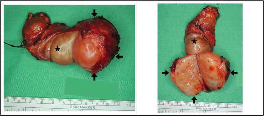
974 Chi-Fang Chen et al.
examination at our out-patient department per 3 months
initially. Our follow-up policy is scrotal echo every 6
months for 2 years and whole abdominal CT annually.
Ethics approval and consent to participate: Not applicable.
Consent for publication: Informed consent was obtained
from the patient for publication of this case report and
any accompanying images.
3 Discussion and conclusion
Most documented cases of AAMs have been reported in
the genitals, perineal, and pelvic regions in women of
childbearing age. In men, only case reports or case series
have been reported. A male to female tumor ratio of 1:6
has been described in some studies [5]. The lesions
usually originate from the scrotum, spermatic cord, peri-
neum, or pelvic cavity [6]. A case of AAM in the right
thigh has been reported [7]. The tumor is slow-growing
[2]. The term “aggressive” was first described in female
patients, and implies a characteristically locally infiltra-
tive tumor with frequent recurrence [4]. Considering its
benign nature, the nomenclature was changed from
“aggressive” to “deep” in the Fourth Edition of the World
Health Organization Classification of Soft Tissue tumors
in 2013. The reason for frequent recurrence may be its
location, which was difficult to resect with definite mar-
gins. Despite the frequent recurrence, distant metastasis
has seldom been reported [2]. A literature review identi-
fied seven male cases in the scrotum and paratestis
(Table 1)
According to literature reviews, the most common
site of AAMs is the scrotum followed by the inguinal
region. Most of the tumors were growing beside the sper-
matic cord [8]. For the paratesticular region, four other
Figure 1: Ultrasound sonography: (a) a well-capsulated heteroe-
cases were presented. Only one patient is younger than
choic round mass over the tail of epididymis, 4.7 × 3.5 cm in size,
and (b) abundant tortuous vessels adjacent to the paratesticular
65 years. Because the tumor was usually asymptomatic,
tumor. (c) The tumor had a rich blood supply on color Doppler most tumors were more than 10 cm in size. All of them
sonography. received orchiectomy for diagnosis and treatment. In the
limited follow-up duration, no local recurrence was men-
tioned after excision with a clear margin.
cells in a myxoid matrix containing delicate vessels. The As the tumor can occur at various sites, the diagnosis
lesion was immunoreactive for CD34, smooth muscle is difficult. It can mimic other non-neoplastic lesions
actin, and desmin and negative for S100, estrogen recep- including hydrocele, inguinal hernia, or testicular neo-
tors (ERs), and progesterone receptors (PRs) (Figure 3). plasm [9]. Scrotal ultrasound is typically the primary
The patient has been followed up for 4 months without imaging modality, and magnetic resonance imaging
disease recurrence. Long-term follow-up is needed for (MRI) can provide further details [3]. Sonography for
AAMs, we will arrange further images and physical AAMs demonstrates mixed echogenicity, lamellated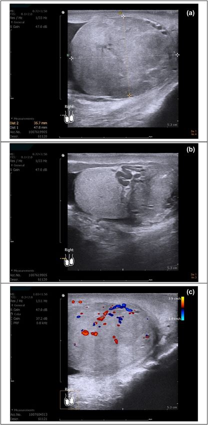
Rare paratesticular AAM mimicking an epididymal tumor 975
Figure 2: A white, grayish, well-circumscribed, solid tumor (4 × 3.5 cm) adherent to atrophic testis and epididymis (arrow: tumor; star:
testis).
Table 1: Clinical details of paratesticular angiomyxoma in a literature review
Source (author) Site Age Size Treatment Immunostaining results Follow-up
(cm) (NED/LR)
Chen et al.* Paratestis 82 4.7 × 3.5 Orchiectomy CD34, Desmin, SMA(+) S100, ER, PR(−) NED (4 months)
Neyaz et al. [2] Paratestis 53 15 × 10 Orchiectomy Vimentin, CD34, SMA(+) Desmin (focal+) S100, NED (12 months)
ER, PR(−)
Serao et al. [6] Paratestis 72 7.7 × 5.6 Orchiectomy CD34, Desmin, Actin, PR(+), ER(−) NED
Aydin et al. [13] Paratestis 66 11 × 7.5 Orchiectomy CD34, ER, PR(+) SMA, S100 (−) NED (6 months)
Ismail et al. [14] Paratestis 65 11 × 8 Orchiectomy Desmin, SMA(+) ER (focal+) PR, CD34, S100(−) NED (24 months)
Durdov et al. [15] Scrotum 37 7×5 Wide excision Vimentin(+) Desmin(−) SMA(−) S100(−) NED (24 months)
Chihara et al. [16] Scrotum 47 17 × 10 Wide excision ER(focal+) PR(−) S100(−) HHF35(−) NED (14 months)
Kirkilessis Scrotum 57 11 Wide excision Desmin, CD34, SMA(+) ER(focal+) PR(focal+) NED (24 months)
et al. [17]
Abbreviations: NED, no evidence of recurrence; LR, local recurrence; SMA, smooth muscle alpha-actin; ER, estrogen receptor; PR, proges-
terone receptor (*our case).
appearance with alternating layers of hypo and hypere- of AAM from other benign and malignant myxoid soft
choic tissues. Color Doppler demonstrates internal vascu- tissue tumors are mainly based on morphology and
larity. On T2-weighted MRI, high signal intensity relative immunohistochemistry. Immunoreactivity for desmin,
to the muscle with “swirled” low signal intensity bands SMA, vimentin, and CD34 is noted in tumor cells, at
within a hyperintense tumor has been noted [10]. How- least focally [2]. A case report mentioned that the dif-
ever, typical ultrasound images to definitively describe ference between males and females is the presence of
AAMs are lacking. The imaging nowadays is not always ERs and PRs. In contrast to females, where ERs or PRs
clear in terms of benign tumors and this imposes a sur- are almost always expressed, ER and PR stainings are
gical excision as a treatment and diagnostic approach usually negative in males. The authors suggested that
[11]. In our cases, MRI was not arranged before the opera- these markers play a role in tumor development and
tion, because AAMs had been diagnosed after excision. It pathogenesis, but do not apply to males [2]. In our
will be an option if we find a similar tumor in the scrotum case, ER and PR immunostaining were negative. It is
in the future. possible that the expressions of ER and PR are related
Since none of these imaging modalities can reliably to tumor development and recurrence, and that fre-
distinguish AAM from malignant tumors including sar- quent recurrences do not apply to males. ERs and
coma, the mainstay of treatment is the exploration and com- PRs can help diagnose the AAMs in the immunohisto-
plete surgical resection [3]. The diagnosis and distinction logical stain. However, the relationship of AAM with976 Chi-Fang Chen et al.
a novel staining with quite sensitivity but not an entirely
specific maker for deep angiomyxoma.
In men, AAMs should be considered in the differen-
tial diagnosis of paratesticular tumors. To the best of our
knowledge, four paratesticular AAMs have been reported.
It is a slowly growing benign tumor, but its locally
aggressive and infiltrative nature makes it difficult to
resect alone without orchiectomy. Although approxi-
mately 70% of cases locally recurred within the first 3
years [4], our review showed a lower rate of local recur-
rence of AAMs in males. Although AAMs in females
occurred frequently in the pelvis area with easy local
recurrence after excision, in men, no local recurrence
was presented during short period follow-up. Wide exci-
sion with free margin is important for any tumor doubted
malignancy. Due to its atypical presentation and rarity in
males, we presented our clinical images and typical his-
tologic findings for future references.
Although AAMs are rarely noted in men, however,
if we find a well-circumscribed tumor in the scrotum,
the diagnosis should be kept in mind. Furthermore,
the infiltrated paratesticular tumor was slowly growing
but it would be severe adhesive to the testis. So that,
before wide excision, informed consent to the patient
about the possibility of orchiectomy for clear margin is
considered.
Funding information: No funding.
Author contributions: C.F.C. made contributions to the
acquisition of history, image, and wrote the manuscript.
T.Y.W. carried out the pathology part interpretation and
description. M.C. performed the treatment and reviewed
the manuscript to give clinical opinions. M.C. and Y.C.L.
made supervision and helped to review the manuscript.
All the authors read and approved the final manuscript.
Figure 3: Typical bland spindle or stellate cells with little or no
nuclear polymorphism and variably elongated cytoplasm set in a Conflict of interest: No potential competing interest was
mucomyxoid stroma. Vascularity was variably composed of delicate reported by the authors.
to more hyalinized vessels (a) (hematoxylin and eosin, magnifica-
tion 200×); typical cytoplasmic desmin (b) and smooth muscle actin
Data availability statement: Records and data pertaining
(c) immunopositivity (magnification 200×).
to this case are in the patient’s secure medical records in
Mackay Memorial Hospital.
other hormones is not well described in the literature.
The hormone profile surveying, such as pituitary, thyroid,
parathyroid, adrenal, and sex hormone, will be consid-
ered, if we found similar AAMs in men with a positive References
result of ERs and PRs. The potential use of HMGA2 immu-
nohistochemistry staining is also quite significant [12]. [1] Steeper TA, Rosai J. Aggressive angiomyxoma of the female
Although, we did not stain the HMGA2 for our case, it is pelvis and perineum. Report of nine cases of a distinctive typeRare paratesticular AAM mimicking an epididymal tumor 977
of gynecologic soft-tissue neoplasm. Am J Surg Pathol. 1983 [10] Tariq R, Hasnain S, Siddiqui MT, Ahmed R. Aggressive angio-
Jul;7(5):463–75. doi: 10.1097/00000478-198307000-00009. myxoma: swirled configuration on ultrasound and MR imaging.
[2] Neyaz A, Husain N, Anand N, Srivastava P. Rare paratesticular J Pak Med Assoc. 2014;64(3):345–8.
aggressive angiomyxoma with negative oestrogen and pro- [11] Cochetti G, Paladini A, Boni A, Silvi E, Tiezzi A, De
gesterone receptors in a male patient. BMJ Case Reports. Vermandois JAR, et al. Robotic treatment of giant adrenal
2018;2018:bcr-2017-222164. myelolipoma: a case report and review of the literature.
[3] Gaunay GS, Barazani Y, Kagen AC, Stember DS. Aggressive Mol Clin Oncol. 2019;10(5):492–6.
angiomyxoma of the scrotum. Clin Imaging. 2013;37(6):1122–4. [12] Harkness R, McCluggage WG. HMGA2 is a useful marker of
[4] Sutton BJ, Laudadio J. Aggressive angiomyxoma. Arch Pathol vulvovaginal aggressive angiomyxoma but may be positive in
Lab Med. 2012;136(2):217–21. other mesenchymal lesions at this site. Int J Gynecol Pathol.
[5] Dehuri P, Gochhait D, Srinivas BH, Sistla SC. Aggressive 2021;40(2):185–9.
angiomyxoma in males. J Clin Diagn Res. [13] Aydin AM, Katipoglu K, Baydar DE, Bilen CY. Long-standing
2017;11(6):Ed01–3. aggressive angiomyxoma as a paratesticular mass: a case
[6] Serao A, Tiranti D, Ferraro M, Malinaric R, Re P, Calamaro P. report and review of literature. SAGE Open Med Case Rep.
Incidental finding of paratesticular aggressive 2017;5:2050313x17712090.
angiomyxoma in a 72-year-old monorchid male. Urologia. [14] Ismail MI, Wong YP, Tan GH, Fam XI. Paratesticular aggressive
2020;87(4):194–8. angiomyxoma: a rare case. Urol Ann. 2017;9(2):197–9.
[7] Liu X, Li X, Yang Y. Aggressive angiomyxoma of the thigh: [15] Durdov MG, Tomić S, Pisac VP, Spoljar MS. Aggressive
a case report and review of the literature. Oncol Lett. angiomyxoma of scrotum. Scand J Urol Nephrol.
2012;4(3):467–70. 1998;32(4):299–302.
[8] Idrees MT, Hoch BL, Wang BY, Unger PD. Aggressive angio- [16] Chihara Y, Fujimoto K, Takada S, Hirayama A, Cho M,
myxoma of male genital region. Report of 4 cases with Yoshida K, et al. Aggressive angiomyxoma in the scrotum
immunohistochemical evaluation including hormone receptor expressing androgen and progesterone receptors. Int J Urol.
status. Ann Diagn Pathol. 2006;10(4):197–204. 2003;10(12):672–5.
[9] Morag R, Fridman E, Mor Y. Aggressive angiomyxoma of the [17] Kirkilessis G, Kakavia K, Bougiouklis D, Papadopoulos A,
scrotum mimicking huge hydrocele: case report and literature Lampropoulos C, Kirkilessis I. Aggressive angiomyxoma to
review. Case Rep Med. 2009;2009:157624. 57-year old man. J Surg Case Rep. 2020;2020(9):rjaa313.You can also read