Rehabilitation Guidelines Following Osteochondral Allograft or Autograft Transplantation (OATS)
←
→
Page content transcription
If your browser does not render page correctly, please read the page content below
Rehabilitation Guidelines
Following Osteochondral
Allograft or Autograft
Transplantation (OATS)
There are two types of cartilage in subchondral bone. This is referred
the knee—meniscus and articular to as an articular cartilage lesion
cartilage. There are two menisci in (Figure 1). When this happens you
the knee—a medial meniscus and lose the normal smooth gliding
a lateral meniscus. These menisci articulation and the ability to resist
are semi-lunar wedges that sit compressive forces at the joint. These
between the femur (thigh bone) and changes can cause pain, swelling,
tibia (shin bone). The menisci are loss of motion, weakness and reduced
primarily composed of fibrocartilage, function or performance.
with about 75% of the dry weight
The osteochondral autograft
being Type I collagen. The function Figure 1 Full thickness articular cartilage lesion on
transplantation (OATS) procedure
of the menisci is to protect the other the femoral condyle of the knee, exposing the
involves transplantation of plugs of subchondral bone plate
type of cartilage in the knee—the
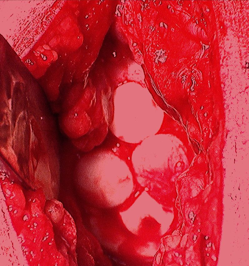
bone with overlying articular cartilage
articular cartilage.
(Figure 3) from areas of relatively no
The articular cartilage is a layer of weight bearing (Figure 2) to weight
hyaline cartilage that covers the end bearing areas of the knee which have
of bones that articulate with other articular cartilage loss.2 An allograft
bones. In the knee you have articular (cadaver) plug is also an option that
cartilage on the end of the femur can be used to fill the lesion. The size
(femoral condyles), the top of the of the harvested plug is sized to match
tibia (tibial plateau) and the back of that of the injury/lesion. These plugs
the knee cap (patella). The articular are then press fit into holes created at
cartilage has a frictional coefficient the lesion. This can be done with a
approximately one fifth of ice on ice single large plug (Figure 4) or several Figure 2 Donor site from area of relatively no weight
(i.e. rubbing articular cartilage on smaller plugs (Figure 5). Initially bearing
articular cartilage would be five times these plugs can be susceptible to
smoother than rubbing ice on ice.) getting pushed in further, thus weight
This allows for a very smooth gliding bearing is restricted for the first six
surface. A large portion of articular weeks to ensure that the cartilage
cartilage is fluid, which provides plug heals “flush” with the rest of the
significant resistance to compressive cartilage surface.2
forces.1 The OATS procedure is currently
During athletic trauma or injury, the only procedure that restores the
focal areas of the articular cartilage normal hyaline articular cartilage
can be damaged or torn, exposing the to the injured knee. Microfracture Figure 3 A harvest bone plug with overlying articular
cartilage (removed from donor site, Figure 2)
6 2 1 S c i e n c e D r i v e • M a d i s o n , W I 5 3 7 1 1 • u ws p o r t sm e d i c i n e . o r g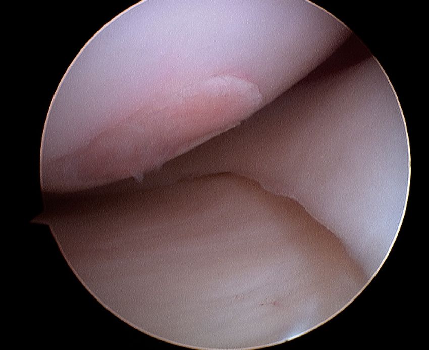
Rehabilitation Guidelines for Osteochondral Allograft or Autograft Transplantation (OATS)

and chondroplasty procedures attempt                  Initially post-operative rehabilitation           time frames are also given for reference
to fill in the chondral defects with                  will focus on regaining range of motion           to the average, but individual patients
fibrocartilage. Research has shown that               and protecting the healing plugs. As the          will progress at different rates depending
fibrocartilage is more likely to deteriorate          rehabilitation progresses the focus shifts        on the size and location of the chondral
over time, and that the chance of                     to regaining strength and movement                lesion, their age, associated injuries, pre-
returning to sports is greater with the               control. Developing the muscular ability          injury health status, and rehabilitation
OATS procedure. A study by Gudas et                   to reduce force will help decrease stress to      compliance. Specific attention must
al3 found that 93% of patients who had                the articular surfaces. In the final phase        be given to impairments that caused
an OATS procedure were able to return                 of rehabilitation the athlete will work           the initial problem. For example
to their pre-injury level of sports versus            on regaining movement control with                if the patient is status post medial
52% who underwent microfracture. The                  change of direction activities, such as           compartment OATS procedure and they
ability to return to sport is also dependent          cutting and pivoting. This is imperative          have a varus alignment, post-operative
on the size of the lesion (or degree of               to prevent increase shear stresses on the         rehabilitation should include correcting
injury), patient age, patient size (BMI),             articular cartilage. The rehabilitation           muscle imbalances or postures that
associated injuries and length of time                guidelines are presented below in a               create medial compartment stress.
that the injury has been present. For                 criterion based progression. Specific time
some patients the goal will be to return              frames, restrictions and precautions are
to daily activities without pain, for others          given to protect healing tissues and the
it may be returning to sports.                        surgical repair/reconstruction. General

Figure 4 A large single plug press fit into a hole created at the site of
the lesion

                                                                              Figure 5 Several smaller plugs press fit into a hole created at the site of
                                                                              the lesion

2                    6 2 1 S c i e n c e D r i v e • M a d i s o n , W I 5 3 7 1 1 • u ws p o r t sm e d i c i n e . o r g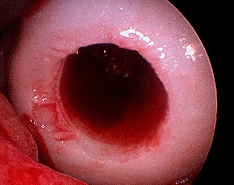
Rehabilitation Guidelines for Osteochondral Allograft or Autograft Transplantation (OATS)

    PHASE I (Surgery to 6 weeks after surgery)

    Appointments                            • Rehabilitation appointments begin within 3-5 days after surgery and meet
                                              about once per week

    Rehabilitation Goals                    •   Protection of the post-surgical knee
                                            •   Restore normal knee range of motion and patellar mobility
                                            •   Eliminate effusion
                                            •   Restore leg control

    Weight Bearing                          • Week 1-3 = non-weight bearing
                                            • Week 4-6 = touchdown to 25% weight bearing
                                            • 0-6 weeks =  locked extension lock splint brace

    Range of Motion Exercises                • Full knee extension
                                               -- Knee extension on a bolster
                                               -- Prone hangs
                                             • Passive Knee Flexion
                                               -- Supine wall slides
                                               -- Assisted heel slides
                                               -- Continuous passive motion machine
                                             • Week 1-2 = 0-90°
                                             • Week 3-4 = 0-110°
                                             • Week 5-6 = 0-125°
                                             • Biking (week 4)– use contra-lateral leg to create ipsalateral passive range of
                                               motion
                                          NOTE: range of motion exercises should be carried out frequently throughout
                                          the day with high repetitions to help remodel and contour the healing cartilage.
                                          The optimal goal during the first 6 weeks is to do 4-6 hours of range of motion
                                          exercises per day.

    Suggested Therapeutic                   •   Quadriceps sets
    Exercise                                •   Straight leg raises
                                            •   Four way leg lifts in standing with brace on for balance and hip strength
                                            •   Patellar mobilizations
                                            •   Soft tissue mobilization

    Cardiovascular Exercise                 • Upper body circuit training or upper body ergometer

    Progression Criteria                    • Patients may progress to Phase II if they are 6 weeks post-operative, have met
                                              the above stated goals, have trace to no effusion and full knee extension

3                6 2 1 S c i e n c e D r i v e • M a d i s o n , W I 5 3 7 1 1 • u ws p o r t sm e d i c i n e . o r g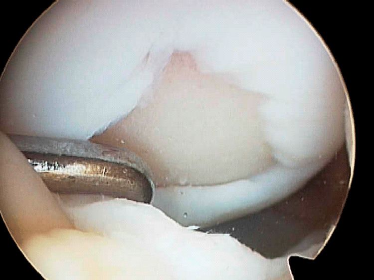
Rehabilitation Guidelines for Osteochondral Allograft or Autograft Transplantation (OATS)

    PHASE II (begin after meeting Phase I criteria, usually 7 to 12 weeks after surgery)

    Appointments                             • Rehabilitation appointments are once a week

    Rehabilitation Goals                     • Single leg stand control
                                             • Normalize gait
                                             • Good control and no pain with functional movements, including step up/
                                               down, squat, partial lunge (staying less than 60° of knee flexion and avoiding
                                               excessive weight bearing at position of the lesion)

    Precautions                              • Avoid post-activity swelling
                                             • Avoid loading knee a deep flexion angles
                                             • No impact activities until 12 weeks after surgery

    Weight Bearing                           • Begin progressive weight bearing as tolerated with axillary crutches and no
                                               brace

    Suggested Therapeutic                    • Weight shifting
    Exercise                                 • Begin pool program – gait drills and initiation of protected weight bearing
                                               strengthening exercises
                                             • Double leg balance and proprioceptive drills
                                             • Stationary bike
                                             • Gait drills (start with pool)
                                             • Protected weight bearing hip and core strengthening
                                             • Stretching for patient specific muscle imbalances
                                             • Quadriceps strengthening – closed chain exercises short of 60° knee flex

    Cardiovascular Exercise                  • Non-impact endurance training, swimming (stiff knee flutter kick), deep water
                                               run, upper body circuits

    Progression Criteria                     • Patients may progress to Phase II if they have
                                               -- Normal gait on level surfaces
                                               -- Full range of motion
                                               -- No effusion
                                               -- Ability to carry out functional movements without unloading affected leg or
                                                  pain, while demonstrating good control
                                               -- Single leg balance greater than 15 seconds

4                 6 2 1 S c i e n c e D r i v e • M a d i s o n , W I 5 3 7 1 1 • u ws p o r t sm e d i c i n e . o r g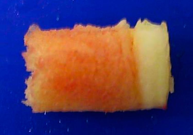
Rehabilitation Guidelines for Osteochondral Allograft or Autograft Transplantation (OATS)

    PHASE III (begin after meeting Phase II criteria, usually about 4 months)

    Appointments                             • Rehabilitation appointments 1 time every 1-2 weeks

    Rehabilitation Goals                     • Good control and no pain with sport and work specific movements, including
                                               impact

    Precautions                              • Post-activity soreness should resolve within 24 hours
                                             • Avoid post-activity swelling
                                             • Avoid knee pain with strengthening

    Suggested Therapeutic                    • Functional leg strengthening
    Exercise                                   -- Squats
                                               -- Lunges – all three planes
                                               -- Step backs
                                               -- Retro step ups
                                               -- Single leg leg press
                                             • Single leg balance and proprioception progression
                                               -- Hip and core strengthening
                                               -- Mini band drills
                                               -- Physioball
                                             • Stretching for patient specific muscle imbalances

    Cardiovascular Exercise                  • Non-impact activities; stationary bike, elliptical, Nordic track, swimming

    Return to Sport/Work Criteria            • Dynamic neuromuscular control with multi-plane activities, without pain or
                                               swelling

5                 6 2 1 S c i e n c e D r i v e • M a d i s o n , W I 5 3 7 1 1 • u ws p o r t sm e d i c i n e . o r g
Rehabilitation Guidelines for Osteochondral Allograft or Autograft Transplantation (OATS)
PHASE IV (begin after meeting Phase III criteria, usually about 6 months after surgery)
Appointments • Rehabilitation appointments 1 time every 1-2 weeks
Rehabilitation Goals • Good control and no pain with sport and work specific movements, including
impact
Precautions • Post-activity soreness should resolve within 24 hours
• Avoid post-activity swelling
• Avoid knee pain with impact
Suggested Therapeutic • Impact control exercises beginning 2 feet to 2 feet, progressing from 1 foot to
Exercise other and then 1 foot to same foot
• Movement control exercise beginning with low velocity, single plane activities
and progressing to higher velocity, multi-plane activities
• Sport/work specific balance and proprioceptive drills
• Hip and core strengthening
• Stretching for patient specific muscle imbalances
Cardiovascular Exercise • Replicate sport or work specific energy demands
Return To Moderate Impact • 8 months post surgery; and
Sport Criteria • Good dynamic neuromuscular control with multi-plane activities, without pain
or swelling
(Jogging, Aerobics)
Return To High Impact Sport • 10 months post surgery; and
Criteria • Good dynamic neuromuscular control with multi-plane activities, without pain
or swelling
(Basketball, Soccer)
These rehabilitation guidelines were developed collaboratively between Marc Sherry, PT, LAT, CSCS
(msherry@uwhealth.org) and Geoffrey Baer, MD.
Updated 03/2011
REFERENCES
1. Pearle AD, Warren RF, Rodeo SA. Basic 2. Reinold MM, Wilk KE, Macrina LC, 3. Gudas R, Kalesinskas RJ, Kimtys V, et al.
science of articular cartilage and Dugas JR, Cain EL. Current concepts A prospective randomized clinical study
osteoarthritis. Clin Sports Med. Jan in the rehabilitation following articular of mosaic osteochondral autologous
2005;24(1):1-12. cartilage repair procedures in the transplantation versus microfracture for
knee. J Orthop Sports Phys Ther. Oct the treatment of osteochondral defects
2006;36(10):774-794. in the knee joint in young athletes.
Arthroscopy. Sep 2005;21(9):1066-1075.
At UW Health, patients may have advanced diagnostic and /or treatment options, or may receive educational materials that vary from this information. Please be aware that this information is not intended to replace
the care or advice given by your physician or health care provider. It is neither intended nor implied to be a substitute for professional advice. Call your health provider immediately if you think you may have a medical
emergency. Always seek the advice of your physician or other qualified health provider prior to starting any new treatment or with any question you may have regarding a medical condition.
SM-27330-11
Copyright 2011 UW Health Sports Medicine Center
6 6 2 1 S c i e n c e D r i v e • M a d i s o n , W I 5 3 7 1 1 • u ws p o r t sm e d i c i n e . o r gYou can also read