Respiratory system Microbiology laboratory section - Microbiology Lab Edited Slides Done by: Dena Kofahi - Doctor 2018
←
→
Page content transcription
If your browser does not render page correctly, please read the page content below
Microbiology Lab Edited Slides
Done by: Dena Kofahi

                  Respiratory system

              Microbiology laboratory section

              In this lecture, we will be taking the most common
            microorganisms (bacteria and fungi) that may affect the
                                respiratory system.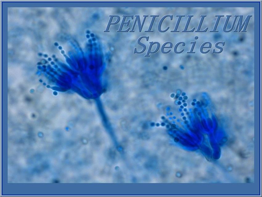
A throat swab culture is a
                Gram Positive                      test commonly used to
                                                   diagnose bacterial

                   Coccus                          infections in the throat.

       These are the common gram positive bacteria that may
     cause throat infection. We will see how we can differentiate
                        between the species.

Staphylococcus                              Streptococcus
     Spp.                                        Spp.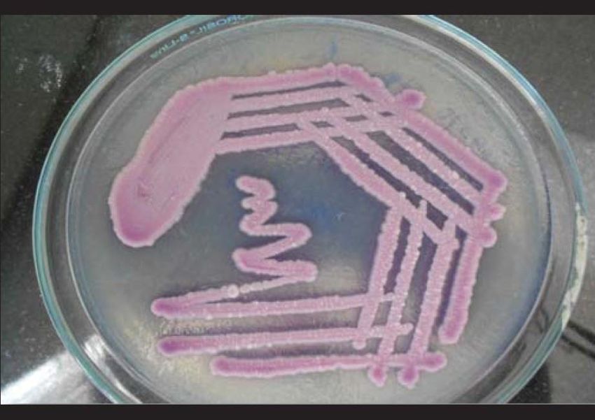
Staphylococcus
• Arranged in grape like clusters.
• Includes at least 40 species. The most common species associated
  with clinical infection are Staph aureus, Staph epidermidis, Staph
  hemolyticus, Staph hominins, and Staph albus.

Streptococcus            Gram Positive
• Arranged in chains or diplococci.
                            Coccus

       Staphylococcus                         Streptococcus
            Spp.                                   Spp.
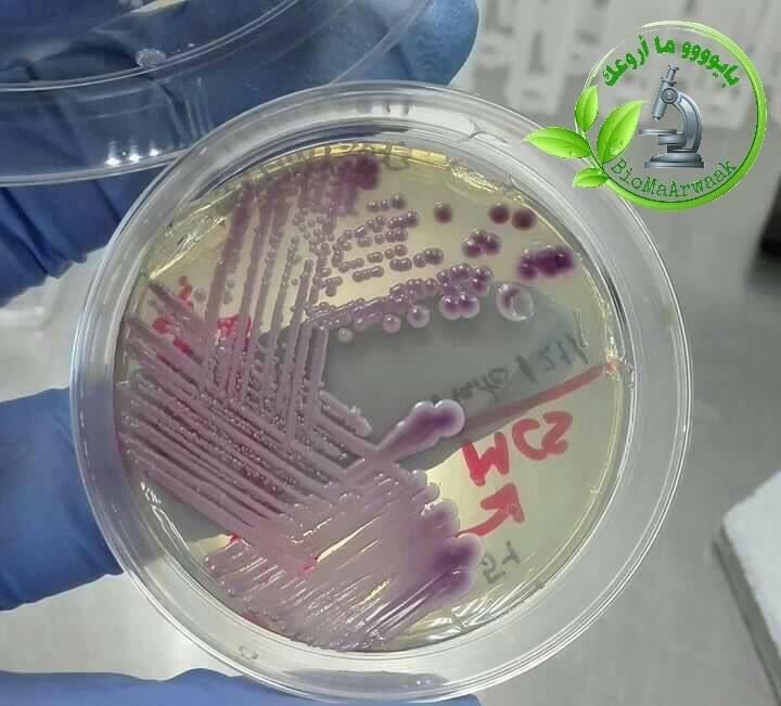
Other staph species such as
staph albus or epidermidis
appear as white colonies on
blood agar.

A- staphylococcus .albus

B- staphylococcus.Aureus

Staph aureus colonies appear yellow-golden and often present with
hemolysis when grown on blood agar plates.               Blood agar

The golden appearance is the etymological root (origin) of the bacteria’s
name “aureus,” as the word means golden in Latin.
Test for differentiation of
                  Staphylococcus species
The major test reaction that should be
used to differentiate between
staphylococcus and streptococcus species
is the catalase test.

To differentiate between staph aureus
and other staph species, the mannitol
salt agar (MSA) and coagulate tests
should be used.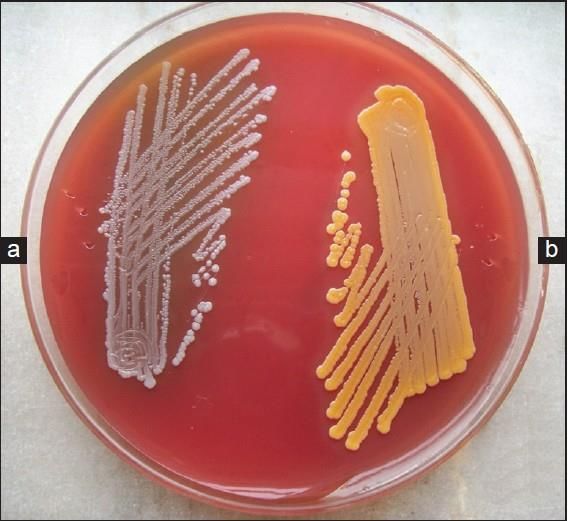
The catalase test is used to
                identify organisms that

Catalase test   produce the enzyme catalase.
                This enzyme detoxifies
                hydrogen peroxide by
                breaking it down into water
                and oxygen gas. Bubbles
                resulting from production of
                oxygen gas clearly indicate a
                catalase positive result.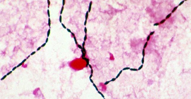
The MSA is a selective
and differential                      MSA
medium. Its high
concentration of salt
(7.5%) selects for
                             Mannitol salt agar media
members of the genus
Staphylococcus since
they can tolerate high                                    l
                                                     nito
saline levels.                                    Man
                                        e   nts
                                   Ferm
Organisms from other
genera may grow but
they grow weakly.

                                            Doe
                                                  s no
                                                       t
                                                  Man fermen
                                                      nito   t
                                                           l

 MSA also contains the sugar mannitol and the pH indicator
phenol red. If an organism can ferment mannitol, an acidic
byproduct is formed that will cause the phenol red in the agar
to turn yellow. Staph aureus can ferment mannitol so its media
will turn yellow while other Staph species will not ferment
mannitol and it will remain red in color.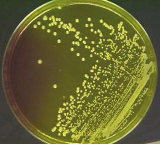
The coagulase test is used to                                 A suspension of the
differentiate staph aureus (+)                                organism is suspended
from other staph species (-).                                 and incubated in
Coagulase is an enzyme                                        plasma at 37C in a
produced by staph aureus to                                   tube. Clot formation
convert soluble fibrinogen in                                 within four hours
plasma to insoluble fibrin.                                   indicates a positive
                                                              test, or the presence of
                                                              staph aureus. Negative
                                                              tubes should be left at
                                                              room temperature
                                                              overnight and re-
                                                              examined the next day.
                                                              This step is essential
                                                              for some strains of
                                                              staph aureus, including
                                 S. Epidermidis   S. Aureus   MRSA strains, as they
                                                              produce a delayed clot
                                                              which is rapidly lysed at
                                                              37C by staphylokinase

                                                                 S.aureus

                                                                  S.albus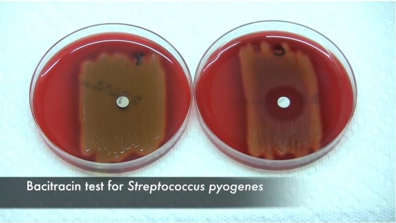
Streptococci are gram positive aerobic organisms that cause many disorders including pharyngitis, pneumonia,
   skin infections, sepsis, and endocarditis. Three different types of strep are initially differentiated by their
                              appearance when they are grown on sheep blood agar.
Streptococcus viridians can be differentiated from S. pneumoniae using an optochin test. Viridans streptococci are optochin resistant; they also lack either the polysaccharide-based capsule typical of S. pneumoniae or the Lancefield antigens of the pyogenic members of the genus.
A zone of inhibition appears on
a strep pneumoniae culture as
it is sensitive to optochin.
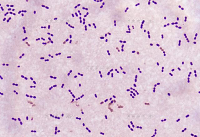
Streptococcus pneumoniae
Strep pneumoniae are gram positive, lancet shaped elongated cocci with a slightly pointed outer
  curvature. Usually they are seen in pairs (diploccoi) but they can be singular or in short chains.
Streptococcus viridans
Strep viridans are gram positive (often elongated) cocci that form short to long chains.
The bacitracin sensitive test is used to distinguish group A S. pyogenes from
other streptococci such as group B S. agalactiae.

When grown on blood agar, S. pyogenes is sensitive to bacitracin and will
exhibit a zone of inhibition. While S agalactiae will not be affected and will
not have a zone of inhibition.
Strep agalactiae            Strep pyogenes
  There is no zone of       We can see the zone
   inhibition as it is     inhibition indicating its
resistant to bacitracin.    bacitracin sensitivity.
Gamma hemolysis
                (Non-hemolytic)
            streptococcus

Enterococcus
                                  Other than
  Group D                         Enterococcus
 - E.feacalis                       group D
• The bile-esculin test is used to differentiate between enterococcus group D and non-enterococcus species. • Enterococcus group D species give a positive test. • The bile-esculent test is based on the hydrolysis of esculin into glucose and esculetin by microorganisms that can produce esculinase. • Esculetin then can interact with an iron salt, ferric citrate, in the medium to form a phenolic iron complex, which produces a dark brown or black color.
Diptheroids are aerobic non-sporulating
pleomorphic gram postive bacilli which are more
uniformly stained than Corynebacterium diptheria.   Diphtheroids
     Gram Positive
     Cocco-bacilli
They lack metachromatic granules and are
arranged as in what is known as the ‘Chinese
letter’ appearance. They are usually
commensals in the skin and mucous
membranes.

    Arrangement as
     Chinese letter
Candida Species
Candidiasis is a fungal
infection caused by a yeast
known as Candida. Some
species of Candida can cause
infections in humans, most
C.albicans
commonly C. albicans.
Candida
C.Krusei Spp. C.tropicalis
Candida normally lives on the
skin and inside the body in
At least 30 Candida
places such as the mouth, throat,
species have been
gut, and vagina, without causing
implicated as causes
any problems. In some
circumstances, such as when
taking a long course of Abx or a C.glabrata of human infection.
The most common
species are those
weakened immune system, the
shown in the figure.
risk of Candida infection
increases.Candida Spp • Larger than Bacteria • Budding • Candida are unicellular fungi. • They may be spherical, elliptical or cylindrical shaped. • Their size varies greatly but they are generally larger than bacteria. • They typically grow asexually by budding.
sabouraud dextrose agar

This agar contains peptones. It is used to cultivate dermatophytes and
other fungi (such as Candida) at 20C. It can also grow filamentous
bacteria such as Nocardia. The pH of the media is adjusted to
approximately 5.6 in order to enhance the growth of fungi, especially
dermatophytes, and to slightly inhibit bacterial growth. Yeast will grow
as creamy white colonies while molds will grow as filamentous colonies
To Differentiate between C.albican and
               other Species

The germ tube test is a
screening test used to
differentiate C. albicans from
other yeasts. When Candida is
grown in human or sheep serum
at 37C for three hours, it forms
germ tubes. These germ tubes
can be detected with wet KOH
films as filamentous outgrowth
                                    Germ tube
extending from yeast cells. In
                                [ Serum + candida ]
this case, the sample is positive
for C. Albicans.
Chrom agar is a novel differential
culture medium that facilitates the
isolation and identification of some
clinically important yeast species. Chrom agar
C.glabrata : violet (dark pink)
glistering C.albicans : GreenChrom agar
C. Krusei isolates forms highly characteristic rough,
spreading colonies with pale pink centers and white edges.
C.Krusei : rough C.Tropicalis
dry pale pink BlueAspergillus Niger Aspergillus niger is a fungus and one of the most common species of Aspergillus. It causes a disease called black mold on certain fruits and vegetables such as grapes, apricots, onions, and peanuts. It is a common contaminant of food. Aspergillus niger is one of the most common causes of otomycosis (fungal ear infection) which can cause pain or temporary hearing loss. In severe cases, it may damage the ear canal and tympanic membrane.
Penicillium Spp.

Penicillium is a genus of ascomycetous fungi that is of major importance in the natural environment, in food
spoilage, and in food and drug production. Some members of the genus produce penicillin, a molecule that is
used as an antibiotic, which kills or stops the growth of certain kinds of bacteria. Penicillium species are
occasional causes of infection in humans and the resultant disease is known generally as penicilliosis. Penicilliums
have been isolated from patients with keratitis, endophtalmitis, otomycosis, necrotizing esophagitis,
pneumonia, endocarditis, peritonitis and urinary tract infections.
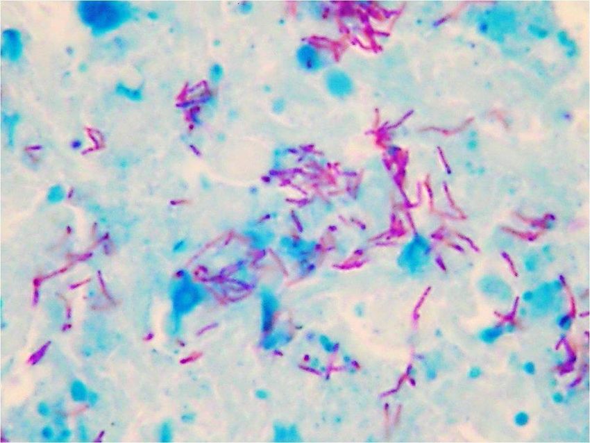
Mycobacterium tuberculosis is a
species of pathogenic bacteria in
the family Mycobacteriacae and is
the causative agent of Tuberculosis.
It has an unusual waxy coating on
its cell surface primarily due to the
presence of mycolic acid. This
coating makes the cell impervious
to gram staining, and as a result M.
Tuberculosis can appear as either
gram negative or gram positive.
So, acid fast stains, such as the
Ziehl-Nielsen stain, are used to
identify it with a microscope.
Lowenstein –Jensen Medium
• Contain malachite
  green and egg albumin
• Media color : green
• Cell show :

 Rough    Tough             Buff

             The LJ medium is a growth medium specially used for the culture of
             mycobacterium species, especially M. Tuberculosis. When grown on LJ medium,
             M. Tuberculosis appears as brown granular colonies sometimes called buffs
             rough, and tough colonies. The medium must be incubated for a significant
             length of time, usually 4 weeks, due to the slow proliferation time of this
             bacteria. The medium appears green, opaque and opalescent. The medium
             consists of malachite green, glycerol (which enhances the growth of M.
             tuberculosis), asparagine, potato starch, coagulated eggs, mineral salt solution,
             potassium dihydrogen phosphate, and magnesium sulfate.
Incubation Period = 4 weeks
Glass
Put the media in covered tubes
to avoid drying of media
It also helps to avoid contamination.ziehl neelsen
Acid fast stain
• Mycobacterium Tuberculosis cell wall are waxed
for that reason do heating while staining .
• Stain made of :
• Carbol fuchsin Primary stain
• Hydrochloric acid alcohol ( 3% HCL )
Works as a de-colorizer
• Methylen blue Counter stainWhen the smear is stained with carbol fuschin, it solubilizes the lipoid always material present in the mycobacterial cell wall. With the application of heat, carbon fuschin further penetrates through the lipoidal wall and enters into the cytoplasm. All cells now appear red. The smear is then decolorized with a decolorizing agent (3% HCl in 95% alcohol) but the acid fast cells are resistant due to the presence of large amounts of lipoidal material in their cell walls which prevents the penetration of decolorizing solution. Non-acid fast organisms lack the lipoidal material in their cell wall and are therefore easily decolorized, leaving colorless cells. Then, the smear is stained with a counter stain, methylene blue. Only decolorized cells absorb the counter stain and take its color and appear blue while acid-fast retain the red color.
T.B
Here, the M, tuberculosis
cells appear red/pink in
color while other cells in

                             Acid fast stain
the background appear
blue.
You can also read