Retrospective analysis of canine monocytic ehrlichiosis in Thailand with emphasis on hematological and ultrasonographic changes
←
→
Page content transcription
If your browser does not render page correctly, please read the page content below

Veterinary World, EISSN: 2231-0916                                                                    RESEARCH ARTICLE
Available at www.veterinaryworld.org/Vol.15/January-2022/1.pdf                                              Open Access

Retrospective analysis of canine monocytic ehrlichiosis in Thailand with
      emphasis on hematological and ultrasonographic changes
            Kris Angkanaporn1     , Jidapha Sanguanwai2, Taratip O. Baiyokvichit2, Pichamon Vorrachotvarittorn2,
                                   Montana Wongsompong2 and Woraporn Sukhumavasi3

   1. Department of Veterinary Physiology, Faculty of Veterinary Science, Chulalongkorn University, Bangkok 10330,
  Thailand; 2. Faculty of Veterinary Science, Chulalongkorn University, Bangkok 10330, Thailand; 3. Parasitology Unit,
 Department of Veterinary Pathology, Faculty of Veterinary Science, Chulalongkorn University, Bangkok 10330, Thailand.
                          Corresponding author: Kris Angkanaporn, e-mail: akris@chula.ac.th
    Co-authors: JS: palm.jidapa@gmail.com, TOB: closefriend_nb@hotmail.com, PV: mp_mint1112@hotmail.com,
                                MW: hammie00@hotmail.com, WS: vetkwan@hotmail.com
                    Received: 30-07-2021, Accepted: 26-11-2021, Published online: 05-01-2022

doi: www.doi.org/10.14202/vetworld.2022.1-9 How to cite this article: Angkanaporn K, Sanguanwai J, Baiyokvichit TO,
Vorrachotvarittorn P, Wongsompong M, Sukhumavasi W (2022) Retrospective analysis of canine monocytic ehrlichiosis in
Thailand with emphasis on hematological and ultrasonographic changes, Veterinary World, 15(1): 1-9.

                                                            Abstract
Background and Aim: Canine monocytic ehrlichiosis (CME) is a tropical endemic tick-borne disease that causes fatality or
chronic infection involving many organs in dogs. This study aimed to examine the prevalence, risk factors, and hematological
and ultrasonographic changes in the liver, gallbladder, kidneys, and spleen following CME infection.
Materials and Methods: This retrospective study used 30,269 samples collected from dogs at the hematology section of
the pathology unit of a university veterinary hospital and 35 samples collected from dogs at the diagnostic imaging unit.
CME was determined using the buffy coat smear method. Data were analyzed using descriptive statistics and odds ratios.
Results: The data revealed that the average yearly prevalence of CME was 1.32%. Risk factors contributing to CME
infection were a tick on the body during physical examination, lack of ectoparasite control, and outdoor living. All 148
dogs with CME infection had low platelet counts. The percentages of CME-infected dogs with elevated serum alanine
aminotransferase, alkaline phosphatase, and both enzymes above the normal range were 33.6%, 65.9%, and 29.8%,
respectively. The rates for elevated serum levels of blood urea nitrogen, creatinine, and both compounds were 33.1%,
19.1%, and 17.3%, respectively. The most common ultrasonographic changes were liver abnormalities (hyperechogenicity
or hypoechogenicity, hepatomegaly, and hypoechoic nodules), hyperechogenicity of the kidneys, and an enlarged spleen.
These ultrasonographic changes were consistent with the hematology results, which showed a greater elevation of serum
liver enzyme levels than renal enzymes.
Conclusion: Ultrasonographic changes during CME infection and after treatment with doxycycline can help to monitor and
identify persistent pathological changes in the target organs resulting from immune response to CME.
Keywords: dog, ehrlichiosis, hematology, monocyte, ultrasound.
Introduction                                                       4-7 days after infection (dai), the dog’s immune sys-
     Canine monocytic ehrlichiosis (CME) is a tick-                tem develops immunoglobulin M and immunoglob-
borne, endemic disease found worldwide that causes                 ulin A antisera, and immunoglobulin G antisera can
animals in the Canidae family to become sick and                   be detected at 15 dai [14]. Following the 8-20 days
die. The disease is caused by rickettsial bacteria,                incubation period, CME infection progresses through
namely, Ehrlichia canis, transmitted by Rhipicephalus              three typical phases; acute, subclinical, and chronic. In
sanguineus (brown dog tick). CME is widely distrib-                the acute phase, which lasts for 3-5 weeks, the clinical
uted in tropical, Mediterranean, and subtropical cli-              symptoms of fever, anorexia, ocular discharge, muco-
mates, including Europe [1,2], the United States [3],              sal and skin petechiae, epistaxis, pale mucous mem-
Costa Rica [4], Brazil [5], and Asia [6-8]. In Thailand,           brane, hemorrhagic tendencies, depression, lymph-
the reported prevalence of E. canis identified using               adenopathy, and neurological signs (from meningitis)
polymerase chain reaction (PCR) in all parts of                    are present [15]. The major hematological changes
the country ranges from 7.6% to 38.3% [9-13].                      are interstitial nephritis and glomerulonephritis [16],
Transstadial transmission occurs in all tick stages, and           whereas pathological changes occur in the cortico-
infection can result while feeding on infected dogs. On            medullary junction, causing a contracted kidney [17].
                                                                   Hyperechogenicity may be present with an enlarged
Copyright: Angkanaporn, et al. Open Access. This article is
distributed under the terms of the Creative Commons Attribution
                                                                   liver, spleen, gallbladder, and ascites [18]. Some dogs
4.0 International License (http://creativecommons.org/licenses/    may recover after the subclinical phase, whereas oth-
by/4.0/), which permits unrestricted use, distribution, and        ers may progress to the chronic phase where severe
reproduction in any medium, provided you give appropriate credit
to the original author(s) and the source, provide a link to the    pancytopenia typically occurs from bone marrow
Creative Commons license, and indicate if changes were made.       hypoplasia and leads to severe leukopenia, anemia,
The Creative Commons Public Domain Dedication waiver (http://
creativecommons.org/publicdomain/zero/1.0/) applies to the data
                                                                   and thrombocytopenia with a high risk of mortal-
made available in this article, unless otherwise stated.           ity [15]. In severe cases, dogs with poor antibiotic
Veterinary World, EISSN: 2231-0916                                                                                       1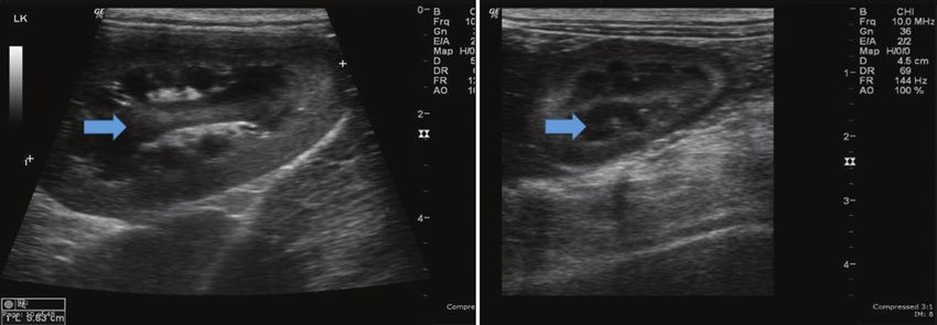
Available at www.veterinaryworld.org/Vol.15/January-2022/1.pdf

response may die from massive hemorrhage, severe             well as the factors involved as in part 1. In part 2, the
debilitation, and/or secondary infection [15]. During        retrospective case-control comparison of dogs that
the chronic phase, pathological lesions occur in the         had data on ultrasonographical and blood analysis was
kidney because of immune complex accumulation                examined.
in the glomerulus that stimulates inflammation, fol-
                                                             Study part 1
lowed by the destruction of cells and tissues in the sur-
rounding area, leading to elevated serum blood urea                We identified a group of CME-positive dogs,
nitrogen (BUN) and creatinine levels. There are also         defined by the presence of the morulae of Ehrlichia
lymphocyte and plasma cell infiltration into the liver       spp. in the buffy coat smear assay and results of the
and kidney parenchyma [15], and moderate increases           Canine SNAP® 4Dx® test kit (IDEXX Laboratories,
in serum levels of the liver alanine aminotransferase        Inc., Westbrook, ME, USA). The prevalence of CME
(ALT) and alkaline phosphatase (ALP) due to hepato-          during the study period was determined. To under-
cyte damage [15,19].                                         stand factors influencing CME risk, we analyzed the
      With the standard doxycycline protocol treat-          following data: Signalment data; historical records;
ment [14], some dogs may not fully recuperate from           complete blood count (CBC) data, including platelet
symptoms related to the immune response, especially          count; and blood chemistry data, including serum lev-
damage to the principal organs involved (liver, kidney,      els of ALT, ALP, BUN, and creatinine. Duplicate data
and spleen). These lasting effects may be missed by          were removed before analysis.
veterinarians that do not provide systematic follow-up             Next, based on the serum platelet count and
after doxycycline treatment. Sarma et al. [18] studied       blood chemistry data (ALT, ALP, BUN, and cre-
pathological changes in the liver and spleen of 101 dogs     atinine), the dogs that were E. canis positive were
positive for infection with tick-borne blood parasites       grouped as below, within, or above the normal range
and found that ultrasound and hematological changes          for these measures. Moreover, the dogs that were
can serve as a useful indicator of the damage status of      E. canis positive were analyzed for (i) the presence
internal organs after infestation with blood parasites.      of ticks on the body during physical examination,
      Although there are several reports [9-12] on           (ii) use of an ectoparasite control program, and (iii)
the prevalence of CME in Thailand, there is scarce           daily indoor or outdoor living. Ectoparasite control
research on the relationship between CME and                 was defined as consistent and routine control using
changes in ultrasound images of dogs during or after         approved products. For daily indoor or outdoor liv-
treatment. Thus, the present study aimed to investi-         ing, only dogs that spent 100% of their time indoors
gate the retrospective prevalence of CME in dogs             were considered indoor living dogs. For comparison,
and examine changes in blood parameters and organs           healthy dogs were randomly chosen from the histori-
(liver and kidney) of infected dogs as revealed by           cal data to serve as a control group. The inclusion cri-
ultrasound images.                                           teria were dogs with no severe diseases or CME. The
                                                             numbers of control dogs were similar to those with
Materials and Methods                                        CME (150, 57, and 40 dogs for small, medium, and
Ethical approval and informed consent                        large breeds, respectively) (Table-1). The odds ratio
      Because of the retrospective nature of this study      (OR) was computed for comparisons between CME
and the use of diagnostic data collected as a part of rou-   and control (healthy) groups.
tine clinical procedures, the need for ethical approval      Study part 2
was waived. All dog owners completed a consent
form giving permission to utilize the data (including              From the data, dogs were selected using
ultrasound images) for clinical research.                    non-probability or non-random sample selection. The
                                                             inclusion criteria did not restrict the gender or breed.
Study period and location
                                                             Nevertheless, the dogs must not be older than seniors
     This study was divided into two parts. part 1 was       because geriatric dogs may show age-related patho-
performed at the Small Animal Teaching Hospital,             physiological changes in the ultrasound appearance of
Faculty of Veterinary Science, Chulalongkorn                 the liver or kidneys unrelated to E. canis infection. We
University from September 2016 to August 2017.               also excluded dogs with a history of severe diseases,
Part 2 of this study was performed on dogs that              including heart, liver, kidney, cancer, and immune
were admitted and underwent examinations at the              system diseases.
Hematology Section, Pathology Unit and Imaging                     Data were divided into a control group and a
Diagnostic Unit of the same Small Animal Hospital            study group. The control group comprised 16 dogs
from January 2017 to September 2018.                         with normal abdominal ultrasound results for their
Study design and analysis                                    internal organs. The study group comprised 19 dogs
     A retrospective, randomized study was per-              positive for E. canis infection that showed abnormal
formed based on hematological and medical records.           abdominal ultrasound results in at least one of the
We divided the study into two parts in order to study        periods before, during, or after infection (treatment
the prevalence of CME based on the yearly data as            with doxycycline at 10 mg/kg/day for 28 days).
Veterinary World, EISSN: 2231-0916                                                                                 2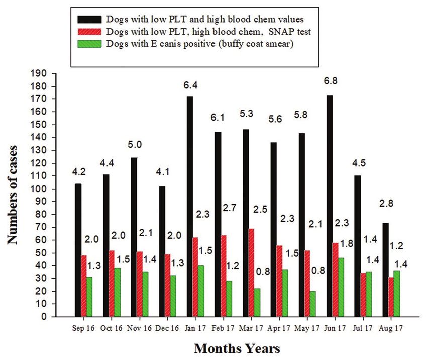
Available at www.veterinaryworld.org/Vol.15/January-2022/1.pdf

Table-1: The OR for E. canis infection (buffy coat smear method) compared to the control group in dogs of different
weight classes (Data from September 2016 to August 2017).

                      Body weight                Tick                Ectoparasite control               Living            n
                         (kg)
                                      Found Not found OR* Control Not control OR* Indoor Outdoor OR*
Control                    25          4         36         ‑        35         5         ‑     24        16       ‑    40
Positive E. canis**        25          10        30        3.0       26         14       3.8    20        20      1.5   40
*Odds ratio, control group compared to the positive group. n=Numbers of dogs in each weight group. The total N for
the positive E. canis group (247 dogs) was less than the data in Tables 1 and 2 due to incomplete history on various
factors examined. The total n for the control group (247 dogs) was randomly selected to match with the positive group
according to weights. **Using the buffy coat smear method. E. canis=Ehrlichia canis

                                                                   Statistical analysis
                                                                          Descriptive statistics were used to analyze and
                                                                   compare all parameters in Parts 1 and 2. The prevalence
                                                                   of E. canis infection was reported as the mean value
                                                                   calculated on a yearly basis. In Part 1, the Odds ratio
                                                                   (OR) was used to measure the association between
                                                                   the control and E. Canis-positive groups in terms of
                                                                   (i) the presence of ticks on the body during physical
                                                                   examination, (ii) ectoparasite control program, and
                                                                   (iii) daily indoor or outdoor living. Statistical analysis
                                                                   was performed using Sigmastat (Systat Software, San
                                                                   Jose, CA, USA). p
Available at www.veterinaryworld.org/Vol.15/January-2022/1.pdf
a b
Figure-2: Hyperechoic parenchyma of the liver compared with the spleen in the (a) Ehrlichia canis-infected and (b) control
groups. The arrow indicates the difference between infected and control dogs.
Table-2: Percentage of dogs that found Ehrlichia canis levels remained elevated, albeit at a lower magnitude,
by buffy coat smear method with platelets counts and
blood chemistry was separated into three groups; below,
after treatment with doxycycline in 7/11 (63.6%)
within, and above from normal range of platelet counts, cases (422±326 IU/L), whereas 4/11 (36.4%) cases
blood chemistry record including SNAP4DX tested from had reverted to normal levels of ALT (Table-4).
September 2016 to August 2017. Similarly, an increased serum ALP level was observed
Parameter Normal range1 Below Within Above in the presence of E. canis in 17/19 (89.5%) cases
(%) (%) (%) (698±449 IU/L). The levels remained elevated in
Platelets (211,000‑600,000) 100 0 0 9/11 (81.2%) cases after treatment with doxycycline,
ALT (10‑109) 0.3 66.4 33.6 but at an even higher extent (948±968 IU/L), with
ALP (1‑114) 0 31.1 65.9 only 2/11 (18.2%) cases returning to within the nor-
ALT and − 0 29.8 29.8
ALP
mal range (Table-4). Hence, the serum levels of ALP
BUN (8‑28) 1.1 65.9 33.0 were in accordance with those for ALT and consistent
Creatinine (0.5‑1.7) 3.0 77.2 19.8 with liver damage.
BUN and − 0 58.5 17.3 Kidney markers (BUN and creatinine)
creatinine
In the presence of detectable E. canis, increased
SNAP4DX positive number 415/626 (66.3%)
tested/total number (percentage) serum levels of BUN (52±12 mg%) were evident in
Reference normal range [20]. E. canis=Ehrlichia canis
1 5/18 (27.8%) cases. Although normal serum levels
of BUN were observed in 8/11 (72.7%) cases after
Part 2
treatment with doxycycline, 3/11 cases (27.3%) still
Hematological and blood data changes in E. canis showed increased BUN levels (50±5 mg%) (Table-5).
infected dogs Normal serum levels of creatinine were found in most
cases (11/14, 78.6%) before detectable infection but
Data from 19 dogs in the study group were included
increased (1.7±0.3 mg%) in the presence of E. canis in
in this analysis. The age was known in all cases, and the
15/18 (83.3%) cases. After treatment, creatinine lev-
mean group age was 7 years (range of 3 months-11 years).
els were within the normal limits in all cases (11/11,
In terms of sex, 47.4% (9 of 19) were female and 52.6%
100%; Table-5).
(10 of 19) were male. The two groups included both entire
and neutered animals. There were 10, 6, and 3 cases of Effects of E. canis infection on ultrasound appear-
small, medium, and large breeds, respectively. ance of the liver, gallbladder, kidneys, and spleen
Hematology Abdominal ultrasonographic examination results
The CME-positive dogs were analyzed in the phase of the liver, gallbladder, and kidneys in all 16 cases in
before the presence of E. canis (Table-3). The platelet the control group were found to be normal. The liver
concentration data from 15 cases revealed 4 dogs (26.7%) showed a normal sharp border with a smooth margin,
with normal levels and 11 (73.3%) with decreased good location, contours with a homogeneous echotex-
platelet concentrations. In the presence of E. canis, all ture, normal appearance of the intrahepatic portal
19 cases (100%) showed a markedly reduced platelet veins, uniform hypoechoic liver parenchyma related
concentration (38,240±23,369/µL). After treatment with to the spleen, and falciform fat with isoechoic to the
doxycycline, 4/11 dogs (36.4%) with available data still right renal cortex. Additional observations included a
had a decreased platelet level (90,000±66,878/µL). normal gallbladder wall thickness and anechoic bile
content; a normal appearance of both kidneys in terms
Serum chemistry profiles of size, shape, location, contour, and echotexture; nor-
Liver markers (ALT and ALP) mal renal cortex echogenicity; a well-defined corti-
Of the 19 E. canis infected dogs, 13 (68.4%) comedullary junction; and a normal renal pelvis and
had elevated serum ALT levels (647±1083 IU/L). The smooth renal capsule.
Veterinary World, EISSN: 2231-0916 4Available at www.veterinaryworld.org/Vol.15/January-2022/1.pdf
Ultrasonographic changes in the liver in the immunofluorescence assay [14,15]. Nevertheless, the
presence of E. canis were noted in all 13 infected buffy coat smear method remains the most common
cases. Hyperechogenicity of the liver was observed method for screening E. canis infection in clinics
in 7 (53.8%) cases, whereas 4 (30.8%) cases revealed because of its convenience and relatively low cost.
hypoechoic hepatic parenchyma. Hepatomegaly was Nevertheless, there are some limitations of the buffy
observed in 10 cases (76.9%), as shown in Table-6 and coat smear method, which has a sensitivity and speci-
Figure-2. After treatment with doxycycline, 4 (30.8%) ficity of 16.1% (confidence interval [CI]=10.7–23.6%)
and 3 (23.1%) cases still showed hyperechogenicity and 89.4% (CI=85.0–92.6%), respectively, resulting
and hepatomegaly, respectively, of the liver (Table-6). in a high chance of false-negative results but a low rate
Ultrasonographic changes in the gallbladder, kid- of false positives. This could be the reason for the low
neys, and spleen prevalence of E. canis infection in our study. A com-
posite study in India reported that the overall prev-
In the presence of E. canis, 2/13 (15.4%) cases
alence rates of ehrlichiosis by microscopic examina-
showed gallbladder distention. This was still evident
tion, commercial dot-ELISA, and nested PCR assay
in 4/6 (66.7%) cases after treatment with doxycy-
were 1.3%, 19.1%, and 5.8%, respectively [21]. The
cline (Table-6). For the kidneys, hyperechogenicity
rate determined by microscopic examination is similar
was evident in 3/13 (23.1%) cases in the presence of
E. canis and persisted in 2/6 (33.3%) cases after treat- to that reported for Thailand, although the occurrence
ment with doxycycline (Table-6 and Figure-3). of CME using the PCR test was higher than that in
Splenomegaly was found in 10/13 (76.9%) cases India [9-13,22]. The sensitivity of an E. canis test also
infected with E. canis, whereas 8/13 (61.5%) cases depends on the stage of infection at the time of sam-
showed hypoechoic spleens (Table-6). After treatment pling. In the acute phase, there is more opportunity to
with doxycycline, there were still 4/6 (66.7%) and find infected leukocytes in the blood smear because
3/6 (50%) cases of splenomegaly and hypoechogenic- of the higher degree of parasitemia. However, in the
ity, respectively (Table-6). subclinical and chronic phases, the chances of finding
infected leukocytes decrease, which can lead to false
Discussion negatives. However, the probability of E. canis detec-
In Thailand, there is no seasonal difference in the tion by specific antibodies, such as through ELISA,
prevalence of E. canis infections. The yearly preva- increases in the chronic phase because the secondary
lence rate of E. canis was found to be 1.32% in the pres- immune system (and thus immunoglobulin levels)
ent study using the buffy coat smear method. There are requires time to respond [23].
several alternative techniques to diagnose CME apart In this study, all E. Canis-positive dogs showed
from buffy coat smears, such as SNAP4DX, PCR, and a serum platelet count below the normal range. The
Table-3: Serum platelet levels in E. Canis‑infected dogs (buffy coat smear method) at three different infection phases.
Phase Normal platelet count Decreased platelet count No data
Before presence of E. canis 4 (295,000±73,289) 11 (83,818±53,842) 4
During presence of E. canis 0 19 (38,240±23,369) 0
After treatment with doxycycline 7 (432,000±231,062) 4 (90,000±66,798) 8
*Number in parentheses represents the mean±SD. E. canis=Ehrlichia canis
Table-4: Numbers of dogs with normal or increased serum levels of liver markers (ALP and ALT) in E. Canis-
infected cases (buffy coat smear method) during different infection phases.
Phase Normal ALT Increased ALT No Normal ALP Increased ALP No
data data
Before E. canis 9 (40±28) 5 (489±372) 5 4 (48±24) 9 (720±451) 6
During E. canis 6 (29±25) 13 (647±1,083) 0 2 (53±18) 17 (698±449) 0
After treatment with doxycycline 4 (53±42) 7 (422±326) 8 2 (161±36) 9 (948±968) 8
*Number in parentheses represents the mean±SD. E. canis=Ehrlichia canis, ALT = Alanine aminotransferase,
ALP = Alkaline phosphatase
Table-5: Numbers of dogs with normal or increased serum levels of kidney markers (BUN and creatinine) in
E. Canis‑infected dogs (buffy coat smear method) during different infection phases.
Phase Normal BUN Increased BUN No data Normal Increased No data
creatinine creatinine
Before E. canis 11 (20±8) 2 (40±8) 6 11 (0.9±0.2) 3 (1.7±0.3) 5
During E. canis 13 (15±3) 5 (52±12) 1 15 (0.7±0.2) 13 (0.95±0.33) 1
After treatment with doxycycline 8 (22±7) 3 (50±5) 8 11 (0.7±0.2) 0 8
*Number in parentheses represents the mean±SD. E. canis=Ehrlichia canis, BUN=Blood urea nitrogen
Veterinary World, EISSN: 2231-0916 5Available at www.veterinaryworld.org/Vol.15/January-2022/1.pdf
a b
Figure-3: Ultrasound images of the kidneys (a) in Ehrlichia canis-infected and (b) control groups. The arrow indicates the
difference between infected and control dogs.
Table-6: Ultrasonographic changes in the liver, gallbladder, kidney, and spleen during detectable E. canis infection (buffy
coat smear) and after doxycycline treatment (10 mg/kg BW, 28 day).
Organ Ultrasonographic changes Control group (n=16) With E. canis (n=13) Post‑infection (n=6)
Liver Hyperechogenicity 0 7 4
Hypoechogenicity 0 4 0
Hepatomegaly 0 10 3
Hypoechoic nodule 0 2 1
Gallbladder Gallbladder distention 0 2 4
Kidney Hyperechogenicity 0 3 2
Spleen Splenomegaly 0 10 4
Hypoechogenicity 0 8 3
E. canis=Ehrlichia canis
magnitude of reduced platelet count has been sug- parasitized by the ticks of R. sanguineus, which are the
gested as a useful screening test for CME in endemic vector of E. canis, showed a higher likelihood of expo-
regions. Although only 1/71 (1.4%) of non-thrombo- sure [3,6,13]. Since R. sanguineus is a three-host tick
cytopenic dogs (platelet count > 200,000/µL) were species, it must complete its life cycle on the ground.
found to be positive for E. canis DNA (16S rRNA) Outdoor living dogs are, therefore, expected to be at
through PCR, 13/62 (21%) dogs with platelet counts higher risk of CME than indoor living dogs [27], as
of 100,000-200,000/µL and 53/84 (63.1%) dogs with demonstrated in this study.
platelet counts ofAvailable at www.veterinaryworld.org/Vol.15/January-2022/1.pdf
infection were still normal in some of the dogs. So the renal cortex [33]. Glomerular lesions were min-
they appeared uninfected in the first phase of infec- imal to absent. These results suggest that a minimal
tion. Similar to our results, the number of platelets change in glomerulopathy can cause proteinuria with-
was previously reported to be normal in the first out histological evidence of renal disease rather than
2 weeks after CME infection and then decreased sig- immune complex glomerulonephritis [33]. The results
nificantly from the 3rd to 5th weeks [29]. In the present of this study show that veterinarians should recognize
study, thrombocytopenia was detected in other dogs the importance of monitoring clinical signs, hema-
in the period before infection, which may be because tology (e.g., hematocrit), platelet counts, and serum
of a false-negative blood smear test result or because chemistry profiles particularly ALT, ALP, BUN, and
some dogs may develop thrombocytopenia because of creatinine levels to identify recurrent or resistant
other causes. All 19 dogs in our study with detectable CME. Increased serum levels of liver enzymes were
E. canis infection had thrombocytopenia. This result found in infected dogs both before and after treatment
is consistent with Bulla et al. [24], who reported that with doxycycline in this study. There was no signifi-
dogs infected with E. canis in the acute and subclinical cant difference between serum liver enzymes changes,
phases had mild thrombocytopenia but showed severe such as ALT, in uninfected dogs and those treated
thrombocytopenia in the chronic stage. Although with doxycycline [30]. The serum renal enzyme lev-
platelet levels returned to normal in 7/11 dogs in the els in some dogs with E. canis detected in the blood-
post-treatment period (after doxycycline treatment stream were higher than normal levels. In dogs treated
for 28 days), 4/11 dogs still showed markedly lower with doxycycline, the serum levels of renal enzymes
platelet levels than normal [20]. This result is in accor- decreased slightly back to normal values, which were
dance with the study of Villaescusa et al. [30], who likely because of the action of tetracycline at nephron
treated CME-infected dogs with doxycycline at a dose sites in the kidneys [30].
of 10 mg/kg/day for 28 days and found that the plate- In the present study, abdominal ultrasonography
let counts increased to the normal level 180 days after of the CME-infected dogs revealed hypoechogenicity
treatment. When doxycycline was administered to of the liver, gallbladder distension, and hepatomegaly.
control group dogs, they also showed increased levels Notably, Sarma et al. [34] reported the same findings.
of platelets. Doxycycline may increase platelet counts; Mylonakis et al. [31] reported enlarged and diffusely
however, the mechanism is unknown. It is common hypoechoic liver in E. canis infected dogs, whereas
and confirmed by our study that thrombocytopenia in severe hepatitis induced by E. canis has been docu-
some dogs persists after treatment to eradicate E. canis mented as a portal infiltration of lymphocytes, plasma
infection. Hence, platelet counts should be examined cells, and macrophages, resulting in a pronounced
routinely after treatment with doxycycline. distortion of the surrounding acinar architecture [34].
In the present study, we found that the rates of This is associated with ultrasonographic changes in
serum hepatic enzymes (ALT and ALP) above the the liver that revealed decreased liver parenchyma
normal range were 33.6% and 65.9%, respectively, echogenicity.
in infected dogs, whereas increased kidney enzymes In the case of tick-borne intracellular diseases,
(BUN and creatinine) were present in 33.1% and hepatomegaly may be due to passive congestion, reticu-
19.8%, respectively, of dogs. Taken together, these loendothelial hyperplasia, or infiltrative diseases medi-
results suggest liver and kidney damage. The liver ated through cytokines [35]. The sonographic changes
histopathology in infected dogs demonstrated infil- observed in the gallbladder included distention with
tration of plasma cells, lymphocytes, and macrophage the presence of sludge/clear bile, which may be due to
cells around the centrilobular veins and in the por- anorexia [35]. Hyperechogenicity of the liver was also
tal triads. Centrilobular fatty degeneration and peri- observed in the present study, which has been previ-
vascular and portal plasmacytosis were previously ously reported in chronic CME infections [36]. Sarma
reported in naturally infected, chronic case of CME et al. [18] also reported hyperechogenicity of the liver,
infected dogs [31]. In addition, dark blue cytoplasmic gallbladder distention, and hepatosplenomegaly con-
inclusions, which are consistent with Ehrlichia moru- comitant with tick-borne disease. Splenomegaly was
lae, have been observed in lymphocytes and macro- also observed in our study, which is consistent with the
phages [32]. Renal protein decreases have also been findings reported by Sarma et al. [34]. Multiplication
reported in E. Canis-infected dogs, resulting in the of E. canis within circulating mononuclear cells and
increased urinary protein to creatinine ratio (average mononuclear phagocytic tissues of the spleen has been
ratio=8.6) during the 3rd and 4th weeks after infection, shown to result in hepatomegaly [32].
which decreased toAvailable at www.veterinaryworld.org/Vol.15/January-2022/1.pdf
suggests that these cells may also play an important References
role in the immunopathogenesis of renal lesions [37]. 1. René-Martellet, M., Lebert, I., Chêne, J., Massot, R.,
Although doxycycline can successfully clear E. canis Leon, M., Leal, A., Badavelli, S., Chalvet-Monfray, K.,
infection when administered for 4 weeks, another Ducrot, C., Abrial, D., Chabanne, L. and Halos, L. (2015)
Diagnosis and incidence risk of clinical canine monocytic
study [38] reported persisting abnormalities of the ehrlichiosis under field conditions in Southern Europe.
liver and kidney through ultrasonography after treat- Parasit. Vectors, 8(3): 1-10.
ment. Mcclure et al. [39] reported that the treatment of 2. Bilgin, B.H., Kirli, P.G., Murat, H. and Turin, K. (2019)
dogs with acute or subclinical CME with doxycycline A retrospective epidemiological study: the prevalence of
for 28 days resulted in them becoming PCR negative Ehrlichia canis and Babesia volgeli in dogs in the Algean
region of Turkey. Acta. Vet. Beograd., 69(2): 164-176.
for E. canis along with improved clinical parameters. 3. Gettings, J.R., Self, S.C.W., McMahan, C.S., Brown, D.A.,
Nevertheless, in the chronic CME cases in this study, Nordone, S.K. and Yabsley, M.J. (2020) Local and regional
there were still abnormalities in hematology, serum temporal trends (2013-2019) of canine Ehrlichia spp. sero-
chemistry profiles, and ultrasonographic changes in prevalence in the USA. Parasit. Vectors, 13(153): 1-11.
4. Barrantes-González, A.V., Jiménez-Rocha, A.E., Romero-
the liver, kidney, and spleen after treatment with dox- Zuniga, J.J. and Dolz, G. (2016) Serology, molecular detec-
ycycline for 28 days. tion and risk factors of Ehrlichia canis infection in dogs in
Given the constraints of this study, it was not Costa Rica. Ticks Tick Borne Dis., 7(6): 1245-1251.
possible to examine ultrasonography data before 5. Paulino, P.G., Pires, M.S., da Silva, C.B., Peckle, M., da
Costa, R.L., Vitari, G.V., Vilela, J.A.R., de Abreu, A.P.M.,
E. canis detection, which limits the ability to explain Massard, C.L. and Santos, H.A. (2018) Epidemiology
changes before, during, and after CME infection. of Ehrlichia canis in healthy dogs from the Southeastern
Nevertheless, the present study has demonstrated the region of the state of Rio de Janeiro, Brazil. Prev. Vet. Med.,
value of ultrasound examination of the liver, kidneys, 159(11 ): 135-142.
6. Rani, P.A.M., Irwin, P.J., Coleman, G.T., Gatne, M. and
and spleen, as these organs are susceptible to change Traub, R.J. (2011) A survey of canine tick-borne diseases in
during CME infection and after doxycycline treat- India. Parasit. Vectors, 4(141): 1-8.
ment. Veterinarians should be aware of the potential 7. Ansari-Mood, M., Khoshnegah, J., Mohri, M. and
need to treat liver and kidney disorders, especially Rajaei, S.M. (2015) Seroprevalence and risk factors of
after 28 days of doxycycline treatment. Ehrlichia canis infection among companion dogs of
Mashhad, North East of Iran, 2009-2010. J. Arthropod
Conclusion Borne Dis., 9(2): 184-194.
8. Nazari, M., Lim, S.Y., Watanabe, M., Sharma, R.S.K.,
CME induces liver and renal pathological Cheng, N.A.B. and Watanabe, M. (2013) Molecular detec-
changes, leading to increased serum ALT, ALP, BUN, tion of Ehrlichia canis in dogs in Malaysia. PLoS Negl.
and creatinine levels. Despite treatment with dox- Trop. Dis., 7(1): e1982.
9. Poolsawat, N., Tazawa, K., Junsiri, W., Watthanadirek, A.,
ycycline at 10 mg/kg/day for 28 days, a persistent Srionrod, N., Chawengkirttikul, R, and Anuracpreeda, P.
increase in serum levels of liver and kidney enzymes (2021) Molecular discrimination and genetic diversity of
was observed in some dogs. Ultrasonographic changes three common tick-borne pathogens in dogs in Thailand.
during and after doxycycline treatment can help moni- Parasitology1-11. doi:10.1017/S0031182021001566.
10. Piratae, S., Senawong, P., Chalermchat, P., Harnarsa, W. and
tor and indicate persistent pathological changes in the Sae-Chue, B. (2019) Molecular evidence of Ehrlichia canis
target organs. and Anaplasma platys and the association of infections
with hematological responses in naturally infected dogs in
Authors’ Contributions
Kalasin, Thailand, Vet. World., 12(1): 131-135.
KA: Designed the study, statistical analysis, and 11. Rucksaken, R., Maneeruttanarungroj, C., Maswanna, T.,
Sussadee, M. and Kanbutra, P. (2019) Comparison of con-
manuscript writing and editing. JS and MW: Assisted
ventional polymerase chain reaction and routine blood
in the ultrasonography, collected and tabulated clinical smear for the detection of Babesia canis, Hepatozoon canis,
data in part 2. TOB and PV: Collected and tabulated Ehrlichia canis, and Anaplasma platys in Buriram province,
data in part 1. WS: Assisted in the manuscript writing. Thailand. Vet. World, 12(5): 700-705.
All authors read and approved the final manuscript. 12. Lorsirigool, A. and Pumipuntu, N. (2020) A retrospective
study of dogs infected with Ehrlichia canis from 2017-2019
Acknowledgments in the Thonburi area of Bangkok province, Thailand. Int. J.
Vet. Sci., 9(4): 578-580.
This work is granted from the senior project fund- 13. Do, T., Phoosangwalthong, P., Kamyingkird, K.,
ing (project number 16-001), Faculty of Veterinary Kengradomkij, C., Chimnoi, W. and Inpankaew, T. (2021)
Science, Chulalongkorn University, Thailand. Molecular detection of tick-borne pathogens in stray
dogs and Rhipicephalus sanguineus sensu lato ticks from
Competing Interests Bangkok, Thailand. Pathogens, 10(5): 561.
14. Mylonakis, M.E. and Theodorou, K.N. (2017) Canine
The authors declare that they have no competing monocytic ehrlichiosis: An update on diagnosis and treat-
interests. ment. Acta. Vet. Beograd, 67(3): 299-317.
15. Waner, T. and Harrus, S. (2013) Canine monocytic ehrlichi-
Publisher’s Note osis-from pathology to clinical manifestations. Isr. J. Vet.
Med., 68(1): 12-18.
Veterinary World remains neutral with regard 16. Oliveira, B.C.M., Ferrari, E.D., Viol, M.A., Andre, M.R.,
to jurisdictional claims in published institutional Machado, R.Z., de Aquino, M.C.C., Inacio, S.V.,
affiliation. Gomes, J.F., Guerrero, F.D. and Bresciani, K.D.S. (2019)
Veterinary World, EISSN: 2231-0916 8Available at www.veterinaryworld.org/Vol.15/January-2022/1.pdf
Prevalence of Ehrlichia canis (Rickettsiales: Ehrlichieae) Rath, S.S. (2014) Molecular prevalence and risk factors for
DNA in tissues from Rhipicephalus sanguineus (Acari: the occurrence of canine monocytic ehrlichiosis. Vet. Med.,
Ixodidae) ticks in areas endemic for canine monocytic ehrli- 59(3): 129-136.
chiosis in Brazil. J. Med. Entomol., 56(3): 828-831. 29. De Castro, M.B., Machado, R.Z., De Aquino, L.P.C.,
17. Behera, S.K., Hoque, M., Sharma, K., Saravanan, M., Alessi, A.C. and Costa, M.T. (2004) Experimental acute
Monsang, S.W. and Mohanta, R.K. (2012) Abdominal ultra- canine monocytic ehrlichiosis: Clinicopathological
sonographic findings in dogs with canine monocytic ehrli- and immunopathological findings. Vet. Parasitol.,
chiosis. Indian Vet. J., 89(9): 148-149. 119(1): 73-86.
18. Sarma, K., Mondal, D.B., Saravanan, M. and 30. Villaescusa, A., García-Sancho, M., Rodríguez-Franco, F.,
Karunanithy, M. (2015) Evaluation of haemato-biochemi- Tesouro, M.A. and Sainz, A. (2015) Effects of doxycycline
cal and oxidative indices in naturally infected concomitant on haematology, blood chemistry and peripheral blood lym-
tick borne intracellular disease in dogs. Asia Pac. J. Trop. phocyte subsets of healthy dogs and dogs naturally infected
Dis., 5(1): 60-66. with Ehrlichia canis. Vet. J., 204(3): 263-268.
19. Mylonakis, M.E., Harrus, S. and Breitschwerdt, E.B. (2019) 31. Mylonakis, M.E., Kritsepi-Konstantinou, M., Dumler, J.S.,
An update on the treatment of canine monocytic ehrlichio- Diniz, P.P.V., Day, M.J., Siarkou, V.I., Breitschwerdt E.B.,
sis (Ehrlichia canis). Vet. J., 246 : 45-53. Psychas, V., Petanides, T. and Koutinas, A.F. (2010) Severe
20. Latimer, K.S. (2011) Generating and interpreting test result hepatitis associated with acute Ehrlichia canis infection in a
test validity. In: Veterinary Laboratory Medicine: Clinical dog. J. Vet. Intern. Med., 24(3): 633-638.
Pathology. Ch. 13. Wiley-Blackwell, New York, United 32. Hildebrandt, P.K., Huxsoll, D.L., Walker, J.S., Nims, R.M.,
States. p365-383. Taylor, R. and Andrews, M. (1973) Pathology of canine
21. Mittal, M., Kundu, K., Chakravartid, S., Mohapatra, J.K., ehrlichiosis. Am. J. Vet. Res., 34(10): 1309-1320.
Nehra, K., Sinha, V.K., Sanjeeth, B.S., Churamani, C.P. and 33. Codner, E.C., Caceci, T., Saunders, G.K., Smith, C.A.,
Kumar, A. (2017) Canine monocytic ehrlichiosis among Robertson, J.L., Martin, R.A. and Troy, G.C. (1992)
working dogs of organised kennels in India: A comprehen- Investigation of glomerular lesions in dogs acute experi-
sive analysis of clinico-pathology, serological and molecu- mentally induced Ehrlichia canis infection. Am. J. Vet. Res.,
lar epidemiological approach. Prev. Vet. Med., 147 : 26-33. 53(12): 2286-2291.
22. Nambooppha, B., Rittipornlertrak, A., Tattiyapong, M., 34. Sarma, K., Mondal, D.B. and Saravanan, M. (2016)
Tangtrongsup, S., Tiwananthagorn, S., Chung, Y.T. and Ultrasonographic changes in dogs naturally infected
Sthitmate, N. (2018) Two different genogroups of Ehrlichia with tick borne intracellular diseases. J. Parasit. Dis.,
canis from dogs in Thailand using immunodominant pro- 40(2): 248-251.
tein genes. Infect. Genet. Evol., 63 : 116-125. 35. Kumar, V., Kumar, A., Varshney, A.C., Tyagi, S.P.,
23. Guedes, P.E.B., Oliveira, T.N.D., Carvalho, F.S., Kanwar, M.S. and Sharma, S.K. (2012) Diagnostic imaging
Carlos, R.S.A., Albuquerque, G.R., Munhoz, A.D., of canine hepatobiliary affections: A review. Vet. Med. Int.,
Wenceslau, A.A. and Silva, F.L. (2015) Canine ehrlichio- 2012 : 672107.
sis: Prevalence and epidemiology in Northeast Brazil. Rev. 36. Mylonakis, M.E., Koutinas, A.F., Billinis, C.,
Bras. Parasitol. Vet., 24(2): 115-121. Leontides, L.S., Kontos, V., Papadopoulos, O., Rallis, T. and
24. Bulla, C., Takahira, R.K., Araújo, J.P. Jr., Trinca, L.A., Fytianou, A. (2003) Evaluation of cytology in the diagnosis
Souza, R.L. and Wiedmeyer, C.E. (2004) The relation- of acute canine monocytic ehrlichiosis (Ehrlichia canis):
ship between the degree of thrombocytopenia and infec- A comparison between five methods. Vet. Microbiol.,
tion with Ehrlichia canis in an endemic area. Vet. Res., 91(2-3): 197-204.
35(1): 141-146. 37. Silva, L.S., Pinho, F.A., Prianti, M.G., Braga, J., Pires, L.V.,
25. Smith, D.R., Ristic, M., Huxsoll, D.L. and Baylor, R.A. Franca, S.A. and Silva, S.M.M. (2016) Renal histopatho-
(1975) Platelet kinetics in canine ehrlichiosis: Evidence for logical changes in dogs naturally infected with Ehrlichia
increased platelet destruction as the cause of thrombocyto- canis. Braz. J. Vet. Pathol., 9(1): 2-15.
penia. Infect. Immun., 11(6): 1216-1222. 38. Eddlestone, S.M., Diniz, P.P., Neer, T.M., Gaunt, S.D.,
26. Sainz, A., Roura, X., Miro, G., Estrada-Peña, A., Kohn, B., Corstvet, R., Cho, D., Hosgood, G., Hegarty, B. and
Harrus, S. and Solano-Gallego, L. (2015) Guideline for vet- Breitschwerdt, E.B. (2007) Doxycycline clearance of
erinary practitioners on canine ehrlichiosis and anaplasmo- experimentally induced chronic Ehrlichia canis infection in
sis in Europe. Parasit. Vectors, 8(75): 1-20. dogs. J. Vet. Intern. Med., 21(6): 1237-1242.
27. Dantas-Torres, F. (2010) Biology and ecology of the brown 39. McClure, J.C., Crothers, M.L., Schaefer, J.J., Stanley, P.D.
dog tick, Rhipicephalus sanguineus. Parasit. Vectors, and Stich, R.W. (2009) Rapid screening and cultivation
3(26): 1-11. of Ehrlichia canis from refrigerated carrier blood. Clin.
28. Milanjeet, S.H., Singh, N.K., Singh, N.D., Singh, C and Microbiol. Infect., 15(2): 72-73.
********
Veterinary World, EISSN: 2231-0916 9You can also read