REVIEW OF THE CURRENT STATE OF GENETIC TESTING - A LIVING RESOURCE - PREPARED BY LIZA GERSHONY, DVM, PHD AND ANITA OBERBAUER, PHD OF THE ...
←
→
Page content transcription
If your browser does not render page correctly, please read the page content below
Review of the Current State of Genetic
     Testing - A Living Resource

                    Prepared by
 Liza Gershony, DVM, PhD and Anita Oberbauer, PhD
                       of the
           University of California, Davis

                 Editorial input by
              Leigh Anne Clark, PhD
                         of
                Clemson University

                      July, 2020
Contents

Introduction .................................................................................................................................................. 1
I.         The Basics ......................................................................................................................................... 2
II.        Modes of Inheritance ....................................................................................................................... 7
      a.   Mendelian Inheritance and Punnett Squares ................................................................................. 7
      b.   Non-Mendelian Inheritance ........................................................................................................... 10
III.       Genetic Selection and Populations ................................................................................................ 13
IV.        Dog Breeds as Populations............................................................................................................. 15
V.         Canine Genetic Tests ...................................................................................................................... 16
      a.   Direct and Indirect Tests ................................................................................................................ 17
      b.   Single versus Multipanel Tests....................................................................................................... 18
VI.        Interpretation of DNA Test Results ............................................................................................... 18
VII.       Diversity Testing ............................................................................................................................. 20
VIII.      Breed Specificity of Genetic Tests.................................................................................................. 22
IX.        Applications of Genetic Tests......................................................................................................... 23
X.         Genetic Test Providers and Oversight of Genetic Testing ............................................................ 26
XI.        Caution Regarding Test Interpretation .......................................................................................... 28
XII.       Impact of Genetic Testing .............................................................................................................. 29
XIII.      Future Advances ............................................................................................................................. 30
XIV.       Closing Thoughts ............................................................................................................................ 31
Glossary ...................................................................................................................................................... 31
References .................................................................................................................................................. 35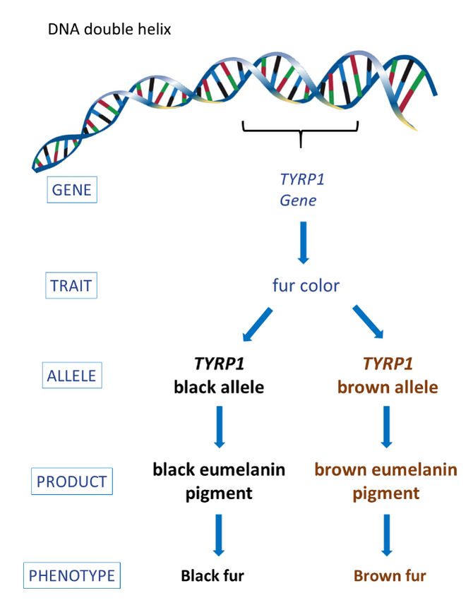
Introduction

     Genetic tests for dog traits, that is their physical characteristics, and diseases have become
increasingly available over the past few years, offering exciting opportunities for improving the health of
dogs at both individual and population levels. Dog owners and breeders have long dreamed of having
genetic tools to allow them to determine if a dog is predisposed to any genetic diseases and/or to make
educated breeding decisions that will improve the health of future generations. While the increasing
availability of these tools is exciting, many genetic test results are difficult to interpret and can lead to
confusion. Proper interpretation of genetic test results and understanding the limitations of a test are
important to avoid their misapplication, which can cause unnecessary concern and expense to dog owners
(e.g., clinical exams, dietary modifications). Moreover, the misapplication of genetic test results in
breeding programs could lead to excessive neutering and unnecessary removal of individuals from the
breeding population, which can negatively impact genetic variability within a breed, rather than improve
its overall health.
     The present document was supported by collaborative funding provided by the Orthopedic
Foundation for Animals and the American Kennel Club Canine Health Foundation with the goal of
providing dog owners, breeders, and veterinarians with a foundation in canine genetics to help interpret
and understand the implications of genetic test results. The information in this document is intended to
help users of genetic tests determine which (if any) changes should be adopted to improve a dog’s quality
of life and wellbeing. The document is designed to be a “living resource” that will be updated as new
information becomes available and is accompanied by a glossary of genetic terms and didactic videos that
will help the reader better understand important genetic concepts. The types of genetic tests currently in
use, their application in breeding programs and their limitations will be discussed as well as potential
detrimental effects of the misapplication of test results in both pet and breeding dogs. Finally, the future
of genetic testing in dogs will be discussed as well as any anticipated limitations.

                                                                                                           1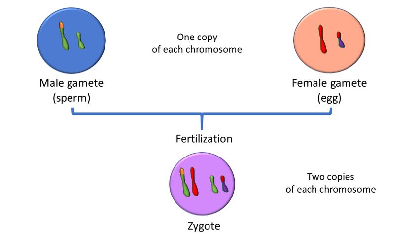
I. The Basics
To understand the nuances of genetic testing, one must have a good grasp of the basics of
genetics and inheritance. This section will address the structure of a dog’s genetic material, how it
determines different physical characteristics, and the different ways that genes are passed on from
parents to their puppies. An accompanying video can be found here.
a. DNA and Chromosomes
A dog is made up of trillions of cells. Within most cells is a compartment (the nucleus) containing
the dog’s genetic material. The genetic material, also referred to as the genome, is in many ways
equivalent to an instruction manual and dictates how that dog will grow, what it will look like, how well it
will process certain food items and, to some extent, how it will behave and reproduce.
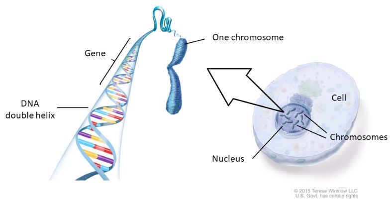
A dog’s genome is made up of long molecules of DNA, composed of two strands wound around
each other in the form of a double helix and tightly packed to form structures called chromosomes (Figure
1). The DNA molecule making up a chromosome is composed of building blocks called nucleotides. There
are four types of nucleotides (sometimes
referred to as bases) and they are
abbreviated A, C, T and G. In total, a dog has
nearly 3 billion of these nucleotides
arranged in a specific sequence that allows
the DNA to be used as an instruction manual.
Figure 1: Localization and structure of a dog’s
genetic material or genome.
There are many distinct chromosomes within a nucleus, and they differ in length and DNA
content. Chromosomes exist in pairs, meaning that every cell has two copies of each of the chromosomes
that make up the dog’s genome: one copy of each chromosome came from the dog’s mother and one
copy came from its father. Dogs have 39 pairs of chromosomes, where one pair makes up the sex
chromosomes and all non-sex chromosomes (i.e., the 38 remaining pairs) are called autosomes. Sex
chromosomes determine the sex of an individual; if the sex chromosomes are XX then the puppy is female
and if XY then the puppy is male.
b. Cell Division and Sexual Reproduction
To maintain healthy tissues and organs, cells of the body are constantly replacing old or damaged cells
through cell division, where a cell will create an exact duplicate of itself. Reproductive cells go through a
specialized form of cell division, called meiosis, to form the egg and sperm (Figure 2). In meiosis, first all
the chromosomes are copied so the cell has four copies of each chromosome. Then, through a process
called recombination, some of the chromosomes swap segments of DNA with each other. After this
process, two of the chromosomes will be the same as the original and two will be different because they
have been “recombined.” The cell then divides those four chromosomes into separate cells to form the
2gametes—the egg, in the case of females, and the sperm, in the case of males. We will use the general
term “gamete” to mean either the egg or the sperm throughout this document.
Figure 2: Simplified illustration of the process of DNA duplication and recombination that
occur in the nucleus of a reproductive cell undergoing meiosis for the formation of
gametes. Only two of the 39 chromosome pairs are represented.
Through the specialized cell division of meiosis, the gametes only receive one copy of each
chromosome. During sexual reproduction, the female’s egg is combined with the male’s sperm to form a
new cell that will then contain two copies of each chromosome, one coming from the mother and one
from the father (Figure 3). This new cell, called a zygote, now has the necessary blueprint to create a
puppy that will be different from either parent [1]. A video about gamete formation is found here.
Figure 3: Simplified illustration of fertilization showing the nucleus
of two gametes (one male, one female) that combine to form a
diploid cell (zygote). The zygote will give rise to a puppy.
c. Traits, Genes and Alleles
Along the chromosome are regions of DNA that represent genes. At this time, there are believed
to be about 19,000 different genes in the dog, but as we learn more, that may change in the future. Each
chromosome contains hundreds of genes and each gene is at a specific location on a chromosome. The
3specific location is referred to as a “locus,” which can be thought of as the street address of the gene. The
specific order of the nucleotides provides instructions for making up a product that composes a particular
trait. For the purpose of this document, a trait is any physical characteristic that can be passed on from
parents to their puppies, and the observable expression of a trait will be referred to as a phenotype
throughout this document because some traits have more than one phenotype (e.g., eye color). Some
phenotypes may not be desirable, such as a disease or a particular coat color that does not comply with
the breed standard.
Let us consider genes and their DNA sequences in the framework of fur traits. Hair/fur has many
different attributes: it can be long or short, straight or curly or wiry, and it could have rich color or pale
color or no color -- all of those characteristics combine into what we observe as a dog’s coat. Yet, in
actuality, what we are seeing is a collection of many different phenotypes, each governed by a different
gene. For example, fur length is a trait and long fur is one possible phenotype of that trait. Similarly, fur
color is a trait and black fur is a possible phenotype of that trait. While all dogs have the same genes, the
observed phenotype can vary.
When a cell duplicates itself, the genetic material of that cell is also duplicated. Though the
machinery responsible for copying the DNA is extremely efficient and accurate, mistakes can happen,
resulting in a change to the original DNA sequence of that cell. These mistakes can be thought of
somewhat like typographical errors: there could be one or more extra nucleotides included in the original
DNA sequence, or one or more nucleotides could be substituted or even missing. These mistakes that
happen during the copying of a cell’s DNA are referred to as mutations or genetic variants. When a variant
is just a single nucleotide difference in the DNA sequence, it is called a single nucleotide polymorphism
(SNP, pronounced “snip”). When parts of the DNA are missing, it is called a deletion (DEL for short) and
when more sequence is added in, it is called an insertion (INS for short). The DNA change is called a variant
because the sequence “varies” from a reference sequence. For the dog, the genome reference sequence
is that of a female boxer sequenced in 2005 [2].
Although some variants may not alter the function of a DNA sequence, others can change the
instructions contained in a gene such that a slightly different protein will be produced and a different
phenotype observed when compared to the original phenotype or “wildtype.” Now going back to the fur
example, one of many genes that can determine fur color is the tyrosinase related protein 1 (TYRP1) gene,
also known as the B-locus. As an aside, the term “locus” is sometimes used when the gene governing a
trait has not yet been discovered. For example, the term B-locus was used before TYRP1 was identified,
and even though the gene is now known, it is still commonly called the B-locus because that name is easier
to remember. The TYRP1 gene provides instructions for producing black pigment that makes the fur look
black. Figure 4 shows the two existing forms of the TYRP1 gene, one resulting in a black fur phenotype
(i.e., the wildtype version of that gene) and another resulting in brown fur (i.e., a variant form of that
gene). The different fur colors are produced by the same gene, but the actual DNA sequence is slightly
different. The different forms of a same gene are called alleles.
4Figure 4: DNA structure, its building blocks and an
example of how different forms of a same gene can
produce distinct phenotypes.
Although there may be many different
alleles for a gene within a population, an individual
dog can have at most two alleles for every gene: one
allele it inherited from its mother and one that it
inherited from its father. The combination of the
two alleles a dog has for a gene is called a genotype.
When a puppy receives the exact same allele from
its father and its mother it is said to be homozygous
for that allele and have a homozygous genotype for
that gene (Figure 5). Alternatively, a puppy that
receives different alleles
from its mother and father, is said to be
heterozygous and have a heterozygous genotype for
that gene.
Since every individual dog carries exactly
two alleles for every one of their genes, what happens when the alleles are different? What color would
a dog’s fur be if it had one brown and one black allele of the TYRP1 gene? The phenotype of dogs with
two different alleles depends on the relationship between those two alleles and the protein products
produced by them. Certain alleles are completely dominant, and their phenotype is always expressed over
other alleles. This is the case for the black allele of the TYRP1 gene. This means that every dog that has at
least one black allele for the TYRP1 gene will display black fur, even if the second allele is the brown allele.
Dominant alleles are represented by an uppercase letter (e.g., B; Figure 5). Note that in this particular
case, the dominant allele is also the wildtype allele,
but this is not always the case. Some genes have
wildtype alleles that behave as recessive alleles.
Figure 5: Pedigree (or family tree) demonstrating the
inheritance of the TYRP1 gene in the dog. Puppies
receiving the same allele from both parents will have a
homozygous genotype; those homozygous dominant
(i.e., BB) will have black fur and those homozygous
recessive (i.e., bb) will have brown fur. Puppies receiving
different alleles from mom and dad will have a
heterozygous genotype and black fur because the black
fur allele is completely dominant over the brown allele.
Heterozygous black dogs carry the recessive allele and
can pass it on to the next generation.
Since the brown color is not seen when the black dominant allele is present, the brown allele is said
to be recessive. Recessive alleles are represented by a lowercase letter (e.g., b), and a dog needs to have two
5recessive brown alleles (i.e., the bb genotype) to have brown fur. Conversely, dogs only need one black allele
to have black fur, meaning that black dogs can have a homozygous dominant genotype (i.e., BB) or a
heterozygous genotype (i.e., Bb). The terms dominant and recessive are also used to describe the mode of
inheritance of a phenotype, which will be addressed in section II.
Some genes like TYRP1 only exist in two forms in the canine population, but other genes (such as the
gene for white spotting or agouti) may have more than two alleles, each resulting in a different phenotype.
The existence of more than two alleles for a gene is often referred to as an allelic series and there may be a
hierarchy of dominance across the different alleles. An example of this is described here.
The alleles of a given gene can determine desirable traits, undesirable traits, or even disease traits.
Though people often talk about a dog having a “bad” gene for a trait, in actuality, the dog has “bad” alleles
for the gene governing that trait. And a group of dogs can have many different alleles. The abundance of the
different possible alleles within a population is referred to as allele frequency. The more abundant an allele
is, the higher the frequency and the more likely the phenotype associated with that allele will be observed in
the population. For example, the allele governing short fur will have a very high frequency within the
population of a dog breed that is characterized by short fur.
During the formation of breeds, breeders purposely increased the frequencies of alleles that caused
breed-defining phenotypes. When there is no allele variability for a breed-defining trait, then all puppies from
any parents of that breed will also have those same breed-defining traits. Alleles are said to be “fixed” in a
breed when there is only one allele in the breed population (i.e., no variation). For example, the B allele of
the TYRP1 gene is essentially fixed in the Schipperke, in which black is a breed-defining phenotype.
Conversely, TYRP1 is variable in the Labrador Retriever. Since both alleles can be found in the Labrador
Retriever breed population, it is possible to have black Labradors that are BB or Bb, and brown/chocolate
Labradors that are bb.
d. Genes, Linkage and Recombination
As described above, a process called recombination takes place during gamete formation, so that
some chromosome copies will swap segments of DNA with each other and become recombined. The DNA
segments swapped correspond to the same region of both chromosomes, and thus contain the same genes;
however, each chromosome of a pair could have a different allele for a swapped gene. Recombination is a
random event that can take place at any location on a chromosome and is an important mechanism for
creating genetic diversity by shuffling the alleles between chromosome pairs.
An important concept to understand though is that the closer two genes are to each other on the
same chromosome, the lower the chances are that recombination will happen in between them to shuffle
their alleles. This is why the alleles of genes that are physically close to one another on a chromosome tend
to be inherited together and are said to be “linked.” Though it is possible that recombination could happen
in between two linked genes, thus breaking that “linkage”, it is very unlikely given their close proximity. A
visual illustration of linkage and recombination can be found here.
6II. Modes of Inheritance
a. Mendelian Inheritance and Punnett Squares
Mode of inheritance (or inheritance pattern) refers to how a trait is transmitted from parents to
offspring across generations. While studying pea plants, the geneticist Gregor Mendel observed phenotype
and characterized aspects of inheritance that are now referred to as Mendelian inheritance. The term
“Mendelian” is typically used to describe traits that are governed by a single gene (i.e. monogenic) where
one allele displays complete dominance over another (like the black and brown fur color described above).
The patterns of Mendelian inheritance are described next. Two accompanying videos describe autosomal
and sex-linked modes of inheritance.
i. Autosomal Dominant
As a reminder, there are 39 pairs of chromosomes and 38 are autosomes and 1 pair is the sex
chromosomes. Autosomal dominant refers to the inheritance of a phenotype that is caused by a completely
dominant allele for a gene located on one of the autosomes. For example, the black fur color described
previously can be caused by an autosomal dominant allele at the TYRP1 gene, or the B-locus, which is located
on canine chromosome 11 – an autosome. When examining a family tree or pedigree that shows the
inheritance pattern of an autosomal dominant phenotype, such as the black fur governed by the TYRP1 gene,
we will see that the dominant phenotype appears in every generation (Figure 6). If at least one of the parents
displays the dominant phenotype, there is at least a 50% chance that one of its puppies will too. How to
determine the statistical probability of dominant phenotypes being present in a litter is discussed here.
ii. Autosomal Recessive
Autosomal recessive refers to the inheritance of a phenotype that is caused by a recessive allele of a
gene located on one of the autosomes. Therefore, the observable phenotype expression requires two of
those same recessive alleles (i.e., a homozygous recessive genotype). This is because recessive alleles are
easily “overridden” by the presence of a dominant allele. As described above, the brown fur color has an
autosomal recessive mode of inheritance, and only dogs possessing two of these recessive alleles at the B-
locus will appear brown. When examining a family tree or pedigree that shows the inheritance pattern of an
autosomal recessive phenotype, such as the brown fur governed by the B-locus, we will notice that, unlike
the dominant phenotype, a recessive phenotype does not usually appear in every generation (“skips a
generation”). As seen in figure 6, there may be litters where both parents have black fur and a brown puppy
is produced. This is because black dogs can have a heterozygous genotype, which means that although they
display a dominant phenotype, they are carriers for the recessive brown allele. How to determine the
statistical probability of recessive phenotypes being present in a litter when we know the parent genotypes
is discussed here.
7Figure 6: A pedigree (family tree) for black and brown fur color as determined by the
TYRP1 gene. Genotypes are noted under each dog: BB – homozygous dominant
(black), Bb – heterozygous (black), and bb – homozygous recessive (brown). Note, in
this example, the dominant phenotype (black fur) can be seen in every generation,
whereas the recessive genotype can skip generations.
iii. Sex-linked
As mentioned earlier, one pair of chromosomes is referred to as sex chromosomes because they are
responsible for determining the sex of a dog: female dogs are XX (i.e., have two X chromosomes) and male
dogs are XY (i.e., have one X and one Y chromosome). Only a few traits have been attributed to genes found
on the sex chromosomes of dogs. The “dominant” and “recessive” terms apply to sex-linked phenotypes in
the same way they do to autosomal traits, but the phenotypic outcome will depend on the dog’s sex.
Since female dogs have two X chromosomes, expression of a dominant or recessive phenotype will
mostly follow the same patterns described for autosomal traits: dominant traits will manifest in females that
are homozygous or heterozygous for the gene, whereas recessive traits will only be seen in females with two
recessive alleles for the gene (i.e. homozygous recessive). Males, however, only have one X chromosome.
This means that, in males, a single allele on the X chromosome will govern expression of the X-linked trait. In
that case a single recessive allele can cause the expression of a trait in males because they will have no other
allele that could “override” that recessive allele.
For example, hemophilia B, an X-linked recessive disorder that causes prolonged bleeding in affected
dogs, is seen in the German Wirehaired Pointer breed. Because the Factor IX gene that governs this disorder
is on the X chromosome, all male dogs having the recessive disease-causing allele will show clinical signs of
the disorder. Females, however, may be heterozygous and have one normal dominant allele and one disease-
causing recessive allele and they will not exhibit the disorder. Though heterozygous females will not develop
the disorder, they may pass a disease-causing recessive allele to their puppies. A female known to have
produced an affected puppy when mated to a healthy male is an obligate carrier of the allele causing
hemophilia B.
8While the Y chromosome also contains genes, it is physically much smaller than the X chromosome
and contains fewer genes. Traits or disorders governed by genes located on the Y chromosome will only affect
males because females lack a Y, and so these genes are only passed on from a father (sire) to its male puppies.
To date, no Y-linked health conditions have been reported in dogs.
It is important to clarify that sex-linked traits are not the same as sex-limited traits. Sex-limited traits
are those that can only be observed in one of the sexes. For example, cryptorchidism, characterized by one
or both testicles failing to descend through the inguinal canal within the first few months of a puppy’s life, is
a disorder that can only be observed in male dogs. Cryptorchidism is a sex-limited trait (and not sex-linked)
because, although it is seen only in male dogs, it has an autosomal mode of inheritance [3]. Genetic studies
indicate that cryptorchidism is governed by more than one autosomal gene, although no causative genes
have been identified to date [4]. Therefore, female dogs can have cryptorchidism-causing alleles, however
their status will not be observable because they do not have testicles.
iv. Punnett Squares and Phenotype Prediction
For Mendelian traits, once we know the inheritance pattern of a trait, we can use the parent
information to predict all possible genotypes and phenotypes of puppies. We can do this with the help of a
table containing four quadrants, commonly referred to as a Punnett square. At the top of the table, we list
the alleles that the mother’s gametes would have, and on the side of the table we list the alleles that the
father’s gametes possess. The quadrants are then filled in with one gamete from each parent (simulating
fertilization) and will reveal all possible combinations a puppy from that breeding could inherit. The
proportion of homozygous dominant, heterozygous, and/or homozygous recessive genotypes that may be
seen in the puppies is then used to estimate the chance of seeing each of the possible phenotypes in the
resulting litter. For example, as shown in Figure 6, if we consider a breeding between a male heterozygous
for a given gene (Aa) and a female homozygous for recessive alleles at that same gene (aa), after simulating
fertilization, two of the quadrants will have a puppy with a heterozygous genotype and two other quadrants
will have a puppy with a homozygous recessive genotype. This means there is a 50% chance of a puppy in a
litter from that breeding exhibiting the dominant phenotype and a 50% chance that a puppy will exhibit the
recessive phenotype. For more information and examples, please refer to the Punnett square video.
Additionally, when we look at more than one gene simultaneously, the number of quadrants expands in the
Punnett square proportional to the number of possible gametes and can get very complicated. For more
examples of how Punnett squares can be used,
refer to this video.
Figure 6: A Punnett square showing all possible
genotype combinations for a breeding between a
heterozygous male (Aa) and a homozygous
recessive female (aa). Puppies obtained from this
breeding would have a 50% chance of being
heterozygous and a 50% chance of being
homozygous recessive.
9b. Non-Mendelian Inheritance
Knowledge of Mendelian inheritance helps us understand how traits can be passed from a father or
mother to their puppies. However, as noted in other mammals, many dog traits do not play by Mendel’s rules
due to various reasons, which makes it difficult to assess their mode of inheritance by looking at pedigrees.
It is also harder to identify the causal genes behind these traits. When available, genetic testing for such traits
need to be interpreted with more caution as they may not be able to capture all determining factors
underlying the trait. The following types of non-Mendelian inheritance have been described and a brief
explanation of each with visual examples can be found here.
i. Incomplete Dominance
Incomplete dominance occurs in the absence of a recessive/dominant relationship between the
different alleles of a gene. However, in this case, heterozygous dogs display a mixture of the two possible
phenotypes (i.e. an intermediate phenotype that appears to be a blend between the two phenotypes caused
by each of the alleles).
The merle coat color is an example of incomplete dominance [5]. Two alleles exist for this gene,
designated as uppercase M and lowercase m. Despite the use of uppercase and lowercase letters to designate
the two alleles, there is no dominant/recessive relationship between the two alleles. Dogs homozygous mm
display full expression of color on their fur whereas dogs homozygous MM are almost all white and can
additionally have associated defects, such as deafness (see the section on pleiotropy below). Dogs
heterozygous for the merle allele (Mm), however, will display an intermediate phenotype with areas of full
pigmentation intertwined with areas of light pigmentation producing a marble-like pattern [6].
ii. Incomplete Penetrance
Penetrance refers to the proportion of the dogs who have a particular genotype that actually display
the associated phenotype. Complete penetrance means that every dog with that genotype will express the
associated phenotype, whereas incomplete penetrance means that only a portion of the dogs carrying that
genotype will display the expected phenotype. The rare condition of a mild and disproportionate dwarfism
in Labrador Retrievers is caused by a mutation in a collagen gene (COL11A2) that has incomplete penetrance
[7]. When known, traits having incomplete penetrance can also be characterized by the percentage of dogs
with the genotype who actually display the trait. For example, a dominant mutation in the gap junction
protein alpha 9 (GJA9) gene has been associated with polyneuropathy in Leonbergers and has an 80%
penetrance, meaning that 80% of dogs with the mutation show clinical signs of the disorder[8].
iii. Multiple Alleles
In many cases, more than two forms of a gene (that is, alleles) exist within a population, resulting in
multiple possible phenotypes depending on which combination of alleles a dog possesses and how those
alleles relate to one another. A good example of this is the S-locus (microphthalmia-associated transcription
factor (MITF) gene), which determines most of the white spotting patterns of dogs. Four alleles have been
identified exhibiting a dominance hierarchy: the most dominant allele is the S allele that causes a solid-
colored coat color; the second in the hierarchy is the si allele, which results in a white underside and white
neck collar that may or may not be accompanied by white face markings, a pattern commonly referred to as
10“Irish spotting”; the third allele in this dominance hierarchy series is the sp allele, which creates a random
distribution of white fur referred to as “piebald spotting”; and the final allele in the series is the sw allele
which is recessive to the three other alleles and dogs carrying two sw alleles are almost entirely white, which
is also referred to as “extreme white” (Figure 7)[9, 10].
Figure 7: Different white spotting phenotypes determined by the S-locus alleles
iv. Variable Expressivity
Variable expressivity refers to a range of phenotypes resulting from the same genotype. This means
that all individuals carrying a particular genotype will display the associated phenotype, but the degree or
extent to which the phenotype is displayed can vary. An
example of variable expressivity is the piebald spotting of
dogs (Figure 8), where the actual amount of white spotting
seen may vary considerably among dogs with the same
genotype [11].
Figure 8: The spsp genotype producing piebald spotting exhibits
variable expressivity as shown in these four dogs.
v. Polygenic and Complex Inheritance
In contrast with Mendelian traits, which are governed by a single gene (i.e., monogenic), complex
traits are those governed by more than one gene (i.e., polygenic) and may also be influenced by the
environment (i.e., multifactorial) [12, 13]. In dogs, eye color is considered a polygenic trait, whereas body size
is a multifactorial trait [14], governed by multiple genes and also influenced by environmental factors such
as exercise and nutrition. Hip dysplasia is another example of a multifactorial trait in dogs [15].
vi. Gene Interactions
In some cases, the phenotype observed for a particular trait can be affected by the alleles present at
other genes. The interaction between genes, where one gene influences the phenotype expression of
another gene, is termed “epistasis.”
Although the term epistasis generally refers to the interaction between genes [16], it is often used
to describe a more extreme case of gene interaction where one gene’s phenotype is completely masked or
hidden by another independently inherited gene [17]. Epistasis can also be dominant or recessive. In
dominant epistasis, the presence of a single dominant allele at one gene is sufficient to completely mask the
phenotype of another gene, whereas in recessive epistasis two recessive alleles at one gene are necessary to
mask the phenotype of another gene. An example of recessive epistasis in dogs is the yellow coat color seen
11in Labrador Retrievers. Yellow Labradors are yellow because they have two recessive alleles at the E-locus,
which we now know to be the melanocyte-stimulating hormone receptor (MC1R) gene, and this homozygous
recessive genotype (ee) overrides the genotype found at the B-locus. So, for example, a Labrador that would
otherwise be black because it has a homozygous dominant genotype at the B-locus (BB) will actually look
yellow because its E-locus genotype (ee) overrides the B-locus. Because of this, it can be difficult to determine
which alleles a yellow Labrador has at the B-locus.
The term “modifying gene” often refers to a less extreme situation where one gene will simply
influence the phenotype of another gene, resulting in a variation of the expected phenotype. For example,
dilute coat color in dogs is determined by a modifying gene called melanophilin (MLPH), also referred to as
the D-locus. At least three different mutations within this gene, having a recessive inheritance pattern, are
associated with dilute coat colors in multiple dog breeds [18-20]. The presence of any two mutated alleles at
the D-locus results in a lighter variation of the coat color, so that dogs with a black coat color, as determined
by the B-locus, will then display a dilute black coat color (blue) and dogs with a brown coat color will display
a dilute brown color often referred to as lilac, fawn or Isabella coat color, as seen in the Doberman Pinscher
and Dachshund. If you are interested in coat colors, please refer to this paper.
vii. Pleiotropy
Pleiotropy refers to a situation where a single gene affects two or more seemingly unrelated traits.
For example, some coat patterns (e.g., piebald white spotting) are associated with congenital hereditary
deafness, blue eyes, and eye defects [21]. This is because the mutations that cause the change in the coat
pattern also impacts the cells that are important for ear and eye development. Another example of pleiotropy
is seen in Rhodesian Ridgebacks, where the gene that governs the characteristic ridge of backward-growing
hair that runs across the dog’s back can also predispose for a neural tube defect called dermoid sinus [22].
viii. Mitochondrial Inheritance
In addition to the genetic material found in a cell’s nucleus, which is composed of DNA inherited from
the dog’s mother and father (i.e., the nuclear genome), a separate and much smaller genome is also present
in the mitochondria of mammalian cells. Mitochondria are organelles within a cell, distinct from the nucleus,
that generate the energy needed for a cell to function. The mitochondrial genome differs from the nuclear
genome in that it is exclusively inherited from the mother, and this mode of inheritance is thus referred to as
maternal inheritance. Although mitochondrial disorders only come from the mother, they can equally affect
male and female puppies because they are not sex-linked (i.e., not located on a sex chromosome) [11]. Two
mitochondrial disorders have been described to date in dogs: canine spongiform leukoencephalomyelopathy
in Australian cattle dogs and Shetland sheepdogs [23], and sensory ataxic neuropathy in Golden Retrievers
[24].
ix. Codominance
Similar to incomplete dominance, codominance also occurs in the absence of a recessive/dominant
relationship between the different alleles of a particular gene. However, unlike incomplete dominance, in
codominance, the products of both alleles in a heterozygous individual are fully expressed and observed at
the same time. Codominance has been described in humans [25], but a clear example has not yet been
identified in the dog.
12III. Genetic Selection and Populations
Genetic variation refers to the differences at the genetic level observed amongst individuals of a
same species. The diversity of alleles, rearrangement of genetic material and parental contribution of
different alleles to their offspring leads to genetically unique individuals, providing the flexibility in traits that
allows for some individuals of a species to adapt, thrive, and survive any changes to their environment.
Certain genetic combinations that enable an animal to survive and reproduce in a changing natural
environment will be favored under natural selection. For example, imagine that dogs with certain genetic
combinations are more resistant to a deadly infectious disease and, should that disease spread through the
population, individuals resistant to the infection will survive and pass on those resistance alleles to the next
generation. The result is a change in the frequency of alleles in the population where resistance alleles will
be more common in the surviving population compared to the original population. Natural selection is based
on the fitness or adaptability of an animal to the environment and is very important in wild animals.
Genetic selection can also occur artificially, when people purposefully select individuals with specific,
desirable traits. Artificial selection, or breeder selection, reflects a breeder’s preference for particular traits.
As an example, in the formation of dog breeds characterized by short legs, breeders hundreds of years ago
found dogs closer to the ground better suited for hunting some prey and, therefore, selected mates having
short legs to produce short-legged puppies. With more dogs bred for functionality and/or companionship,
artificial selection now plays a larger role than natural selection [26]. Artificial selection, while enhancing
certain traits, does not necessarily increase the overall fitness of a species.
In extreme situations, both natural and artificial selection can result in a drastic reduction in the
number of individuals that will breed and contribute to the next generation, a situation referred to as
population bottleneck. When few individuals contribute to future generations, genetic diversity of the
resulting population is reduced [27], leading to individuals with a more homogenous genetic background and
reduced gene pool (Figure 9).
Figure 9: Illustration of how a bottleneck event can affect the genetic diversity (gene pool) of a
population. The different colors used symbolize dogs with the different alleles of a same gene. Three of
the six alleles of that gene conferred survival advantage, so that individuals with the green, red and blue
allele survived the bottleneck event. The new population now reflects a reduced genetic diversity
compared to the original population.
13All dogs have undergone two major population bottlenecks, the first occurred when dogs were
domesticated from wolves and the second, more recently, when modern dog breeds were created [27]. Note
that natural selection can also occur within artificially selected populations. For example, in the early 1900’s,
a subpopulation of Nova Scotia Duck Tolling Retrievers (NSDTRs) survived two distemper outbreaks [28],
resulting in the breed undergoing an additional population bottleneck cause by natural selection; the small
number of distemper survivors gave rise to the modern NSDTR population. It should also be noted that,
following a population bottleneck, any disease mutations present in the surviving individuals will also become
more prominent in future generations. The bottleneck suffered by NSDTRs in the early 1900’s is thought to
be associated with the high prevalence of disease in the breed.
The genetic diversity of a population can also be affected by chance, that is, when only some
individuals within the population reproduce successfully. That means, the alleles present in the limited
number of dogs used to produce the next generation will increase in frequency in the resulting population,
even those alleles not being actively selected. This is called genetic drift and is extremely important in small
populations, such as dog breeds, where often only a limited number of dogs are selected for breeding. For
example, the overuse of a particular stud dog can dramatically change the frequency of all alleles in a small
population, and then subsequent generations will have a predominance of his alleles, both the good and the
bad. Popular sires have been shown to spread deleterious alleles much more profoundly throughout a
population than other breeding practices, such as inbreeding [29]. An outcome of genetic drift is reduced
genetic variability in the population. The following link provides an overview of the effect of genetic drift,
founder individuals and population bottlenecks.
As a consequence of artificial or natural selection, alleles for other traits may also be inadvertently
selected for due to genetic linkage; this circumstance underlies the concept of a selective sweep. Selective
sweep refers to different traits being inherited together because of the physical proximity of genes governing
them such that selective pressure for one trait will unintentionally cause selection for the linked trait. In other
words, when traits are being actively selected for, other traits will “piggyback” or “hitchhike” along merely
because of the physical adjacency of genes underlying those traits on a chromosome. For example, genes
that govern breed-specific or desirable temperament traits may be adjacent to a different gene that controls
an undesirable trait. In this case, if a dog was used for breeding because of its desirable traits, yet it has “bad
alleles” for an adjacent linked gene, the “bad alleles” will piggyback and be passed on to the next generations
because of linkage. The closer the genes are on a chromosome, the less likely that recombination will take
place in between them disrupting their genetic linkage. What this means is that when genes are tightly linked,
such as the hypothetical example above, when you select for a desirable trait you are also unintentionally
selecting for the undesirable one.
An interesting phenomenon that also affects the frequency of alleles in a population is called
balancing selection. In natural selection, one allele if often favored over another because it provides better
adaptation and survival of individuals in that environment. With time, the favored allele will increase in
frequency and the frequency of the other allele will be greatly reduced or eliminated. Balancing selection,
however, works to maintain the genetic variation within a population instead of favoring one allele over
another. In balancing selection, the benefits of maintaining multiple alleles offset the potentially damaging
effects of any individual allele. For example, the mutation that causes sickle cell anemia in humans affects an
individual’s ability to thrive. Yet, the mutation seems to be maintained in the population because it can
14benefit individuals in areas where malaria is endemic. Humans who have one sickle cell disease allele and
one normal allele are more resistant to malaria infection than those with two normal alleles. Examples of
balancing selection have also been described in dogs. For example, a mutation in the myostatin gene causes
double-muscling in Whippets; dogs with two mutant alleles are over-muscled, described as “bully” Whippets,
and have episodes of muscle cramping, whereas dogs that have one mutant and one normal (wildtype) allele
are more muscular than those with two normal alleles and are able to race faster [30]. The mutated allele
provides an advantage in the heterozygous state and is, therefore, maintained in the population. Another
example was described in the Belgian Malinois, where dogs heterozygous at the Cadherin 2 (CDH2) gene,
having one mutated and one normal (wildtype) allele, display an intermediate level of circling behavior that
allows them to have better work performance in confined spaces, whereas dogs with two mutated alleles
exhibit excessive circling behavior that is characteristic of obsessive compulsive disorder [31].
IV. Dog Breeds as Populations
There are over 350 dog breeds in the world [11, 32], 193 of which are officially recognized by the
American Kennel Club [33]. A breed consists of individuals that conform to a particular set of physical
descriptors that include size, coat color, fur length, and skull shape, among others. When mated, individuals
within a breed will always produce offspring with the same physical traits that characterize that breed [34].
Centuries of selective breeding to produce dogs with specific characteristics has reduced genetic diversity of
individuals within a breed, yet substantial variation remained across breeds, so that each individual breed
consists of a genetically distinct population [11, 35].
As mentioned above, different alleles can cause different phenotypes of a particular trait. During
breed creation, a small number of founder dogs with desirable traits and features was selected for breeding
with the purpose of homogenizing the resulting population. This allowed breeders to eliminate alleles
responsible for phenotypes that were undesirable. Selective breeding within a small starting population
inevitably leads to the mating of closely related individuals, also known as inbreeding. The positive
consequence of inbreeding is the concentration of desirable traits and their corresponding alleles within the
population, resulting in more homogenous litters where all puppies exhibit those desirable traits breeders
sought to emphasize. This ability to replicate breed-defining traits down through the generations is due to
the reduction in genetic diversity and fixation of alleles causing breed-defining traits (that is, homozygosity
at breed-important genes). However, another consequence of inbreeding is the increase of observed genetic
disorders.
Most genetic disorders in dogs are believed to be controlled by recessive alleles, and by increasing
the frequency of desirable alleles, which leads to homozygosity of breed-defining genes, inbreeding also
inadvertently increases the frequency and homozygosity of recessive disease alleles present in the dogs
selected for breeding. Note that inbreeding does not introduce disease mutations, but rather reveals their
presence in the population due to increased frequency of any disease alleles. Despite selecting mating pairs
that are not as closely related as seen with inbreeding, line breeding employed to concentrate and maintain
desirable breed traits [29, 36], along with the preferential and widespread use of specific individuals within
a breed (i.e. popular sire effect), also contributes to reducing genetic diversity within individual dog breeds
[29].
15When a breed is fixed for certain alleles, meaning all dogs of that breed only have one type of allele,
and that allele confers an advantage, then fixation is beneficial to the breed. However, when the allele that
is fixed causes disease, then fixation becomes detrimental to breed health. Although deleterious recessive
alleles are not intentionally selected for, they become more frequent in the population due to genetic drift
and selective sweeps, as described in section IV. When deleterious alleles are fixed within a dog breed,
selection for a healthy allele becomes impossible because no alternate alleles exist within the breed
population. An example of this has been reported in Dalmatians: all Dalmatians are homozygous for a
recessive allele that causes hyperuricosuria and hyperuricemia (HUU), predisposing them to bladder stones.
Since Dalmatians lack diversity for that gene, breeders cannot selectively breed to reduce disease. In contrast,
although that same recessive allele causing HUU has been identified in other breeds, those other breeds are
not fixed for the recessive HUU allele and breeders can use genetic testing and selective breeding to avoid
HUU [37]. [Note, since the discovery of the mutant HUU allele being fixed in Dalmatians, in 2011 the American
Kennel Club (AKC) and Dalmatian Club of America permitted the careful incorporation of another breed to
introduce a normal allele in the breed population].
When we think of a breed as a population, the physiological characteristics that define a breed should
be considered. Certain phenotypes that are characteristic and/or definitive of a breed may also be associated
with higher risk for particular disorders [38], especially if previous artificial breeder selection exaggerated
those breed-specific features. For example, current efforts to address heat and exercise intolerance in
brachycephalic breeds is in response to concerns of exaggerated selection for skull shape in brachycephalic
breeds. If there is sufficient genetic variability within a breed to purposely select away from the extreme
characteristics that may predispose to disorders, breeders should do so.
When genetic tests are available to help select for improved health, those can be used provided the
selection is applied gradually so as not to depopulate the breed, causing another population bottleneck. Dog
breeders must be mindful of the “big picture” and how their choices will impact the population. When
selecting mating pairs, one should not just think about the characteristics of their individual dogs and the
puppies they hope to produce, but also consider making choices that can improve the breed as a whole for
future generations. For example, when a dog is not used for breeding, none of its alleles for any of its genes
will be passed on to the next generation, which may shrink the gene pool of available alleles for the future.
Breeders must balance their desire for certain attributes with breed health concerns, all while avoiding
decisions that could contribute to a loss of genetic diversity in the population (i.e., a reduced gene pool).
V. Canine Genetic Tests
Breeders and owners want to maximize desirable traits and minimize traits that are undesirable or
negatively impact a dog’s health. Hundreds of desirable traits and genetic disorders have been compiled and
curated in the dog. Punnett squares can be used to predict the possible genotypes, and thereby phenotypes,
of puppies from a particular breeding pair when dealing with single-gene traits. However, Punnett square
predictions require knowledge of parental genotypes and determining the true genotype of every dog can
be difficult, since those exhibiting dominant phenotypes may have a homozygous dominant or a
heterozygous genotype. In these cases, it may not be possible to determine an individual’s true genotype
16until it is bred enough times and the puppies evaluated, unless a genetic test exists to determine which alleles
the dog possesses.
Another issue is seen with traits that are governed by the interaction of different genes, making it
difficult, if not impossible, to accurately infer a dog’s genotype solely based on their observable phenotype.
Moreover, some traits, especially undesirable disease traits, are expressed later in life, long after a dog
reaches the appropriate breeding age, making it difficult to select against these conditions. Genetic testing,
offers the opportunity to accurately determine a dog’s genotype at a very young age, before they are ever
bred, allowing breeders to make important decisions regarding which dogs to breed to enhance desirable
traits, and reduce undesirable ones.
Among the many canine genetic tests currently available, some are used in parentage verification
(e.g., genetic identity), such as the AKC DNA profile. Other tests are used to predict breed ancestry
composition, detect alleles responsible for particular phenotypes, such as coat color, or indicate whether a
dog is at risk for developing a particular trait, the latter often associated with undesirable disease traits.
Genetic tests can also identify the carrier status of an individual dog; carrier status is a term often used to
describe individuals with a heterozygous genotype that carry an allele for a recessive phenotype. That is, dogs
who do not exhibit a specific phenotype or disease but, since it has a heterozygous genotype, could pass on
a recessive allele to their puppies [39].
a. Direct and Indirect Tests
When the actual DNA sequence responsible for a trait has been identified, a direct, or mutation-
based, genetic test can be developed. Genetic tests that use direct analysis identify the presence of the exact
variation in the DNA that causes the phenotype. Direct tests are highly accurate because they determine
whether or not a dog’s DNA contains the causal mutation.
In some cases, however, the actual gene or precise mutation has not been identified. There may be
a region of the dog’s genome that clearly contains the causal variation, but amongst the many variations, the
exact one is not yet known. As segments of DNA physically close to one another tend to be inherited together
as a block, or “linked”, indirect (or linkage-based) DNA tests rely on genetic variants (markers) linked with a
phenotype to predict the dog’s alleles at the causal variant [40]. An example of a linkage-based test is the
one developed for cerebellar ataxia in the Italian Spinone. Indirect tests may not be 100% accurate because
recombination during gamete production can separate linked markers from the causal variant. In this way,
linkage-based tests are less reliable than direct mutation-based tests.
With few exceptions, genetic tests currently available for dogs are based on the analysis of a dog’s
DNA. An owner will either take a swab sample of cheek cells from inside the dog’s mouth with a tiny brush,
collect saliva on a special sponge, or submit a blood sample for testing; some testing providers even accept
dew claws. Regardless of the sample submitted, the DNA is purified from the cells and then used in the testing
scheme.
17b. Single versus Multipanel Tests
Both direct and indirect tests can be performed either as a single test, where we look for one segment
of DNA, or in combined tests, where multiple segments of DNA are being looked at simultaneously. These
are also referred to as singleplex or multiplex testing, respectively. That is the essential key piece for
understanding how the tests are done. However, for those interested in more specifics and some of the
challenges of DNA testing, please read on.
Singleplex tests are most often done through a process called polymerase chain reaction (PCR). In
PCR, a short guiding sequence of DNA, called a primer, specifically targets the chromosomal region being
evaluated and is used to create thousands of copies of the target DNA sequence. This amplification of the
DNA is necessary so the DNA sequence present in an individual dog can be accurately determined. Each single
gene test requires its own reagents and reaction time to analyze, as well as a separate sample of DNA [41].
Multiplex testing also uses PCR but bundles the reactions. Multiplex or panel testing can be more
cost-effective and time-efficient, allowing for more information to be obtained with fewer reagents and a
single DNA sample. However, multiplex panels can be harder to develop and validate because all the
components necessary for each individual test must work well together in a combined test. The primers for
each chromosomal sequence targeted must be able to work efficiently and not interfere with each other.
Errors in primer design, whereupon an incorrect chromosomal region is targeted or where more than one
region of the DNA is being unintentionally amplified, can result in erroneous results. Moreover, some target
chromosomal sequences may be easier to amplify than others, such that reagents could get preferentially
used up for one target sequence at the expense of amplifying the other targets, leading to incomplete or
inaccurate results [42].
An alternative to PCR multiplexing that simplifies the process of panel testing is the microarray
genotyping technology offered by companies that specialize in technology specific for DNA sequence
determination. Microarray genotyping technology allows for the simultaneous testing of hundreds of
thousands of single nucleotide variations, producing high-quality and accurate SNP detection [43].
Commercial companies that offer testing through microarray panels assess hundreds of genetic mutations
that have been associated with traits, and company advertisements often highlight the number of different
tests run at one time. The mutations that are being tested may have been rigorously validated or they may
have been incorporated into the testing scheme based upon small and/or recently published studies from
the scientific literature. Similar to what we see in humans, with 23&me and other direct-to-consumer genetic
testing, large-scale panel testing can be very cost-effective for dog owners and breeders, yielding information
on multiple traits, and may contribute to new genetic variants being discovered. Furthermore, just as being
done for humans, some dog genetic testing companies also offer to identify relatives of the tested dog.
VI. Interpretation of DNA Test Results
The dog owner who requests a DNA test should understand what the testing service actually
provides, how the test results will be shared, and how to interpret them. The DNA test result provided by
most testing laboratories usually includes a dog’s genotype for the investigated gene(s) and/or trait(s), an
interpretation of that genotype in terms of whether the dog is expected to exhibit a particular phenotype,
18You can also read