Rhinosinusitis in Companion Animals - Mini Review - Austin Publishing Group
←
→
Page content transcription
If your browser does not render page correctly, please read the page content below

Open Access
Austin Journal of Allergy
Mini Review
Rhinosinusitis in Companion Animals
Galler A*
Department for Companion Animals and Horses,
University of Veterinary Medicine, Austria
*Corresponding author: Alexandra Galler,
Department for Companion Animals and Horses,
University of Veterinary Medicine, Vienna, Austria
Received: April 12, 2021; Accepted: May 08, 2021;
Published: May 15, 2021
Introduction
Nasal disease is a frequently encountered problem in companion
animals, especially in cats. Typical signs include nasal discharge,
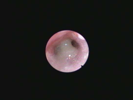
sneezing, nasal stridor, dysphagia, mouth-breathing and cough [1- Figure 1: Unilateral purulent nasal discharge.
4]. While acute feline nasal disease is commonly caused by infections
with herpes or calicivirus, the diagnostic work up for chronic nasal
discharge is far more laborious.
A common diagnosis in cats with chronic nasal discharge is
Idiopathic Chronic Rhinosinusitis (ICRS). This final diagnosis is
based on the exclusion of all known possible reasons for chronic nasal
discharge and can only be settled after a thorough diagnostic work-up
[1,3-5].
Chronic nasal discharge in cats can be caused by many different
diseases like e.g. nasal neoplasia, mycotic rhinitis, foreign body
rhinitis, dental disease, congenital malformations (cleft palate),
oronasal fistulae, nasopharyngeal polyps, nasopharyngeal stenosis
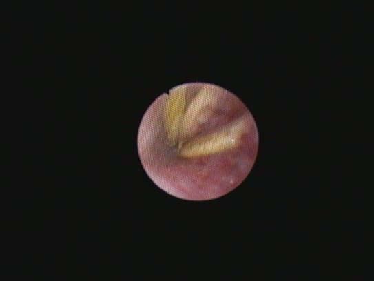
or traumata [1,4-6]. As a targeted therapy is dependent on the Figure 2: Nasal foreign body srucking in the nasopharynx of a cat.
underlying disease, systematic diagnostic work-up is crucial.
ICRS and it is the second most common reason for feline chronic
Nasal Neoplasia nasal disease [2-4,7]. ICRS is a chronic inflammatory disorder. The
In about 30-50 % of cats with chronic nasal discharge a nasal aetiology of this disease is by definition unknown. It is therefore
neoplasia can be diagnosed. 90% of nasal neoplasia are malignant in important to rule out other causes of chronic rhinitis. Although
cats (Figure 1). Although they grow invasive, they rarely metastasize herpesvirus and calicivirus infections commonly cause acute rhinitis,
and are often locally restricted. In 90% of cats with nasal lymphoma active or re-activated infections do not play a role in chronic nasal
the nose is the solitary location. Nasal lymphoma is the most often disease [11]. However, previous infections with herpes virus and the
encountered nasal neoplasia, followed by adenocarcinoma. Other resulting anatomical alterations in the nasal cavity with partial loss
tumours (e.g. fibrosarcoma, chondrosarcoma, squamous cell of mucociliary clearance capacity are under discussion as a possible
carcinoma) may also be found [2-4,7]. Cats with nasal neoplasia cause of ICRS development [1,2]. Histological examination of
are commonly older (8-10years), more often have unilateral, samples obtained from ICRS patients mostly reveals a mixed-cell and
haemorrhagic discharge, facial deformations and have a shorter in rare cases a lymphoplasmacytic infiltration of the nasal mucosa [1].
duration of signs then cats with other nasal disorders. However Treatment is symptomatic with anti-inflammatory medications and
there is strong overlap especially regarding cats with ICRS [2-5]. The antibiotics. Anti-herpes-virus therapies are not effective. . Recurrence
diagnosis of nasal neoplasia can only be confirmed by histopathology. of signs is common and should be expected. The prognosis for healing
If staging (thoracic radiographs and abdominal ultrasound) confirms is poor but the disease can be satisfactorily managed in many cats.
local disease radiation therapy is the preferred treatment. If radiation Others
is not available or the disease is not restricted to the nose cats are
Juvenile nasopharyngeal polyps are benign pedunculated masses
treated by systemic chemotherapy (e.g. COP-based protocols in nasal
originating from the Eustachian tube or the tympanic bulla, growing
lymphoma). The prognosis is fair, with complete remission in 70%
into the nasopharynx (Figure 2). Young cats up to 2 years are most
and median survival times of up to 30 months [8-10].
commonly affected by polyps; congenital defects or reactions to
ICRS infections are being discussed as causes [12,13]. Polyps can be
Up to 30% of cats with chronic nasal discharge suffer from removed by traction-avulsion, ventral bulla osteotomy or total ear
Austin J Allergy - Volume 7 Issue 1 - 2021 Citation: Galler A. Rhinosinusitis in Companion Animals. Austin J Allergy. 2021; 7(1): 1036.
Submit your Manuscript | www.austinpublishinggroup.com
Galler. © All rights are reserved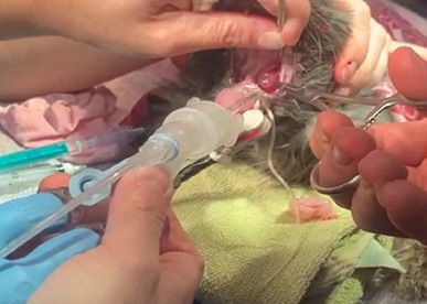
Galler A Austin Publishing Group
Figure 3: Mucupurulent discharge, obstructing the nasopharynx. Figure 5: Removal of nasopharyngeal polyps in cats. A histopathology should
always be performed to secure the diagnosis.
Figure 4: Nasal mass protruding in the nasopharyneal area of a dog.
Figure 6: Removal of nasopharyngeal polyps in cats. A histopathology should
always be performed to secure the diagnosis.
ablation with lateral bulla osteotomy. Traction-avulsion is the most
gentle procedure for removing a polyp. However, recurrence is
possible and a transient Horner´s syndrome or vestibular signs may
develop [14,15].
Nasopharyngeal stenoses may develop as a sequela of
inflammatory diseases or be present as a congenital defect [16,17],
they can be treated by balloon dilatation.
Nasal foreign bodies (most often grass awns or other plant-
parts), which travel to the nasopharynx or the nasal cavities via the
retrograde route, may also cause chronic rhinitis [3,4] (Figure 3).
While fungal rhinitis is very common in dogs, it is more rare in
cats. Nasal cryptococcosis is the most common fungal nasal disease in
cats. It is quite rare in central Europe but its incidence is dependant
of the geographic regions [5]. Contrary to dogs, nasal aspergillosis is
uncommon in cats [3,4,18] (Figure 4).
Figure 7: Tranient Horner syndrome after removal of a nasopharyngeal polyp
Diagnosis in a cat.
Since a targeted therapy is dependent on the underlying disease,
systematic diagnostic work-up is crucial. The history (age, indoor/ prevalence in healthy cats (Figure 7). Cytology of nasal is usually
outdoor cat, duration of signs, character of nasal discharge, former unspecific, but fungal organisms or neoplastic cells may be found.
episodes of “cat flu”), physical examination, imaging techniques and Bacterial culture of nasal discharge is of no value as it commonly
rhinoscopy as well as cytological and histological examination of yields a mixed growth of normal commensal microflora. Culture and
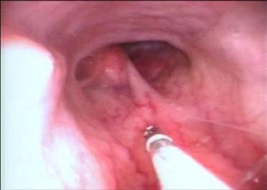
biopsy samples may shed light on the aetiology of the disease (Figure sensitivity of nasal flushes or nasal biopsies resemble the mucosal
5,6). bacterial infiltration more closely but the majority of the spectrum
of microorganisms observed in cats with chronic rhinitis can also
Blood work is usually unspecific. Testing for feline calicivirus, be identified in healthy cats. The role of Mycoplasma spp. in feline
feline herpesvirus-1, FELV and FIV is of no value due to the equal chronic rhinitis is under debate.
Submit your Manuscript | www.austinpublishinggroup.com Austin J Allergy 7(1): id1036 (2021) - Page - 02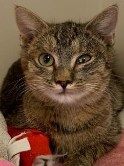
Galler A                                                                                                                                    Austin Publishing Group

    Imaging may reveal the extent (sinus affection) and character                     6. Van Pelt DR, Lappin MR. Pathogenesis and treatment of feline rhinitis. Vet
                                                                                         Clin North Am Small Anim. Pract. 1994; 24: 807-823.
of the disease and help direct biopsy sampling. Disadvantages of
radiographs include the need for proper patient positioning and the                   7. O´Brien RT, Evans SM, Wortman JA, Hendrick MJ. Radiographic fi ndings in
superimposition of structures. Superimposition can be prevented by                       cats with intranasal neoplasms or chronic rhinitis: 29 cases (1982-1988). J
                                                                                         Am Vet Med Assoc. 1996; 208: 385-389.
obtaining intraoral views. CT/MRI are superior, providing detailed
images without the problem of superimposition. However there is                       8. Theon AP, Peaston AE, Madewell BR, Dungworth DL. Irradiation of
                                                                                         nonlymphoproliferative neoplasms of the nasal cavity and paranasal sinuses
substantial overlapping of imaging findings between cats with nasal                      in 16 cats.J Am Vet Med Assoc. 1994; 204: 78-83.
neoplasia and those with ICRS [3,4,7,19-21].
                                                                                      9. Mellanby RJ, Herrtage ME, Dobson JM. Long-term outcome of eight cats with
     Rhinoscopy can be performed with flexible or rigid endoscopes.                      non-lymphoproliferative nasal tumours treated by megavoltage radiotherapy.
The nasopharynx can easiest be assessed with a flexible endoscope                        J Feline Med Surg. 2002; 4: 77-81.

and should be performed first. Lymphomas are often visible in the                     10. Sfiligoi G, Théon AP, Kent MS. Response of nineteen cats with nasal
nasopharynx and samples can be taken under sight in this location.                        lymphoma to radiation therapy and chemotherapy. Vet Radiol Ultrasound.
                                                                                          2007; 48: 388-393.
Rhinoscopy can be diagnostic (foreign bodies, polyps, nasopharyngeal
stenosis, fungal plaques) as well as therapeutic (removal of foreign                  11. Johnson LR, Foley JE, De Cock HE, Clarke HE, Maggs DJ. Assessment of
                                                                                          infectious organisms associated with chronic rhinosinusitis in cats. J Am Vet
bodies, fungal plaques, flushing/suctioning excessive secretions) [3,4].                  Med Assoc. 2005; 227: 579-585.
It allows for tissue sampling under direct visualization. If rhinoscopy
                                                                                      12. Baker GJ. Nasopharyngeal polyps in cats. Vet Rec. 1982; 111: 43.
is not available a thorough examination of the oral cavity with digital
palpation of the soft palate an inspection of the nasopharynx, intraoral              13. Anderson DM, Robinson RK, White RAS. Management of infl ammatory
radiographs, nasal flush and blind biopsies can be performed. For                         polyps in 37 cats. Vet Rec. 2000; 147: 684-687.

blind nasal biopsies it is important not to pass the instrument further               14. Muilenburg RK, Fry TR. Feline nasopharyngeal polyps. Vet Clin North Am
than the eye level to avoid damage to the cribriform plate.                               Small Anim Pract. 2002; 32: 8398-8449.

                                                                                      15. Veir JK, Lappin MR, Foley JE, Getzy GM. Feline inflamatory polyps: historical,
Summary                                                                                   clinical, and PCR findings for feline calicivirus and feline herpes virus-1 in 28
                                                                                          cases. J Feline Med Surg. 2002; 4: 195-199.
    Chronic rhinitis may have different aetiologies. Prognosis and
treatment differ with the aetiology. A thorough work up is mandatory                  16. Mitten RW. Nasopharyngeal stenosis in four cats. J Small Anim Pract. 1988;
                                                                                          29: 341-345.
to reach a diagnosis. Feline ICRS is a common final diagnosis.
Treatment of ICRS may be frustrating, since there is no cure.                         17. Khoo AML, Marchevsky AM, Barrs VR, Beatty AB. Choanal atresia in a
                                                                                          Himalayan cat – fi rst reported case and successful treatment. J feline med
References                                                                                Surg. 2007; 9: 346-349.
1. Michiels L, Day MJ, Snaps F, Hansen P, Clercx C. A retrospective study of on-      18. Schulz B, Unterer S, Hartmann A, Werckenthin C, Brühschwein A, Hartmann
   specific rhinitis in 22 cats and the value of nasal cytology and histopathology.       K. Diagnostik und Therapie der nasalen Aspergillose bei einer Katze-
   J Feline Med Surg. 2003; 5: 279-285.                                                   Fallbeschreibung und Literaturubersicht. Tierarztliche Praxis Kleintiere. 2007;
2. Henderson SM, Bradley K, Day MJ, Tasker S, Caney SM, Hotston Moore A,                  35: 281-286.
   Gruffydd-Jones TJ. Investigation of nasal disease in the cat – a retrospective     19. Lamb CR, Richbell S, Mantis P. Radiographic signs in cats with nasal
   study of 77 cases. J Feline Med Surg. 2004; 6: 245-257.                                disease. J Feline Med Surg. 2003; 5: 227-235.
3. Galler A, Shibly S, Bilek A, Hirt RA. Chronic diseases of the nose and nasal       20. Schoenborn WC, Wisner ER, Kass PP, Dale M. Retrospective assessment
   sinuses in cats: a retrospective study. Schweiz Arch Tierheilkd. 2012; 154:            of computed tomographic imaging of feline sinonasal disease in 62 cats. Vet
   209-216.                                                                               Radiol Ultrasound. 2003; 44: 185-195.
4. Galler A, Shibly S, Bilek A, Hirt RA. Chronic nasal disease in cats - A            21. Karnik K, Reichle JK, Fischetti AJ, Goggin JM. Computed tomographic fi
   retrospective study. European Journal of Companion Animal Practice. 2013;              ndings of fungal rhinitis and sinusitis in cats. Vet Radiol Ultrasound. 2009;
   23: 271-277.                                                                           40: 65-68.
5. Demko JL, Cohn LA. Chronic nasal discharge in cats: 75 cases (1993-2004).
   J Am Vet Med Assoc. 2007; 230: 1032-1037.

Submit your Manuscript | www.austinpublishinggroup.com                                                                   Austin J Allergy 7(1): id1036 (2021) - Page - 03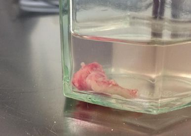
You can also read