Scar-Free Wound Healing Following Full-Thickness Cutaneous Wounding in the Tail and Body of Scincella tsinlingensis
←
→
Page content transcription
If your browser does not render page correctly, please read the page content below

Int. J. Morphol.,
39(4):1139-1146, 2021.

      Scar-Free Wound Healing Following Full-Thickness Cutaneous
       Wounding in the Tail and Body of Scincella tsinlingensis

                            Cicatrización de Heridas sin Cicatrices Luego de Heridas Cutáneas
                            de Espesor Total en la Cola y el Cuerpo de Scincella tsinlingensis

                                                Chun Yang1,2; Xin Wang2; Huihui Zhang2 & Lin Li2

YANG, C.; WANG, X.; ZHANG, H. & LI, L. Scar-free wound healing following full-thickness cutaneous wounding in the tail and
body of Scincella tsinlingensis. Int. J. Morphol., 39(4):1139-1146, 2021.

         SUMMARY: The cutaneous wounds of trunk and tail healing scar-free or with scar were different in lizard species. Full-
thickness cutaneous injuries of tail and body of Scincella tsinlingensis were examined by histomorphological and immunohistochemistrical
methods. The results showed that all injuries healed without scarring. The process of the wound healing of S. tsinlingensis involved
hemostasis, re-epithelialization, proliferation and remodelling, which also could be further subdivided into six stages. Stage I, 0-2 day
post wound (dpw), the blood oozed gradually, no obvious wound contraction, minimal blood loss. Stage II, 2-10 dpw, the wound bed
covered by the fibrin clot of blood, tissue fluid and tissue debris. Stage III, 7d-15 dpw, the wrinkled wound epitheliums was gradually
stratified, and its surface was keratinized and exfoliated. Stage IV, 10-28 dpw, pigment cells were distributed at the boundary between
epidermis and dermis, with few blood vessels and no granulation tissue formation. Stage V, 20-70 dpw, opaque scales covered the wound
epithelium with randomly scattered melanophores in the base of the epidermis. Stage VI, 45-135 dpw, the epidermis and dermis restored
to the thickness of the original skin. Regenerated scales were similar to scales of the uninjured dermis. The positive immunostaining of
matrix metalloproteinases-9, cytokeratin 6, alpha smooth muscle actin, caspase 3 and transforming growth factor-β3 showed the specificity
of healing period and different stages, which participated in skin wounds healing of S. tsinlingensis.

           KEY WORDS: Scincella tsinlingensis; Skin wound healing; Histology; Immunohistochemistry.

INTRODUCTION

        Skin is composed of epidermis and dermis in animals                     2011). Investigation of skin regeneration using various
and humans, which covers the entire surface of the body                         vertebrate models, including lizards, could lead to the
with frequently occurring injuries (Abe et al., 2020). Skin                     realization of scar-free skin regeneration in humans
wound healing involves an overlapping cascade of tissue                         (Alibardi & Toni, 2005).
events, i.e., hemostasis, inflammation, proliferation and
remodeling (Jourdan et al., 2019; Korntner et al., 2019).                               During wound repair, the quantity and quality of
The tissue events of the last three stages determine whether                    new blood vessels formation is imperative for efficient
the wound is scar-free or whether the abnormal healing                          tissue restoration (Yu et al., 2014; Naito et al., 2020).The
process causes extensive angiogenesis and collagen                              inhibition or acceleration of angiogenesis can suppress the
deposition to form a scar (Poetschke & Gauglitz, 2016;                          spontaneous regenerative response of mice and zebrafish.
Stoica et al., 2020). In non-amniote vertebrates, the wound                     Comparative studies of skin regeneration together with the
bed involve a more rapid closure of the wound epithelium,                       tail and body of non-mammalian amniotes is represented
a delay vasculogenesis and collagen deposition without                          by the lizard (Alibardi & Toni). Scar-free wound healing
scarring (Peacock et al., 2015; Abe et al.). All mammals                        is not a universal feature of lizard reptiles. Scar-free skin
but a few species are unable to regenerate dermis, and form                     regeneration is reported in the leopard gecko (Eublepharis
a scar after a deep injury (Seifert et al., 2012). Skin                         macularius) after a deep injury (Peacock et al.; Payne et
regeneration of the lizard tail and body has garnered much                      al., 2017), while only the tail wound of Anolis carolinensis
attention as wound healing models (McLean & Vickaryous,                         can regenerate without scars and produce keratinized scales,

1
    Prof., Modern College of Arts and Sciences, Shanxi Normal University, Linfen, Shanxi Province, P.R. China, 041004.
2
    School of Life Sciences, Shanxi Normal University, Linfen, Shanxi Province, P.R. China, 041000.

                                                                                                                                      1139
YANG, C.; WANG, X.; ZHANG, H. & LI, L. Scar-free wound healing following full-thickness cutaneous wounding in the tail and body of Scincella tsinlingensis. Int. J. Morphol., 39(4):1139-1146, 2021.

but the color of the regenerated skin cannot be restored                                            intersection of the dorsal midline and the hind limbs,
(Wu et al., 2014). The Scincella tsinlingensis, an endemic                                          respectively. Then, the animals were kept under the same
lizard species to China, can regenerate their tails after                                           conditions and the healing process was observed.
autotomy (Yang et al., 2021). The studies to investigate
the pattern of expression of markers associated with wound                                          Tissue collection. After receiving the full-thickness excision
healing in S. tsinlingensis help to clarify elucidation of the                                      biopsies, the lizards were permitted to heal. At select time
mechanisms underlying the skin regeneration.                                                        points (0dpw, 2dpw, 7dpw, 14dpw, 25dpw, 45dpw). Animals
                                                                                                    were humanely killed at -20 ºC, the tissues from body and
        Matrix metalloproteinases-9 (MMP-9) play                                                    tail was fixed in 4 % paraformaldehyde for 1-3 days,
important role in extracellular matrix (ECM) remodeling                                             decalcified for 1-3 days, and changed every 24h. Then,
in a variety of animals. In the early wound healing phase                                           samples were dehydrated with graded alcohols, cleared in
during amphibian regeneration, MMP-9 is upregulated and                                             xylenes, embedded in paraffin wax. Sections were cut at 10
act as an important factor to initiate the dedifferentiation                                        µm on a rotary microtome and stained by hematoxylin-eosin
of the mesenchymal tissues. Activity levels of MMP-9 are                                            (H-E) or Masson's trichrome. The sections were observed
evaluated during tail regeneration in Hemidactylus                                                  with BX-51 light microscope.
flaviviridis (Pillai et al., 2020). Cytokeratin 6 is produced
in activated reptilian keratinocytes (Alibardi & Toni).                                             Immunohistochemistry. Slides were dewaxed with xylene,
Caspase 3(C3), the effector of the caspase-mediated                                                 rehydrated, incubated dark in 3 % H2O2 for 30 minutes at a
apoptotic program, is widely recognized as a definitive                                             room temperature, then rinsed three times with PBS (0.1M,
marker of programmed cell death. Elevated levels of                                                 PH 7.4), and blocked with 5 % BSA to prevent non-specific
transforming growth factor-β3(TGF-β3) promote scar-free                                             binding. Sections were incubated with primary antibodies
wound healing (Delorme et al., 2012). Alpha smooth muscle                                           overnight at 4 ºC. Following primary antibodies and were
actin(α-SMA) belongs to the family of the six conserved                                             used: anti rabbit forMMP-9, cytokeratin 6, α-SMA, C3 and
actin isoforms. α-SMA is essential for the formation of new                                         TGF-β3(1:100, Booster). Negative control slides were
blood vessels (Rafii et al., 2016) and abundant in vascular                                         replaced primary antibody with PBS. After rinsing in the
smooth muscle cells (SMCs) (Skalli et al., 1989). In light                                          buffer, sections were incubated with biotinylated goat anti-
of this information, localization of MMP-9, cytokeratin 6,                                          mouse/rabbit IgG (1:100, Booster) for 4 h at room
α-SMA, C3 and TGF-β3 are examined by                                                                temperature, subsequently incubated with SABC and
immunohistochemistry after full-thickness cutaneous                                                 visualized using DAB. All pictures were obtained with
wounding in the tail and body of S. tsinlingensis.                                                  Olympus BX-51digital imaging system and conducted by
                                                                                                    Photoshop CS6.

MATERIAL AND METHOD
                                                                                                    RESULTS

Experimental animal. The S. tsinlingensis were captured
from the hilly areas of Qiliyu forest in Taiyue Mountain,                                           Stage I (0-2 dpw). During stage I, the trunk and tail wounds
Shanxi, China (110º40′-112º 21′E, 36º21′-36º 45′N).                                                 showed open wounds, the tissues were exposed, there was
Experiments were carried out according to protocols                                                 no obvious bleeding on the surface of the wound at the
approved by animals care guidelines of the Ethical                                                  beginning of the wound. The wound edge collapsed slowly
Committee of Shanxi Normal University. The lizards were                                             and began to contract, the open wound area decreased and
kept in a terrarium with a 14:10 photoperiod, a room                                                the wound edge began to appear melanosis, which could be
temperature of 24 ºC and an ambient humidity of 40-50 %,                                            the initial mechanism to protect the wound surface. Full-
fed with a diet of insects and water in daily.                                                      thickness excision cutaneous wounds of the tail and trunk
                                                                                                    were confirmed by histology, and sometimes the underlying
Biopsies. All adult individuals of both sexes of the lizards                                        muscles were involved (Figs. 1-A and H).The wound of the
were randomly selected as the experimental object. The                                              tail and trunk began to reepithelialize, and the wound
animals were placed at 4 ºC for 20 minutes for hypothermic                                          epithelium was incomplete and consisted of 1-4 layers of
anesthesia, then laid on pre-cooled culture plate and full-                                         round or squamous cells on 2dpw (Figs. 1-D and K). The
thickness excision cutaneous wounds were created using a                                            epithelial cells stained with H.E were pink and the underlying
1.5 mm disposable biopsy tool (Integra TMMiltex®). The                                              muscle tissue was blue-purple (Figs. 1-C and J). Masson's
skin wounds of the trunk and original tail were distributed                                         trichrome showed that the wound epithelium was red, while
along the dorsal midline, 1cm before and after the                                                  the lower muscle tissue was turquoise (Figs. 1-B and I).
1140
YANG, C.; WANG, X.; ZHANG, H. & LI, L. Scar-free wound healing following full-thickness cutaneous wounding in the tail and body of Scincella tsinlingensis. Int. J. Morphol., 39(4):xxx-xxx, 2021.

       At the end of the stage I, cytokeratin 6 (Figs. 1-G                                         were present at the wound (Figs. 1-E and L). Isolated cells
and N) and MMP-9 (Figs. 1-F and M) showed                                                          from the original epidermis and dermis were immunopositive
immunopositive, and the expression of MMP-9 in wound                                               for C3. No TGF-β3 immunopositive cells were observed at
epithelium were strong. a-SMA expressed myofibroblasts                                             this stage.

Fig. 1. Stage I of the tail and body scar-free wound healing of S. tsinlingensis. A and H. Transverse section of the wound area of the body
and the tai; B and I. Higher magnification image of the wound area of the body and the tai, stained with Masson’s trichrome; C and J.
Closer view of the region identified in panel (A) and (H); D and K. Closer view of the region identified in panel (C) and (J); E-G and L-
N. The body and tail wound areas, stained with a-SMA, MMP-9 and cytokeratin 6 by immunohistochemistry. The Arrows indicate
immunopositive cells. epidermis (e), dermis (d), muscle (m), wound area (w), wound epithelium (we).

Stage II (2-10 dpw). During stage II, blood clots formed                                           Stage III (7-15 dpw). During stage III, the gross
at the wound bed of the trunk and tail, which gradually                                            morphological results showed that the epithelium of trunk and
thickened and covered the wound surface (Figs. 2-A and                                             tail wound was wrinkled. The clot of the trunk disappeared,
I). The histological results showed that the wound                                                 and the wound was completely covered with epithelium (Figs.
epithelium began to form beneath the clot and gradually                                            3-A). Small round cells accumulated in the matrix of loose
deepened into the clot, and the wound epithelium was                                               tissue, with clear boundaries between undamaged dermis (Figs.
wrinkled evenly (Figs. 2-D and L), and re-                                                         3-D). However, the clot of the tail still exists (Fig 3-9 I), the
epithelialization was not completed until the third stage.                                         number of cells in the wound matrix is less than that of the
Blood clots were easily lost during tissue remodeling,                                             trunk, and the extracellular matrix is less (Fig .3-9 L).
indicating poor adhesion to the wound bed and high                                                 Inflammation of the wound site ended at this stage. Wound
melanin content at the wound site. The clots and epithelial                                        epithelium began to keratinize and melanin decreased, luteal
cells of the wound stained with H.E were pink and the                                              layer appeared below epidermis, tail and trunk wound
underlying muscle tissue was blue-purple (Figs. 2-C and                                            epithelium gradually stratified, wound epidermis exfoliated
K). Masson's trichrome showed that the clot and wound                                              cuticle (Fig .3-9 D). The H.E. staining results showed that the
epithelium were red and the muscle tissue below it was                                             exfoliated wound epidermis and the cuticle of the wound epi-
turquoise (Figs. 2-J).                                                                             dermis were pink (Figs. 3-C and K), while Masson's trichrome
                                                                                                   showed a mixture of green and red (Figs. 3-B and J).
        There were cytokeratin 6 immunopositive cells (Figs.
2-H and P) and MMP-9 immunopositive cells (Figs. 2-G                                                        Mesenchymal cells at the wound site were weakly
and O) in the wound epithelium, and α–SMA                                                          immunopositive for MMP-9(Figs. 3-G and O). The regenerated
immunoreactive myofibroblasts were more abundant than                                              epithelial fibroblasts and perivascular cells were immunopositive for
the first stage stage I (Figs. 2-F and N). C3 immunopositive                                       α-SMA (Figs. 3-F and N). The wound epithelial cells maintained
cells were observed at the wound surface (Figs. 2-E and M),                                        cytokeratin 6 expression (Figs. 3-H and P). A large number of C3
without TGF-β3 immunostaining.                                                                     immunopositive cells were observed in the dermis (Figs. 3-E and M).
                                                                                                                                                                                       1141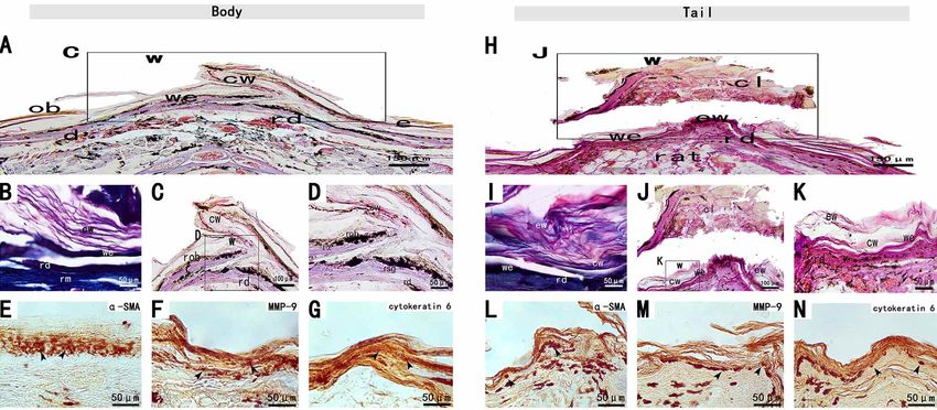
YANG, C.; WANG, X.; ZHANG, H. & LI, L. Scar-free wound healing following full-thickness cutaneous wounding in the tail and body of Scincella tsinlingensis. Int. J. Morphol., 39(4):1139-1146, 2021.

Fig. 2. Stage II of the tail and body scar-free wound healing of S. tsinlingensis. Exudate clot (cl), regenerated dermis (rd), adipose
tissue (at).

Fig. 3. Stage III of the tail and body scar-free wound healing of S. tsinlingensis. Exfoliating wound (ew), epidermis (e), corneous layer
of the wound epidermis (cw), xanthophore layer (x).

1142
YANG, C.; WANG, X.; ZHANG, H. & LI, L. Scar-free wound healing following full-thickness cutaneous wounding in the tail and body of Scincella tsinlingensis. Int. J. Morphol., 39(4):1139-1146, 2021.

Stage IV(10-28 dpw). During stage IV, the caudal and trunk                                          blue and purple, and the regenerated dermis was pink. (Figs.
wounds exposed a smooth and tight wound epithelium                                                  4-D and K). The H.E. staining or Masson's trichrome staining
without squamous coverage. The caudal clot detached from                                            results showed extensive escalation and reticular collagen
the wound, showing intact wound epithelium (Figs. 4-H). A                                           deposition, with low cellularity (Figs. 4-B and I).
exfoliated cuticle was observed in the wound epidermis of
the tail and trunk cadres (Figs. 4-A and H), and the boundary                                              Immunohistochemical results showed that wound
between the epidermis and the undamaged dermis was                                                  epithelium was consistently immunopositive for cytokeratin
obvious, thicker than the original epidermis, and the blood                                         6 (Figs. 4-G and N). The wound epithelium with a small
vessels in the regenerated tissue were obvious (Figs. 4-C and                                       number of apoptotic C3 positive cells was still MMP-9
J). Pigment cells were observed at the junction of epidermis                                        immunopositive (FigureFigs. 4-F, M). Perivascular cells
and dermis (Figs. 4-D and K). The regenerated horn cortex                                           were still immunopositive for α-SMA (Figs. 4-F and L), but
was yellow and brown, the regenerated β-layer, middle layer                                         myofibroblasts showed negtive. No TGF-β3 positive cells
and α-layer were pink, the regenerated germinal layer was                                           were observed.

Fig. 4. Stage IV of the tail and body scar-free wound healing of S. tsinlingensis. regenerated muscle (rm), regenerated oberhautchen
(rob), regenerated beta layer (rb), regenerated alpha layer (ra), regenerated dermis (rd), regenerated stratum germinativum (rsg).

Stage V(20-70 dpw). During stage V, the wound epithelium                                            MMP-9(Figs. 5-F and M) and TGF-β3 (Figs. 5-E and L)
keratinizes and begins to form scales. Histological results                                         were positive in fibroblasts.
showed that the wound epithelium differentiated and formed
a cuticle (Figs. 5-A and H). The epidermis of regenerated                                           Stage VI(45-135 dpw). During Stage VI, the pigmentation
skin was relatively thin and covered by the cuticle (Figs. 5-                                       on the wound surface was obvious, and the scales gradually
C and J). The wound epithelium invaginated into a hinged                                            formed from the front edge of the wound to the inside over
area, which in turn gave rise to scales. The wound epithelium                                       time. This stage was characterized by complete
of the caudal and trunk showed similar histomorphology                                              differentiation of the epidermis, dermis and scales. In the
(Figs. 5-A and D). The interface between the intact dermis                                          trunk and tail wounds, the regenerated epidermis and the
and the provisional tissue was not clear (Figs. 5-H). The                                           undamaged epidermis without obvious boundary (Figs. 6-A
wound epithelium was four to seven cell layers thick (Figs.                                         and H) were composed of the angular cortex, β-layer, middle-
5-D and K). Both H.E. staining and Masson's trichrome of                                            layer, α-layer, and germinal layer in the same way, and the
wound bed revealed the regenerated horn cortex, β-layer,                                            epidermis folded into a stranded region. The regenerated
middle-layer, α-layer, and the regenerated germinal layer                                           dermis was composed of fibroblasts, loose connective tissue,
(Figs. 5-B and I).                                                                                  and collagen (Figs. 6-D and K).

       The trunk and tail wounds showed abundant reticular                                                 Only the basal layer of regenerated epidermis was
collagen, with low cellularity. Only regenerative epithelium                                        immunopositive for cytokeratin 6 (Figs. 6-G and N).
had an immune response to cytokeratin 6(Figs. 5-G and N).                                           Fibroblasts from the regenerated skin of the tail and trunk
                                                                                                                                                                                         1143
YANG, C.; WANG, X.; ZHANG, H. & LI, L. Scar-free wound healing following full-thickness cutaneous wounding in the tail and body of Scincella tsinlingensis. Int. J. Morphol., 39(4):1139-1146, 2021.

were immunopositive to MMP-9 (Figs. 6-F and M) and TGF-β3 (Figs. 6-E and L).

Fig. 5. Stage V of the tail and body scar-free wound healing of S. tsinlingensis. Oberhautchen (ob), regenerated mesos layer (rml),
regenerated stratum germinativum (rsg), regenerated superficial dermis (rsd), regenerated deep dermis (rdd), corneous layer of the
wound epidermis (cw).

Fig. 6. Stage VI of the tail and body scar-free wound healing of S. tsinlingensis. regenerated superficial dermis (rsd), regenerated deep
dermis (rdd), regenerated hinge region (rh).a

DISCUSSION

        The skin of the trunk and tail in S. tsinlingensis showed                                   24 h. There were no signs of infection in the wound sites of
scar-free cutaneous wound healing following full-thickness                                          the tail and trunk. Compared with scar healing, caudal and
excisional injury, which was consistent with E. macularius.                                         trunk wounds were characterized by rapid re-epithelialization,
After wound formation, the caudal wound had less bleeding                                           delayed collagen deposition and angiogenesis, and no
and was covered by blood clot within 12 h, while the trunk                                          granulation tissue formation. Eventually, pigmented scales
cadre had more bleeding and was covered by blood clot within                                        were formed, and the skin heals without scar (Peacock et al.).
1144
YANG, C.; WANG, X.; ZHANG, H. & LI, L. Scar-free wound healing following full-thickness cutaneous wounding in the tail and body of Scincella tsinlingensis. Int. J. Morphol., 39(4):1139-1146, 2021.
The whole skin of S. tsinlingensis was removed as The MMP-9 immunopositive cells in wound
an open wound, which could be quickly closed during the epithelial cellssuggested that MMP-9 involved in the
wound healing process. The wound closure and re- remodeling of the extracellular matrix. During stages II-III,
epithelialization of the trunk and tail could be completed cytokeratin 6 immunopositive keratinocytes were increased,
within 7 dpw, and the closure time of the wound was 7-15 the data revealed that the epidermis were differentiating.
dpw due to the large wound surface in the regeneration During stages II-III, the α-smooth muscle actin
process of tail severing. The healing time range of this species immunopositive myofibroblasts were presented in wound
was roughly the same as that of other lizards. For example, surfaces to promote wound close. And then, the neutrophils,
re-epithelialization could be completed within 5 dpw for the macrophages and fibroblast demonstrated immunoreactivity
body and tail wounds of leopard print keeper, and within for C3 that can inhibition scare formation. During stages V-
10dpw after tail amputation (Delorme et al.). The 2 mm x VI, the fibroblast and vascular endothelial cells demonstrated
5mm full-thickness skin biopsy wound of the green anile immunoreactivity for transforming growth factor-β3 that
lizard could be re-epithelialized within 7 dpw and healed suggested the transforming growth factor can inhibition
without scar within 45 dpw (Wu et al.). Wound healing in granulation tissue formation. The expression of α-SMA
ectoderms was affected by the external environment and within the healing wound was similar to that of other
temperature, so its closing rate varied with the experimental regeneration competent species (Desmoulière et al., 2005),
environmental conditions (Smith et al., 1988). Therefore, but it was different from that in the leopard gecko (E.
rapid wound closure was a hallmark of scarless wound macularius), no myofibroblasts were found in the leopard
healing. Although the role of rapid wound closure epithelium gecko (E. macularius), whereas α-SMA positive
in scarless wound healing was unclear, it provided a reference myofibroblasts were present at the wound site following tail
for reducing scar formation. loss (Peacock et al.). Myofibroblasts also secreted abundant
type I and type III collagen to the wound bad, hence persistent
Dermal repair not only played an important role in and abundant presence of myofibroblasts led to scar
wound healing, but also contributed to the regeneration of formation. During the healing wound in most mammals,
skin appendages (Driskell et al., 2013). Dermal repair myofibroblasts were continuously and abundantly expressed
involved collagen deposition and fibroblast proliferation. The (Desmoulière et al.).
experimental results showed that the beginning time of
collagen deposition in the process of scarless healing of S. In conclusion, the results of this study showed that
tsinlingensis was almost the same as that of other scarless the skin of the trunk and tail in S. tsinlingensis showed scar-
species. The collagen content in the skin wound of S. free cutaneous wound healing following full-thickness
tsinlingensis increased further from 14 dpw to 25 dpw. The excisional injury. The localization of MMP-9, cytokeratin6,
process of fiber proliferation was similar to that of cicatricial α-SMA, C3 and TGF-β3 positive cells showed the specificity
wound healing in the African spinyrat (Seifert et al.). In of healing period and different stages, and participated in
contrast, fibrosis in mammalian scar wounds began at 3-4 skin wounds healing of S. tsinlingensis.
dpw (Greaves et al., 2013). Therefore, early collagen
deposition caused tissue fibrosis to form scar, while delayed
collagen deposition led to scarless wound healing. ACKNOWLEDGEMENTS
Subcutaneous tissue was consisted of adipocytes and
ECM. A large number of adipocytes were found in the This research was supported by a grant from the
regenerated tail of S. tsinlingensis. Hypodermic tissue in the Scientific Research Project of Modern College of Arts and
tail of a lizard was an important part of fat storage, and well- Sciences, Shanxi Normal University (Project No.
nourished individuals produced a large number of adipocytes 2020JCYJ15).
(Lynn et al., 2013). During the healing process of reptile skin
wound, fibroblasts migrated from the skin surface. Therefore,
adipose tissue repair after skin trauma in lizards was associated YANG, C.; WANG, X.; ZHANG, H. & LI, L. Cicatrización de
heridas sin cicatrices luego de heridas cutáneas de espesor total en
with deep dermis. ECM provided a suitable microenvironment
la cola y el cuerpo de Scincella tsinlingensis. Int. J. Morphol.,
for the expression and separation of growth factors, which
39(4):1139-1146, 2021.
made white blood cells secrete protease to decompose clots
and extracellular matrix, and promoted cell division and RESUMEN: En las diferentes especies de lagartos las he-
migration. The deposition of ECM began during stage III of ridas cutáneas del tronco y la cola sin cicatrices, o con algún tipo
wound healing in S. tsinlingensis, which was consistent with de cicatriz son diversas. En este estudio se examinaron las heridas
other scarless healing species. cutáneas de espesor total de la cola y el cuerpo de Scincella
1145YANG, C.; WANG, X.; ZHANG, H. & LI, L. Scar-free wound healing following full-thickness cutaneous wounding in the tail and body of Scincella tsinlingensis. Int. J. Morphol., 39(4):1139-1146, 2021.
tsinlingensis mediante métodos histomorfológicos e McLean, K. E. & Vickaryous, M. K. A novel amniote model of epimorphic
inmunohistoquímicos. Los resultados indicaron que todas las le- regeneration: the leopard gecko, Eublepharis macularius. BMC Dev.
siones sanaron sin cicatrices visibles. El proceso de cicatrización Biol., 11:50, 2011.
Naito, H.; Iba, T. & Takakura, N. Mechanisms of new blood-vessel formation
de heridas de S. tsinlingensis implicó hemostasia, reepitelización,
and proliferative heterogeneity of endothelial cells. Int. Immunol.,
proliferación y remodelación, que también podrían subdividirse 32(5):295-305, 2020.
en seis etapas. Etapas I, 0-2 días después de la herida (dph), la Payne, S. L.; Peacock, H. M. & Vickaryous, M. K. Blood vessel formation
sangre filtraba gradualmente, sin contracción evidente de la heri- during tail regeneration in the leopard gecko (Eublepharis macularius):
da, con pérdida mínima de sangre. Etapa II, 2-10 dph, el lecho de The blastema is not avascular. J. Morphol., 278(3):380-9, 2017.
la herida estaba cubierto por el coágulo de sangre, líquido tisular y Peacock, H. M.; Gilbert, E. A. B. & Vickaryous, M. K. Scar-free cutaneous
restos tisulares de fibrina. Etapa III, 7-15 dph, los epitelios de la wound healing in the leopard gecko, Eublepharis macularius. J. Anat.,
herida se estratificaron gradualmente y su superficie se queratiniza 227(5):596-610, 2015.
Pillai, A.; Patel, S.; Ranadive, I.; Desai, I. & Balakrishnan, S. Fibroblast
y exfolia. Etapa IV, 10-28 dph, las células pigmentarias se distri-
growth factor-2 signaling modulates matrix reorganization and cell cycle
buyeron en el límite entre la epidermis y la dermis, con pocos va- turnover rate in the regenerating tail of Hemidactylus flaviviridis. Acta
sos sanguíneos y sin formación de tejido de granulación. Etapa V, Histochem., 122(1):151464, 2020.
20-70 dph, escamas opacas cubrieron el epitelio de la herida con Poetschke, J. & Gauglitz, G. G. Current options for the treatment of
melanóforos dispersos al azar en la base de la epidermis. Etapa VI, pathological scarring. J. Dtsch. Dermatol. Ges., 14(5):467-77, 2016.
45-135 dph, la epidermis y la dermis restauradas al grosor de la piel Rafii, S.; Butler, J. M. & Ding, B. S. Angiocrine functions of organ-specific
original. Las escamas regeneradas eran similares a las escamas de endothelial cells. Nature, 529(7586):316-25, 2016.
la dermis sin herida. La inmunotinción positiva de metaloproteinasas- Seifert, A. W.; Kiama, S. G.; Seifert, M. G.; Goheen, J. R.; Palmer, T. M. &
Maden, M. Skin shedding and tissue regeneration in African spiny mice
9 de matriz, citoqueratina 6, actina de músculo liso alfa, caspasa 3 y
(Acomys). Nature, 489(7417):561-5, 2012.
factor de crecimiento transformante-β3 mostró la especificidad del Skalli, O.; Pelte, M. F.; Peclet, M. C.; Gabbiani, G.; Gugliotta, P.; Bussolati,
período de curación y las diferentes etapas, que participaron en la G.; Ravazzola, M. & Orci, L. Alpha-smooth muscle actin, a
curación de heridas cutáneas de S. tsinlingensis. differentiation marker of smooth muscle cells, is present in
microfilamentous bundles of pericytes. J. Histochem. Cytochem.,
PALABRAS CLAVE: Scincella tsinlingensis; Cicatri- 37(3):315-21, 1989.
zación cutanea; Histología; Immunohistoquimica. Smith, D. A.; Barker, I. K. & Allen, O. B. The effect of ambient temperature
and type of wound on healing of cutaneous wounds in the common
garter snake (Thamnophis sirtalis). Can. J. Vet. Res., 52(1):120-8, 1988.
Stoica, A. E.; Grumezescu, A. M.; Hermenean, A. O.; Andronescu, E. &
REFERENCES
Vasile, B. S. Scar-free healing: current concepts and future perspectives.
Nanomaterials (Basel), 10(11):2179, 2020.
Abe, G.; Hayashi, T.; Yoshida, K.; Yoshida, T.; Kudoh, H.; Sakamoto, J.; Wu, P.; Alibardi, L. & Chuong, C. M. Regeneration of reptilian scales after
Konishi. A.; Kamei, Y.; Takeuchi, T.; Tamura, K.; et al. Insights wounding: neogenesis, regional difference, and molecular modules.
regarding skin regeneration in non-amniote vertebrates: Skin Regeneration (Oxf.), 1(1):15-26, 2014.
regeneration without scar formation and potential step-up to a higher Yang, C.; Zhang, H.; Kou, Z.; Zhang, Y.; Gao, Z. & Liu, B. Histology and
level of regeneration. Semin. Cell Dev. Biol. Apr.,100:109-21, 2020. immunocytochemical localization of glial fibrillary acidic protein in
Alibardi, L. & Toni, M. Wound keratins in the regenerating epidermis of the inner ear of Scincella tsinlingensis. Int. J. Morphol., 39(2):497-
lizard suggest that the wound reaction is similar in the tail and limb. J. 505, 2021.
Exp. Zool. A Ecol. Genet. Physiol., 303(10):845-60, 2005. Yu, L.; Yan, M.; Simkin, J.; Ketcham, P. D.; Leininger, E.; Han, M. &
Delorme, S. L.; Lungu, I. M. & Vickaryous, M. K. Scar-free wound healing Muneoka, K. Angiogenesis is inhibitory for mammalian digit
and regeneration following tail loss in the leopard gecko, Eublepharis regeneration. Regeneration (Oxf.), 1(3):33-46, 2014.
macularius. Anat. Rec. (Hoboken)., 295(10):1575-95, 2012.
Desmoulière, A.; Chaponnier, C. & Gabbiani, G. Tissue repair, contraction,
and the myofibroblast. Wound Repair Regen., 13(1):7-12, 2015.
Corresponding author:
Driskell, R. R.; Lichtenberger, B. M.; Hoste, E.; Kretzschmar, K.; Simons,
B. D.; Charalambous, M.; Ferron, S. R.; Herault, Y.; Pavlovic, G.;
Chun Yang, Assoc. Prof.
Ferguson-Smith, A. C.; et al. Distinct fibroblast lineages determine dermal Modern College of Arts and Sciences
architecture in skin development and repair. Nature, 504(7479):277-81, Shanxi Normal University
2013. Linfen
Greaves, N. S.; Ashcroft, K. J.; Baguneid, M. & Bayat, A. Current Shanxi Province
understanding of molecular and cellular mechanisms in fibroplasia and CHINA
angiogenesis during acute wound healing. J. Dermatol. Sci., 72(3):206-
17, 2013.
Jourdan, M.; Madfes, D. C.; Lima, E.; Tian, Y. & Seité, S. Skin care
management for medical and aesthetic procedures to prevent scarring.
E-mail: yangchun774@163.com
Clin. Cosmet. Investig. Dermatol., 12:799-804, 2019.
Korntner, S.; Lehner, C.; Gehwolf, R.; Wagner, A.; Grütz, M.; Kunkel, N.;
Tempfer, H. & Traweger, A. Limiting angiogenesis to modulate scar Received: 26-04-2021
formation. Adv. Drug. Deliv. Rev., 146:170-89, 2019. Accepted: 29-05-2021
Lynn, S. E.; Borkovic, B. P. & Russell, A. P. Relative apportioning of
resources to the body and regenerating tail in juvenile leopard geckos
(Eublepharis macularius) maintained on different dietary rations.
Physiol. Biochem. Zool., 86(6):659-68, 2013.
1146You can also read