Xylem dysfunction in Ficus carica infected with wilt fungus ceratocystis Ficicola and the role of the vector beetle euwallacea interjectus - Brill
←
→
Page content transcription
If your browser does not render page correctly, please read the page content below

Kajii
                               IAWA et Journal
                                       al. – Wilt34disease
                                                    (3), 2013:
                                                           in Ficus
                                                               301–312
                                                                    xylem                             301

Xylem dysfunction in Ficus carica infected with wilt
  fungus Ceratocystis ficicola and the role of the
       vector beetle Euwallacea interjectus

       C. Kajii1, T. Morita2, S. Jikumaru 2, H. Kajimura3, Y. Yamaoka 4 and
                                    K. Kuroda1, *
1 Graduate  School of Agriculture, Kobe University, 1-1 Rokkodai, Nada-ku, Kobe 657-8501, Japan
 2Agricultural  Technology Research Center, Hiroshima Prefectural Technology Research Institute,
                     2835 Mitsu, Akitsu-cho, Higashihiroshima 739-2402, Japan
     3 Graduate School of Bioagricultural Sciences, Nagoya University, Furo-cho, Chikusa-ku,

                                     Nagoya 464-8601, Japan
       4 Faculty of Life and Environmental Sciences, University of Tsukuba, 1-1-1 Tennodai,

                                     Tsukuba 305-8572, Japan.
                    *Corresponding author; e-mail: kurodak@garnet.kobe-u.ac.jp

                                              Abstract

    Ceratocystis ficicola causes serious wilt disease in many fig orchards in Japan.
    The transmission of this pathogen is thought to occur via soil to host roots, and
    an ambrosia beetle, Euwallacea interjectus, has been reported as a vector of
    the pathogen. Anatomical investigations were made on the disease development
    process with a particular focus on the responses of host tissue to the activities
    of the vector beetle and the pathogen. Living 26- and 8-year-old Ficus carica
    trees that were naturally infected with C. ficicola and had holes excavated by
    E. interjectus were used for analysis. Dark brown discoloration was observed
    in the sapwood of specimens with poor shoot elongation and slight leaf wilt at
    harvest. Discolored sapwood coincided with the distribution of hyphae of the
    pathogen, which was verified by the presence of conidiophores. Most of the
    beetle’s gallery was distributed inside the discolored area. In the non-discolored
    sapwood adjacent to the border of the discolored area, some galleries were elon-
    gated and contained living new generation adults and larvae of E. interjectus.
    Hyphae of the pathogen and colored substances were identified also around
    those new galleries.
    The present study showed that elongation of galleries by E. interjectus in the
    functional sapwood induces the wide distribution of the pathogen and contributes
    to the expansion of the discolored area in which vessels were dysfunctional.
    This process causes a shortage of water supply and wilting in the infected trees.
    Euwallacea interjectus must be contributing to the symptom development of
    this wilt disease.
    Keywords: Wilt disease, sapwood, defense reaction, discoloration, ambrosia
    beetle.
© International Association of Wood Anatomists, 2013                  DOI 10.1163/22941932-00000025
  Published by Koninklijke Brill NV, Leiden

                                                                       Downloaded from Brill.com08/06/2021 09:08:52PM
                                                                                                        via free access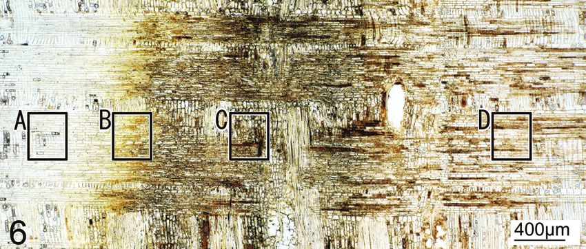
302                             IAWA Journal 34 (3), 2013

                                    INTRODUCTION

The fungus Ceratocystis includes pathogens of many economically important tree dis-
eases all over the world. In Japan, Ceratocystis ficicola Kajitani et Masuya, attacks culti-
vars of fig trees (Ficus carica L.) in orchards. This disease is a vascular wilt, and the
pathogen was first reported as Ceratocystis fimbriata Ellis et Halsted in Japan (Kato et al.
1982). Kajitani and Masuya (2011) later identified it as a new species of the same genus.
During the growing season of fig trees, which occurs from May to October in Japan,
trees infected with this fungus start wilting from the top of the shoots, the leaves gradu-
ally turn yellow, defoliation occurs leaving just fruits, and the plants finally die (Kato
et al. 1982; Shimizu et al. 1999; Nitta et al. 2005). Today, this disease occurs widely
across fig plantation areas, most likely because of the distribution of infected saplings
from diseased orchards (Kajitani et al. 1992; Shimizu et al. 1999; Togawa et al. 1999).
Some farms abandoned their orchards because of the extensive damages caused by this
wilt disease (Shimizu et al. 1999; Kajitani & Masuya 2011; Morita et al. 2012).
   The dispersal process of C. ficicola has not been fully clarified. This pathogen is
primarily recognized as a soil-borne plant disease because fig trees planted in soil pol-
luted with the pathogen C. ficicola are easily infected (Kato et al. 1982). Therefore,
application of fungicide to polluted soil and the breeding of resistant rootstocks have
been tried during this decade (Hirota et al. 1984; Shimizu et al. 1999; Togawa et al.
1999; Yakushiji et al. 2012), but are not always effective to reduce the damage.
   Kajitani (1996) found that an ambrosia beetle, Euwallacea interjectus (Blandford),
contributed as a vector of this pathogen because it carries C. ficicola on its elytron (Kaji-
tani 1999). This beetle species has a sib-mating system (inbreeding) and an extremely
female-biased sex ratio: mating occurs after emergence in their galleries (nests in the
host trees), and several male adults (brothers) of the new generation (offspring) copulate
with many female adults (sisters) and then die without dispersal flight in search of new
host trees because of their dwarf wings (H. Kajimura, personal observation). Female
adults fly from dead trees in late March and mid-July (Kajitani 1999) and invade healthy
tree trunks near the ground (Nitta et al. 2005). Morita et al. (2012) reported from field
researches that this disease became epidemic in relation to the activity of vector beetles
in Hiroshima Prefecture in western Japan. However, many researchers and orchard
owners do not believe that the beetle is a vector of this disease, supposing instead that
E. interjectus is a “secondary pest” and cannot affect healthy trees. Because the inter-
action between invasion by the beetle and development of the disease is still unclear,
precautions against pathogen transmission by the insect are not taken in orchards.
   The objective of this study is to clarify the host responses against the activities of
E. interjectus and C. ficicola with anatomical techniques, and tracing the role of this
insect in the process of disease development in fig trees.

                            MATERIALS AND METHODS
Specimens
  Specimens of Ficus carica L. cv. Horaishi were obtained from two trees (tree A
& tree B) in two adjacent orchards in Hiroshima Pref., Japan (latitude: 34° 16 ' 58 ",

                                                               Downloaded from Brill.com08/06/2021 09:08:52PM
                                                                                                via free access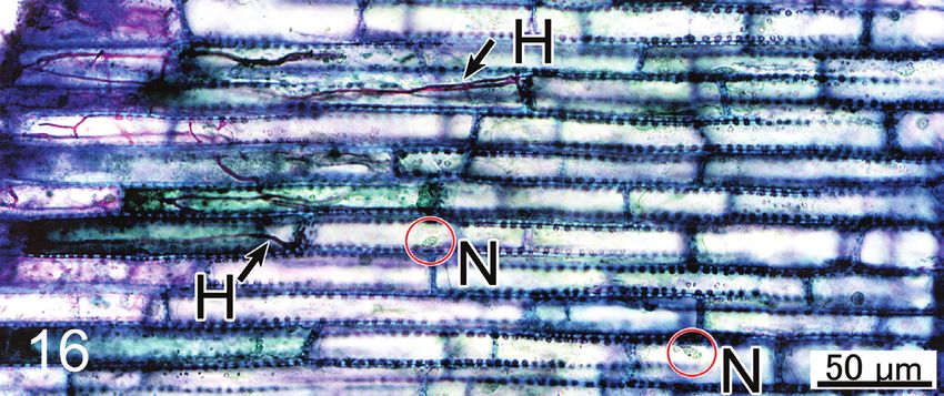
Kajii et al. – Wilt disease in Ficus xylem                             303

longitude: 132° 45' 21"). Tree A (with a main trunk height of 43 cm, and a basal stem
diameter of 29 cm) was 26 years old and harvested on 22 July, 2011. Many pinholes
made by insects assumed to be E. interjectus were found on the lower trunk near the
base on 10 August, 2010. The leaves of tree A had started wilting, but the tree was still
alive. Tree B (main trunk height 39 cm, basal stem diameter 14 cm) was 8 years old
and harvested on 17 July, 2012. Tree B had 6 pinholes on the trunk on 6 July, 2012,
but did not show any wilt symptoms at harvest time.
   Tree A was cut from the base and was dissected into discs of c. 7 cm thick (Fig. 1)
just after the harvest. Tree B was cut in the same way as tree A. Discs from tree A and B
were then brought back to the laboratory. The proportions of discoloration on the cross-
cut areas were measured with software ImageJ (National Institutes of Health).

Microscopic observation
   The surfaces of the discs obtained from the trunks and branches were observed under
a binocular microscope (Nikon, SMZ1500). For light microscopy (Nikon, ECLIPSE
80i), xylem blocks (2×2×3 cm) that contained lesions or discoloration were cut from
the discs. These xylem blocks were fixed in FAA (formalin, acetic acid, 50% ethyl alco-
hol; 5:5:90, v/v) for one week and subsequently washed for one day under tap water.
   With a sliding microtome, 20-µm-thick sections (transverse, tangential, and radial)
were cut. Parts of the sections were mounted onto slides without staining to observe
natural colors and cytological changes. Some sections were stained with safranin-fast
green (Johansen 1940) for the observation of xylem cells, and others were stained with
periodic acid-Schiff (PAS) and toluidine blue O (Feder & O’Brien 1968) for the obser-
vation of fungal hyphae.
   The following aspects were given primary focus: a) the range of discolored xylem
in the sapwood in relation to the invasion and gallery formation by the beetle, b) the
distribution of fungal hyphae in the host tissue and the reaction of host cells, and c)
the necrosis of parenchyma cells around the beetle galleries.

Fungal identification
   Xylem blocks (length × radial depth × width = 5 cm × 5 cm × 5 mm) were cut from
infected and discolored xylem of tree A and B with disposable knife blades and then
washed with 70% ethanol. Blocks were sterilized by short-period heating of the surface
with a flame, kept moist in plastic bags with wet tissue paper, and incubated at room
temperature (c. 25 °C). The perithecia formation of Ceratocystis ficicola was checked
every day for two weeks.

                                        RESULTS

External symptoms and macroscopic observation of infected fig trees
   Pinholes found on the lower trunk of tree A were confirmed to be galleries made
by Euwallacea interjectus. The fruits had not grown well since the spring of 2011,
although this tree had not indicated any symptoms of wilt and defoliation during the
previous year. Current-year shoots of tree A poorly elongated during spring, and were
much shorter than those of unaffected trees. Leaves on the current-year shoots indi-

                                                                 Downloaded from Brill.com08/06/2021 09:08:52PM
                                                                                                  via free access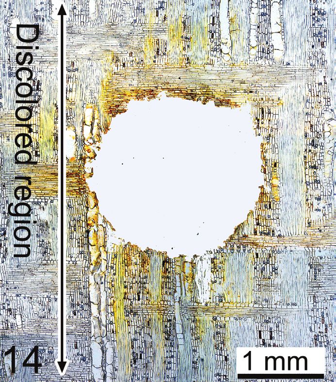
304                                IAWA Journal 34 (3), 2013

Figure 1. Trunk and basal branches of Ficus carica, with sampling positions indicated by the
letters a–e. — Figure 2. Cross section of F. carica tree at 7 cm above ground naturally infected
with Ceratocystis ficicola. (tree A, Fig. 1e). — Figure 3. A gallery of Euwallacea interjectus
in the sapwood infected with F. carica (tree B, cross section). Section I and II: see Fig. 12. —
NS = normal sapwood; D = discolored area; G = gallery of E. interjectus; B = border of dis-
coloration.

cated slight wilting at harvest. These are characteristic symptoms of fig wilt caused by
Ceratocystis ficicola.
   Dark brown discoloration could be seen with the naked eye in a wide area of sap-
wood and a part of the phloem in the basal area of the trunk (Fig. 2). The proportion of
discoloration on the crosscut area of the main stem was the greatest near the base of the
trunk and reached 75% at 7 cm, 59% at 15 cm, and 44% at 22 cm above the ground.

                                                                                                     →
Figure 4–11. Macro- and microscopic views of Ceratocystis ficicola and Ficus carica sapwood. –
4: Perithecia of C. ficicola formed on the xylem block 5 days after sample collection. – 5: Trans-
verse view of the darker band (arrow B) between discolored sapwood (D) and normal sapwood
(NS). – 6: Radial section of the darker band B in Fig. 5. Enlarged images of areas B to D are cited
in Fig. 7 to 9, respectively. – 7: Cells with colored substances (arrow CS) in the outermost part
of the border, the area B of Fig. 6. – 8: Deeply pigmented non-nucleated parenchyma cells (ar-
row) in area C of Fig. 6. – 9: Hyphae (arrow H) observed in the discolored sapwood in area D of
Fig. 6, conidiospoes (C), and colored substances (CS). – 10 & 11: Conidiospores of C. ficicola
in the discolored sapwood in area D of Fig. 6.

                                                                   Downloaded from Brill.com08/06/2021 09:08:52PM
                                                                                                    via free access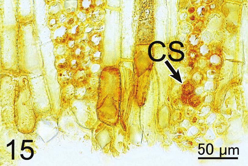
Kajii et al. – Wilt disease in Ficus xylem                             305

                                        Downloaded from Brill.com08/06/2021 09:08:52PM
                                                                         via free access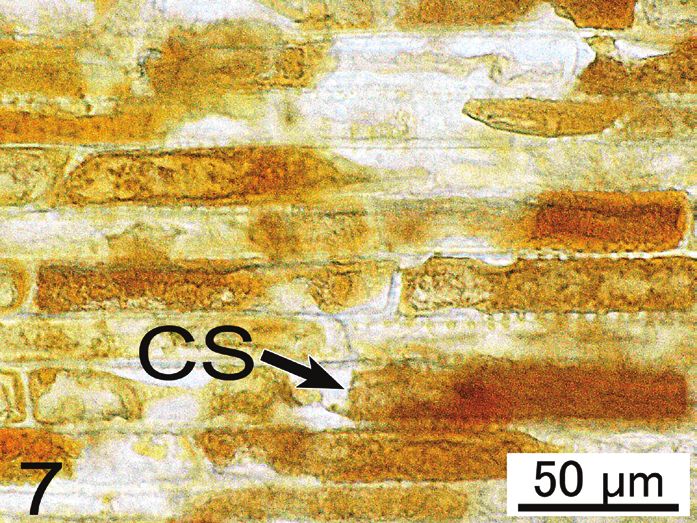
306                              IAWA Journal 34 (3), 2013

                                       Figure 12. Schematic diagram of a newly-formed gal-
                                       lery of Euwallacea interjectus in the normal sapwood
                                       (tree A). Microscopic view of Section I and Section II
                                       are shown in Fig. 14 to 17. – N: normal sapwood; E:
                                       living adult females of E. interjectus; F: area of fungal
                                       distribution; S: area of slightly yellow-stained tissue;
                                       G: gallery of E. interjectus; B: border of discoloration;
                                       D: discolored area.

At the base of two boughs, the discoloration was 31% and 41%. In the disc obtained
from the main stem from 15 cm to 22 cm above ground, 39 entrance holes excavated
by E. interjectus were found. Discolored sapwood indicated in Figure 2 covered the
area of most galleries made by the beetle. Some of the galleries were found to penetrate
through the border of discolored and non-discolored sapwood (Fig. 3).
   The trunk of tree B, which was obtained from a different orchard, adjacent to the
harvest site of tree A, had 6 holes made by E. interjectus. The beetle must have invaded
the tree during the period of early March to late July of 2012 because no beetle attack
was found in early March. Internodes of the current year’s branch were shorter in this
specimen than they were in tree A. However, tree B did not show wilt symptoms at
harvest time. The proportion of discoloration on the crosscut area was up to 52% in
the main stem.
   In tree A and B, cambial necrosis was observed adjacent to the discolored xylem area.
Cambial necrosis was not observed in the branches showing slightly wilting leaves.

Distribution of Ceratocystis ficicola in the host tissue
   On the xylem blocks from the discolored xylem of both trees, perithecia of C. fici-
cola formed about four to seven days after incubation at room temperature (Fig. 4). It
confirmed that the harvested trees had been infected with C. ficicola in the orchards.
   On the crosscut surface of trunk discs from both trees, dark bands measuring 5 to
20 mm wide were observed with the naked eye between the discolored sapwood and
the normal, functional sapwood of whitish color (Fig. 5). Detailed microscopic obser-
vation focused on the black band and the areas on both sides toward the discolored

                                                                                                   →
Figure 13–17. Host reaction associated with the activities of Euwallacea interjectus. – 13: A
new generation of female adult E. interjectus with bright colour. – 14: Beetle’s gallery in the
functional sapwood, close to its end (Section I in Fig. 12). – 15: Accumulation of brownish-
yellow substances in parenchyma cells (arrow CS) and staining of surrounding cell walls. –
16: Hyphae elongating into ray parenchyma cells (arrows H) and living cells with nucleus
(circled N). The section was stained with periodic acid-Schiff (PAS) and toluidine blue O. –
17: Older parts of the same gallery with wider discoloration (= Section II in Fig. 12).

                                                                 Downloaded from Brill.com08/06/2021 09:08:52PM
                                                                                                  via free access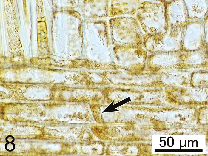
Kajii et al. – Wilt disease in Ficus xylem                             307

                                        Downloaded from Brill.com08/06/2021 09:08:52PM
                                                                         via free access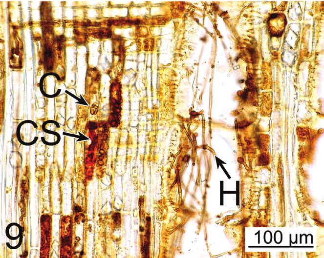
308                            IAWA Journal 34 (3), 2013

area and the normal whitish sapwood. As indicated in Figure 6, detailed microscopic
observations were made along the radius from A to D, based on the condition of the
discoloration. Area A is normal sapwood in which ray parenchyma cells contain big
round nuclei similar to the host tissue without infection. Area B is the outermost area
of discoloration (Fig. 6 B) located just outside the black bands. The ray parenchyma
cells were occluded by pale yellow droplets (Fig. 7, arrow) and some cells have nuclei
in this area. Area C is the center of the black bands (Fig. 6 C). Ray parenchyma cells
in this area contained a small amount of dark brown substance (Fig. 8, arrow), had no
nuclei, and were necrotic. Area D is inside the discolored xylem (Fig. 6 D). All paren-
chyma cells in area D contained no nuclei and were necrotic. Colored substances had
accumulated in some of the dead cells and fiber lumina (Fig. 9). In areas A and B, no
fungal hyphae were observed. In areas C and D, hyphae were observed in vessels, fibers,
and axial and ray parenchyma cells (Fig. 9, arrow). In area D, cylindrical or slightly
lageniform conidia of C. ficicola occurred (Fig. 10 & 11). Tyloses were not observed
in the vessels occupied by the fungal hyphae, although they were found in the vessels
near the boundaries between functional sapwood and infected wood.

Xylem discoloration associated with gallery elongation and fungal distribution
   One of the new galleries found in the pale-colored (sound) sapwood in tree A
is shown diagrammatically in Figure 12. Living, newly-emerged adult females of
Euwallacea interjectus (Fig. 13) were found at the end of new galleries under formation
in the normal sapwood of tree A. In tree B, living females and larvae of E. interjectus
were found in galleries that extended from the discolored area into normal sapwood
(Fig. 3). Section I in Figure 12 indicates the end of the gallery where the females of
E. interjectus occurred. Under the light microscope, a yellowish stain was observed in
the vessel lumina, fiber walls, and axial and ray parenchyma cell walls surrounding the
gallery wall (Fig. 14, arrow). Dark yellow-colored droplets were observed scattered in
the cytoplasm of some parenchyma cells in this area (Fig. 15, arrow). Inside the stained
area (Fig. 14), hyphae were observed in the lumina of vessels, fibers, and parenchyma
cells that had been mechanically broken by the beetle. In the ray tissue, fungal hyphae
are invading living parenchyma cells that contain big round nuclei similar to cells
in the unaffected tissue (Fig. 16). In section II (Fig. 12), the cell walls were stained a
darker brown color (Fig. 17. arrow), and the parenchyma cells contained darker brown
droplets than the cells of section I. The ranges of stained areas and fungal distribution
were larger in section II and formed earlier than in section I.

                                     DISCUSSION

The mortality or survival of trees infected with wilt disease is determined by the degree
of the blockage of xylem sap ascent in the trunk (Kuroda 2001, 2005; Kuroda et al.
2006). The xylem discoloration occurs in the hardwood xylem as a defense mechanism
of trees for protection against microbial invasion (Shigo & Hillis 1973; Hillis 1987)
and is called wound heartwood or pathological heartwood (Hillis 1987). It is well
known that xylem dysfunction progresses as the discolored sapwood expands in the

                                                             Downloaded from Brill.com08/06/2021 09:08:52PM
                                                                                              via free access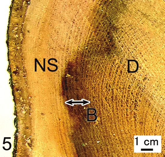
Kajii et al. – Wilt disease in Ficus xylem                             309

diseased hosts (Kuroda 1996). All parenchyma cells are necrotic and all vessels are
dysfunctional in wound heartwood as in the case of normal heartwood (Holbrook &
Zwieniecki 2005). In the present investigation, the trunks of the fig trees infected with
Ceratocystis ficicola had discolored up to 78% of the crosscut area at the base when
wilt symptoms began. This suggests that water conduction had decreased severely
and the wilt symptom started due to the deficit of water supply during the hot and dry
summer season. This progression is similar as in other wilt diseases of Quercus, Picea
and Pinus species (Kuroda 2001, 2005, 2008). Cambial necrosis, which was observed
in a part of the trunk circumference and was not observed in branches around slightly
wilting leaves, is not the direct cause of the wilt symptom as confirmed in other wilt
diseases caused by Ceratocystis species (Kuroda 2005).
    In the part of the xylem without gallery formation, successful compartmentaliza-
tion, a boundary-setting to minimize further damage by the microorganisms (Shigo &
Marx 1977; Shigo 1984) was observed. The boundary between discolored and sound
sapwood, where the parenchyma cells occluded by pale yellow droplets correspond
to the ‘reaction zone’ defined by Shain: parenchyma cells in the ‘reaction zones’ are
filled with antibiotic compounds, such as polyphenols, that are effective in preventing
the spread of pathogens in living trees (Shain 1967, 1971, 1979). Hyphae were not
observed in the normal sapwood outside this zone. The present observations suggest
that the boundary between discolored and sound sapwood is effective to prevent the
spread of pathogens as hypothesized by Shain.
    It was very significant to note that an ambrosia beetle, Euwallacea interjectus was
found excavating galleries from discolored xylem into normal sapwood devoid of
fungi. Euwallacea interjectus adults could enlarge their activity range from discolored,
necrotic xylem into sound sapwood that contains living parenchyma cells. This type
of gallery extension must have assisted wider distribution of C. ficicola in the trunk.
When a microorganism invades a tree, parenchyma cells synthesize and accumulate
antibiotic compounds consisting of phenolic substances such as terpenoids, stilbe-
noids, and alkaloids (Kemp & Burden 1986; Hillis 1987) and prevent the distribution
of pathogens. However, this defense system could not block the extension of beetle
galleries. Around the newly-extended galleries in the living sapwood, hyphae of the
pathogen were found distributing in the lumina of broken cells from the gallery. The
extension of a gallery sets up a new entrance for hyphae into sound tissue that has not
yet experienced a defense reaction. Ceratocystis ficicola is thus successful in crossing
through the defensive boundary zone by using the galleries of E. interjectus.
    As a result of fungal invasion from necrotic and dysfunctional areas into living
sapwood through the beetle’s gallery, the areas of defense reaction expanded in the
sapwood and induced enlargement of the “wound heartwood”. This relationship be-
tween vector beetle E. interjectus and C. ficicola is similar to that of ambrosia beetle,
Platypus quercivorus (Murayama), a vector of the Japanese oak wilt pathogen Raffaelea
quercivora Kubono & Ito. Wilting of oak trees also occurs because of water deficit
due to the expansion of discolored sapwood following the dense gallery formation by
P. quercivorus (Kuroda 2001; Esaki et al. 2004; Kinuura & Kobayashi 2006). In the
case of Japanese oak wilt, survival or mortality of oak trees infected with the pathogen

                                                                 Downloaded from Brill.com08/06/2021 09:08:52PM
                                                                                                  via free access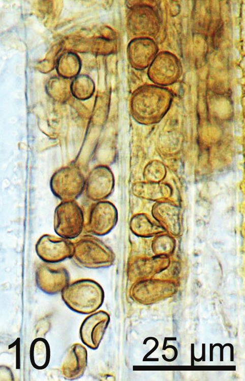
310                              IAWA Journal 34 (3), 2013

is determined during one summer (Kobayashi & Ueda 2005). In contrast, E. interjec-
tus, once colonizing the trunk of a fig tree continues to reside in the same living tree
for a few years as long as the condition in the trunk is suitable for their reproduction
(H. Kajimura, personal observation). Therefore, in the fig trees, enlargement of discolor-
ed and dysfunctional sapwood gradually progresses in association with the continuous
activity of E. interjectus. Morita et al. (2012) reported that fig trees did not die for two
years after the first invasion of E. interjectus into the stems and the simultaneous in-
fection of the pathogen. The delay of wilt symptoms in fig trees of more than one year
from the first infestation of E. interjectus seems to have induced a negative opinion
on the beetle’s contribution to the disease development. The present study showed the
role of the vector beetle in spreading pathogen in the healthy sapwood and its contri-
bution to the enlargement of the dysfunctional area. Judging from the observation of
Japanese oak wilt (Kuroda & Yamada 1996), wilt symptoms do not always occur as
long as water is supplied to the shoots, even though sap flow has diminished compared
to healthy trees. It is logical that infected trees take years from the initial attack by E.
interjectus to reach mortality, judging from the slow and gradual enlargement of xylem
discoloration until the dysfunctional area covers most of the sapwood in the basal area of
trunks. Although many ambrosia beetles have been classified as secondary beetles that
use dead, almost dead, or fallen trees for their reproduction (Furniss & Carolin 1977),
E. interjectus female adults were evidently using living trees for their reproduction.
Therefore, it does not seem correct that E. interjectus is classified as a typical second-
ary attacker. In recent years, some excavations in living trees by beetles were reported,
like Platypus quercivorus (Kühnholz 2001). It suggests that some beetles associated
with pathogenic microorganisms may utilize the trees that look healthy but are physi-
ologically stressed. For an analysis of host conditions suitable for the reproduction of
E. interjectus, detailed investigations on the invasion strategy of mother beetles into
fig trees will be necessary.
   The present investigation showed that xylem dysfunction caused by the wide dis-
tribution of C. ficicola in the sapwood is the cause of the wilt symptom of fig trees.
Although this disease is sometimes called Ceratocystis canker, the pathogen does not
form a canker but causes a wilt. In addition, the results support previous reports by
Kajitani (1996) and Morita et al. (2012) that claimed that E. interjectus is a vector of
C. ficicola. In order to clarify the responses of host cells to C. ficicola, we are currently
conducting inoculation experiments on fig saplings.

                                       REFERENCES
Esaki K, Kato K & Kamata N. 2004. Stand-level distribution and movement of Platypus quer-
    civorus adults and patterns of incidence of new infestation. Agric. For. Entomol. 6: 71–82.
Feder N & O’Brien TP. 1968. Plant microtechnique: some principles and new methods. Amer.
    J. Bot. 55: 113–142.
Furniss RL & Carolin VM. 1977. Western forest insects. U.S.D.A. Forest Service, Misc. Publ.
    1339. 655 pp.
Hillis WE. 1987. Heartwood and tree exudates. Springer-Verlag, Berlin, Heidelberg, New York,
    London, Paris, Tokyo. 268 pp.

                                                                 Downloaded from Brill.com08/06/2021 09:08:52PM
                                                                                                  via free access
Kajii et al. – Wilt disease in Ficus xylem 311
Hirota K, Kato K & Miyagawa T. 1984. Chemical control for fig-shoot blighting disease, Cerato-
cystis canker. Res. Bull. Aichi Res. Cent. 16: 211–218 [in Japanese with English sum-
mary].
Holbrook NM & Zwieniecki MA. 2005. Vascular transport in plants. Elsevier Academic Press,
Burlington. 592 pp.
Johansen DA. 1940. Plant microtechnique. McGraw-Hill, New York. 523 pp.
Kajitani Y. 1996. The possibility of transmission of fig Ceratocystis canker disease by an ambro-
sia beetle (Xyleborus interjectus Eichhoff). Ann. Phytopathol. Soc. Jpn. 62: 275 [in Japa-
nese].
Kajitani Y. 1999. The dispersal period of the Xyleborus interjectus (Coleoptera, Scolytidae), a
vector of the fig Ceratocystis canker, and the organ carrying the causal fungus. Ann. Phyto-
pathol. Soc. Jpn. 65: 377 [in Japanese].
Kajitani Y & Masuya H. 2011. Ceratocystis ficicola sp. nov., a causal fungus of fig canker in
Japan. Mycoscience 52: 349–353.
Kajitani Y, Tsutumi T & Yamada K. 1992. Occurrence of Ceratocystis canker of fig caused by
Ceratocystis fimbriata Ellis et Halsted in Fukuoka prefecture. Kyushu Agric. Res. 54: 89
[in Japanese, tentative translation from Japanese title].
Kato K, Yokota K & Miyagawa T. 1982. A new disease, Ceratocystis canker of fig caused by
Ceratocystis fimbriata Ellis et Halsted. Plant Prot. 36: 55–59 [in Japanese].
Kemp MS & Burden RS. 1986. Phytoalexins and stress metabolites in the sapwood of trees.
Phytochemistry 25: 1261–1269.
Kinuura H & Kobayashi M. 2006. Death of Quercus crispula by inoculation with adult Pla-
typus quercivorus (Coleoptera: Platypodidae). Appl. Entomol. Zool. 41: 123–128.
Kobayashi M & Ueda A. 2005. Wilt disease of Fagaceae trees caused by Platypus quercivorus
(Murayama) (Coleoptera : Platypodidae) and the associated fungus: aim is to clarify the
damage factor. Jpn. For. Soc. 87: 435–450 [in Japanese with English summary].
Kühnholz S, Borden JH & Uzunovic A. 2001. Secondary ambrosia beetles in apparently heal-
thy trees: Adaptations, potential causes and suggested research. Integrated Pest Management
Reviews 6: 209–219.
Kuroda K. 2001. Responses of Quercus sapwood to infection with the pathogenic fungus of a
new wilt disease vectored by the ambrosia beetle Platypus quercivorus. J. Wood Sci. 47:
425– 429.
Kuroda K. 2005. Xylem dysfunction in Yezo spruce (Picea jezoensis) after inoculation with the
blue-stain fungus Ceratocystis polonica. For. Path. 35: 346–358.
Kuroda K. 2008. Physiological incidences related to symptom development and wilting
mechanism. “Pine Wilt Disease”. Zhao, Futai, Sutherland, Takeuchi (eds.). pp. 204–222.
Springer.
Kuroda K, Kanbara Y, Inoue T & Ogawa A. 2006. Magnetic resonance micro-imaging of xylem
sap distribution and necrotic lesions in tree stems. IAWA J. 27: 3–17.
Kuroda K & Yamada T. 1996. Discoloration of sapwood and blockage of xylem sap ascent in
the trunks of wilting Quercus spp. following attack by Platypus quercivorus. Jpn. For. Soc.
78: 84 –88 [in Japanese with English summary].
Morita T, Hara H, Mise D & Jikumaru S. 2012. A case study of Ceratocystis canker epidemic in
relation with Euwallacea interjectus infestation. Ann. Rept. Kansai Pl. Prot. 54: 29–34 [in
Japanese with English summary].
Nitta H, Morita T, Wakasaki Y & Kakogawa K. 2005. Relationship between Ceratocystis
canker and ambrosia beetle in fig orchards. Ann. Rept. Kansai Pl. Prot. 47: 95–98 [in Japa-
nese].
Downloaded from Brill.com08/06/2021 09:08:52PM
via free access312 IAWA Journal 34 (3), 2013
Shain L. 1967. Resistance of sapwood in stems of loblolly pine to infection by Fomes annosus.
Phytopathology 57: 1034–1045.
Shain L. 1971. The response of sapwood of Norway spruce to infection by Fomes annosus. Phyto-
pathology 61: 301–307.
Shain L. 1979. Dynamic responses of differentiated sapwood to injury and infection. Phyto-
pathology 69: 1143–1147.
Shigo AL. 1984. Compartmentalization: A conceptual framework for understanding how trees
grow and defend themselves. Ann. Rev. Phytopathology 22: 189–214.
Shigo AL & Hillis WE. 1973. Heartwood, discolored wood and microorganisms in living trees.
Ann. Rev. Phytopathology 11: 197–222.
Shigo AL & Marx HG. 1977.Compartmentalization of decay in trees. U.S.D.A. Forest Service
Agriculture Information Bull. 405. 73 pp.
Shimizu S, Miyoshi T, Ochi M & Tachibana Y. 1999. Occurrence of fig Ceratocystis canker in
Ehime prefecture and its control by thiophanate-methyl triflumizole fungicide. Bull. Ehime
Fruit Tree Exp. Stn. 13: 27–35 [in Japanese with English summary].
Togawa M, Masui S, Nomura A & Masui H. 1999. Occurrence and control of stem rot on fig.
Bull. Shizuoka Citrus Exp. Stn. 28: 51–62 [in Japanese with English summary].
Yakushiji H, Morita T, Jikumaru S, Ikegami H, Azuma A & Koshita Y. 2012. Interspecific
hybridization of fig (Ficus carica L.) and Ficus erecta Thunb., a source of Ceratocystis
canker resistance. Euphytica 183: 39– 47.
Accepted: 25 June 2013
Downloaded from Brill.com08/06/2021 09:08:52PM
via free accessYou can also read