Sevelamer hydrochloride, a phosphate binder, protects against deterioration of renal function in rats with progressive chronic renal insufficiency
←
→
Page content transcription
If your browser does not render page correctly, please read the page content below
Nephrol Dial Transplant (2003) 18: 2014–2023
DOI: 10.1093/ndt/gfg309

Original Article

Sevelamer hydrochloride, a phosphate binder, protects against
deterioration of renal function in rats with progressive chronic
renal insufficiency

Nobuo Nagano1, Sonoe Miyata1, Sachiko Obana1, Nami Kobayashi1, Naoshi Fukushima2,
Steven K. Burke3 and Michihito Wada1

                                                                                                                             Downloaded from https://academic.oup.com/ndt/article/18/10/2014/1807542 by guest on 01 July 2022
1
 Pharmaceutical Development Laboratories, Kirin Brewery Co., Ltd, Takasaki, 2Fuji Gotenba Research Laboratory,
Chugai Pharmaceutical Co., Ltd, Shizuoka, Japan and 3GelTex Pharmaceuticals, Inc., MA, USA

Abstract                                                         ing kidney calcium at a low level as a result of reducing
Background. Dietary phosphate restriction prevents               serum phosphorus and PTH.
renal function deterioration in animal models. This
study examined whether sevelamer hydrochloride                   Keywords: nephrocalcinosis; parathyroid hormone;
(RenagelÕ; ‘sevelamer’ hereafter), a non-calcaemic               phosphate binder; renal function; serum phosphorus;
phosphate binder could slow deterioration of renal               sevelamer hydrochloride (RenagelÕ)
function in rats with progressive renal insufficiency.
Methods. Wistar Kyoto male rats were singly injected
with normal rabbit serum or rabbit anti-rat glomerular
basement membrane serum. Three days later, rats were             Introduction
fed a powder diet containing 0, 1 or 3% sevelamer for
58 days. Time course changes of serum levels of blood            Phosphate is one of many factors that can promote
urea nitrogen (BUN), creatinine, calcium, phosphorus             progression of renal failure. There is ample evidence
and parathyroid hormone (PTH) were measured                      that dietary phosphate overload accelerates, and
throughout, and creatinine clearance (CCr), kidney               dietary phosphate restriction prevents, the progression
calcium content and renal histology examined at the              of chronic renal insufficiency (CRI) in experimental
end of the study.                                                animal models [1–6]. Although the mechanism of
Results. Sevelamer partially inhibited elevation of              phosphate toxicity in renal failure has not been fully
BUN and serum creatinine, and completely inhibited               established, nephrocalcinosis, renal haemodynamics
increases in serum phosphorus, PTH and calcium                  and intrarenal hypermetabolism are considered to be
phosphorus product. Sevelamer significantly prevented             involved [5,6].
the decrease in CCr and kidney calcium content ele-                 Calcium carbonate and acetate have been widely
vation. Kidney calcium content and BUN and serum                 used as phosphate binders in patients with end-stage
creatinine were strongly positively correlated, and              renal disease. However, there is a limit on the use of
kidney calcium content and CCr strongly negatively               these calcium-containing phosphate binders in pre-
correlated. Kidney calcium content correlated well               dialysis patients because of the risk of causing
with serum phosphorus, serum calcium  phosphorus                deterioration of renal function by nephrocalcinosis.
product and PTH, but not serum calcium. Sevelamer                Sevelamer hydrochloride (RenagelÕ; hereafter referred
treatment partly prevented histological deterioration            to as sevelamer) is a calcium-free and aluminum-free
of both glomerular and tubulointerstitial lesions of             phosphate-binding polymer marketed for the treatment
the kidney.                                                      of hyperphosphataemia in patients undergoing haemo-
Conclusions. The results suggest that sevelamer pro-             dialysis. Many beneficial effects of sevelamer have
tects against renal function deterioration by maintain-          been demonstrated in experimental animals [7,8] and
                                                                 in the clinic [9–11], such as a prevention of parathy-
                                                                 roid hyperplasia and lowering of serum phosphorus,
Correspondence and offprint requests to: Nobuo Nagano, PhD,
Pharmaceutical Development Laboratories, Kirin Brewery Co.,
                                                                 calcium  phosphorus product, parathyroid hormone
Ltd., 3 Miyahara-cho, Takasaki-shi, Gunma 370-1295, Japan.       (PTH), and low-density lipoprotein (LDL) cholesterol
Email: n-nagano@kirin.co.jp                                      without increasing serum calcium levels. It is thought

ß 2003 European Renal Association–European Dialysis and Transplant Association
Phosphate binder protects renal function 2015
that the reduction of calcium phosphorus product and MCP-1 levels and both kidneys were immediately
and LDL cholesterol levels could reduce the incidence dissected out.
of metastatic calcification. Indeed, long-term studies
employing electron beam computed tomography have
demonstrated beneficial effects of sevelamer, as com- Serum and urinary chemistries
pared with calcium-based phosphate binders, on cardiac Serum phosphorus, calcium, blood urea nitrogen (BUN),
and aortic calcification in patients receiving haemo- total cholesterol and urinary phosphorus, calcium, and
dialysis [11]. protein levels were measured with commercial kits (Wako
It is known that Wistar Kyoto (WKY) rats are more Pure Chemical Industries, Ltd, Osaka, Japan) and standard
susceptible to anti-glomerular basement membrane calorimetric methods employing a U-2000 spectrophotometer
(GBM) serum than other strains. WKY rats injected (Hitachi Ltd, Tokyo, Japan). Serum and urinary creatinine
with a small volume of anti-GBM serum rapidly were measured by enzymatic assay (CRE-EN, Kynos, Tokyo,
progress to CRI with glomerulosclerosis and tubulo- Japan) and creatinine clearance (CCr) was calculated from a
interstitial scarring [12]. It has been suggested that standard formula. Serum PTH, 1,25(OH)2D3 and MCP-1
macrophages as well as CD8-positive T lymphocytes levels were measured with a rat PTH-(1–34) immunoradio-
Downloaded from https://academic.oup.com/ndt/article/18/10/2014/1807542 by guest on 01 July 2022
are involved in the initiation and subsequent progres- metric assay (Nichols Institute Diagnostics, CA, USA), radio
sion of CRI in this model [13,14]. Wada et al. [13] receptor assay (SRL, Yamasa, Tokyo, Japan) and enzyme
have reported that injection of antibody against mono- immunoassay (Immuno-Biological Laboratories, Fujioka,
cyte chemoattractant protein-1 (MCP-1), a monocyte Japan), respectively.
recruiting and activating factor, prevents glomerulo-
sclerosis and renal dysfunction with inhibition of Preparation of antibody
macrophage infiltration.
In the present study, we examined whether dietary Rat GBM was prepared from 54 male Sprague–Dawley (SD)
treatment with sevelamer could prevent the progression rats (10–12 weeks of age, SLC Japan, Tokyo, Japan) by the
of CRI in this animal model. In addition, the protective method of Shibata [15]. GBM was digested with trypsin at
effect of sevelamer on renal function was analysed with 37 C for 3 h. After heating at 60 C for 30 min, the mixture
particular attention to nephrocalcinosis. was centrifuged at 27 000 r.p.m. at 3 C for 35 min and the
supernatant lyophilized. The lyophilized sample was mixed
with complete Freund’s adjuvant (Difco Laboratories, MI,
USA) and injected subcutaneously four times at intervals
Subjects and methods of 1–2 weeks into four male rabbits (New Zealand White,
13 weeks of age, SLC Japan). Two weeks after the last
Experimental protocol subcutaneous (s.c.) immunization, blood was collected from
the cervical arteries under pentobarbital sodium anaesthesia
The experimental protocol was approved by the Experimental (20 mg/kg, i.v.). Sera were separated and immobilized at 56 C
Animal Ethical Committee of Kirin Brewery Co., Ltd. Male for 30 min and frozen at –80 C until use. Specificity was
WKY rats, 8 weeks of age, were purchased from Charles confirmed by the Ouchterlony gel diffusion method: a clear
River Japan (Tokyo, Japan) and fed a standard powder diet common precipitate line was observed between antigen and
containing 0.85% phosphorus, 1.12% calcium, 25.3% crude diluted antiserum. In addition, anti-GBM antibody gave a
protein and 2.5 IU/g vitamin D3 (CE-2, CLEA Japan, Tokyo, strong positive finding of the GBM on frozen sections of
Japan). Rats were kept singly in cages and allowed free access normal SD rat kidney when followed by incubation with
to food and water. After an acclimatization period of 7 days, fluorescein isothiocyanate conjugated anti-rabbit IgG
a blood sample was collected from the tail artery to measure (Seikagaku Kogyo Co., Ltd, Tokyo, Japan).
serum phosphorus, calcium and PTH levels (day –4). The rats
were divided into five groups of 12 animals each, matched
with respect to body weight, serum phosphorus and calcium Kidney calcium content
levels. The following day (day –3), three of the groups
The right kidneys were frozen at –20 C for subsequent
received a single injection, via the tail vein, of 0.1 ml/kg rabbit
analysis. After lyophilization, dried kidneys were delipidized
anti-rat GBM serum produced in our laboratory as described
with a mixed solution of chloroform and methanol (2:1) for
below (CRI group). The fourth group received the same
48 h, and dehydrated by acetone for 3 h. Samples were burned
volume of normal rabbit serum (Funakoshi, Tokyo, Japan)
to ash at 550 C for 12 h using an electric muffle furnace (KM-
(control group). On the same day, the fifth group (untreated
600, Advantec Toyo Seisakusho Co., Ltd, Tokyo, Japan), and
rats) was anaesthetized with ether and blood and bilateral
then dissolved in hydrochloric acid. Before measuring calcium
kidneys were sampled (baseline group). Three days later
by means of a commercial kit (OCPC method, Wako Pure
(day 0), rats of the CRI groups were given free access to a diet
Chemical Industries, Ltd.), the solution was diluted with an
containing 0, 1 or 3% sevelamer for the following 58 days
appropriate volume of distilled water.
while the control group was fed a normal diet. Body weight
and volume of food intake were measured every week, and
blood samples collected from the tail artery on days 3, 10, 17,
Histological evaluation
24, 31, 38, 45, 52 and 58. On days 55–56, the rats were held
singly in a metabolic cage to collect urine over 24 h. On day The left kidneys were fixed in Bouin’s fixative, composed of
58, blood was collected by abdominal aortic puncture under picric acid, formalin and acetic acid, at 4 C overnight. After
ether anaesthesia to measure serum creatinine, 1,25(OH)2D3 dehydration by passage through an ethanol/xylene series,2016 N. Nagano et al.
tissues were embedded in TissuePrep (Fisher Scientific Co., Results
NJ, USA) and cut into 1 mm sections. After sequential
dewaxing and rehydration, slides were stained with Body weight and food intake volume
haematoxylin–eosin and periodic acid–Schiff for histological
evaluation. Body weight was significantly lower in the CRI rats and
A semi-quantitative scoring procedure was carrried out the difference increased as the study progressed (Figure
at the Takasaki Pathology Center of Nippon Experimental 1). The body weights in the sevelamer treated groups
Medical Research Institute Co. Ltd (Takasaki, Japan) to were larger than those of the CRI rats receiving a
evaluate the degree of tissue damage. 100 glomeruli in each normal diet and the differences between the normal diet
specimen were examined and the severity of the lesion was group and the 3% sevelamer group on day 44 and 51
graded from 0 to 4þ. Thus, a 1þ lesion represented a one- were statistically significant. Mean food intake volumes
quarter area of the glomerulus revealing sclerosis while a 4þ (g/day), measured weekly throughout the study, were
lesion indicated four-quarters of the area of the glomerulus as follows: control, 18.6 ± 0.2; CRI rats receiving
revealing sclerosis. The injury scores of 100 glomeruli were normal diet, 14.0 ± 0.3; 1% sevelamer, 15.3 ± 0.3;
summed. Tubulointerstitial lesions were similarly scored 3% sevelamer, 16.5 ± 0.3. The mean administered
Downloaded from https://academic.oup.com/ndt/article/18/10/2014/1807542 by guest on 01 July 2022
according to severity. Thus, a 1þ represented a 0–25% mild
sevelamer doses (mg/day) throughout the study were
lesion and a 4þ represented a 75–100% severe lesion.
calculated as 153.2 ± 2.7 and 496.0 ± 10.2 in the
1 and 3% sevelamer groups, respectively. One animal
died at day 45 in the 3% sevelamer group and one
Drugs animal in the CRI group receiving a normal diet died at
Sevelamer (cross-linked poly[allylamine hydrochloride], the day 53.
active ingredient of RenagelÕ) was synthesized by The Dow
Chemical Company (Midland, MI, USA) and supplied via
Chugai Pharmaceuticals Co., Ltd (Tokyo, Japan). Serum and urinary chemistries
Hypocalcaemia was observed in the CRI groups from
days 3 to 38 and treatment with sevelamer had no
Statistics obvious effect on serum calcium levels at any time
All values are expressed as means ± SEM. The data obtained (Figure 2A). Serum phosphorus progressively increased
from control and CRI rats receiving a normal diet were in the CRI rats receiving a normal diet as the study
compared using the Student’s t-test. Multiple comparisons progressed. The 1% sevelamer treatment significantly
were performed among the three CRI groups using the inhibited the occurrence of hyperphosphataemia, and
parametric Dunnett’s test. The correlation between kidney the 3% sevelamer treatment kept serum phosphorus
calcium content and serum parameters and CCr was analysed levels below control levels from days 3 to 38 and then
using Pearson’s correlation test. P < 0.05 was taken to maintained normal levels (Figure 2B). The changes
indicate statistical significance. in serum calcium phosphorus product of all groups
Fig. 1. Effect of dietary treatment with sevelamer on body weight changes in rats with CRI. Values are mean ± SE (n ¼ 11–12) and the
missing SEM bars are hidden within the symbols. Control rats were fed a normal diet (filled circle) and rats with CRI were fed either a normal
diet (open circle), or a diet containing 1 (open triangle) or 3% sevelamer (open square) for 58 days. #P < 0.05, ###P < 0.001 vs control rats fed
a normal diet. *P < 0.05 vs rats with CRI fed a normal diet.Phosphate binder protects renal function 2017
Downloaded from https://academic.oup.com/ndt/article/18/10/2014/1807542 by guest on 01 July 2022
Fig. 2. Effect of dietary treatment with sevelamer on levels of serum calcium (A), phosphorus (B) and calcium phosphorus product (C) in
rats with CRI. Values are mean ± SE (n ¼ 11–12) and the missing SEM bars are hidden within the symbols. Control rats were fed a normal
diet (filled circle) and rats with CRI were fed either a normal diet (open circle) or a diet containing 1 (open triangle) or 3% sevelamer (open
square) for 58 days. #P < 0.05, ##P < 0.01, ###P < 0.001 vs control rats fed a normal diet. *P < 0.05, **P < 0.01, ***P < 0.001 vs rats with
CRI fed a normal diet.
were similar to the changes in serum phosphorus rats receiving a normal diet while they were within
throughout the study (Figure 2C). the normal ranges in the control group. One per cent
BUN and serum creatinine levels increased gradually sevelamer treatment appeared to inhibit the elevation
from days 3 to 38 and sharply after day 38 in the of BUN and serum creatinine levels but the differences2018 N. Nagano et al.
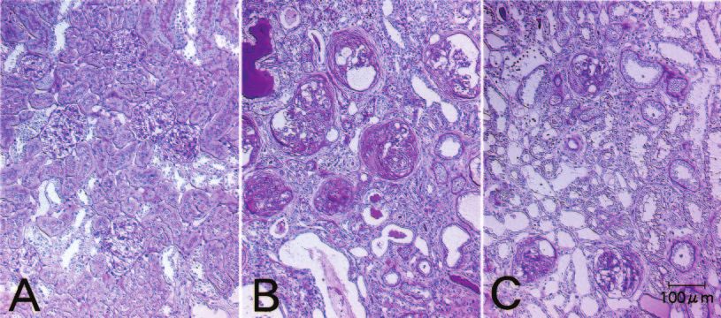
were not statistically significant, whereas 3% sevelamer Histology
treatment inhibited these increases to a statistically
Each photograph shown in Figure 7 is of the renal
significant extent (Figure 3A and B). Serum PTH rose
cortex of the rat whose serum creatinine level was
progressively and steeply parallel to the elevation of
closest to the mean of each group. Control animals
BUN and serum creatinine. Both 1 and 3% sevelamer
displayed normal renal histology (Figure 7A). In
treatments maintained serum PTH levels at control
contrast, many kinds of glomerular lesions, such as
levels until day 52. At the end of the study, a slight
glomerular hypertrophy, glomerular atrophy, focal seg-
elevation was observed in the 1% sevelamer group
mental sclerosis, crescentic formation and Bowman’s
while there was still no increase in the 3% sevelamer
capsule thickening occurred extensively in the kidneys
group (Figure 3C).
of the CRI rats receiving a normal diet (Figure 7B).
Serum total cholesterol increased markedly in all
Tubulointerstitial changes, such as dilated tubules with
three CRI groups. This was not affected by 1%
deciduation of the brush border membrane, tubular
sevelamer, but 3% sevelamer tended to cause a decrease
atrophy, infiltration of mononuclear cells, fibrosis and
throughout the study (Table 1). A marked reduction in
cast formation also dominated in this group. The
serum 1.25(OH)2D3 levels was observed in CRI
Downloaded from https://academic.oup.com/ndt/article/18/10/2014/1807542 by guest on 01 July 2022
extent of histological deterioration was almost the same
rats receiving a normal diet at the end of the study
in the CRI group receiving a normal diet and the
(Table 1). This was unaffected by 1% sevelamer, but
1% sevelamer group (data not shown). In contrast,
3% sevelamer tended to reverse this effect to an extent,
glomerular sclerosis and tubulointerstitial lesions
although the level remained much lower than in the
tended to be reduced in the 3% sevelamer group
controls. A 3.1-fold elevation of serum MCP-1 levels
(Table 2) although some degree of crescentic formation
was observed at the end of the study in the CRI rats
and thickening of Bowman’s capsule still remained
receiving a normal diet (Table 1). Sevelamer treatment
(Figure 7C). Markedly increased mononuclear cell
did not affect this increase.
infiltration in the periglomerular and tubulointerstitial
CRI rats receiving a normal diet showed marked
regions was observed in the CRI animals receiving a
polyuria and massive proteinuria at the end of the
normal diet. Sevelamer treatment did not significantly
study (Table 1), and neither 1 nor 3% sevelamer treat-
affect the number of mononuclear cells in the tubulo-
ment affected these parameters. Significant decreases
interstitial regions (Table 2).
in urinary calcium, phosphorus and calcium
phosphorus product were observed in CRI rats
receiving a normal diet (Table 1). Sevelamer increased
urinary calcium concentration and decreased urinary Discussion
phosphorus concentration in a dose-dependent manner.
Urinary calcium phosphorus product was not sig- Sevelamer treatment increased both food intake
nificantly affected by the sevelamer treatment. volume and body weight gain in the CRI rats. It is
likely that this effect is due, in part, to an improvement
in renal function. Hence, although it is widely accepted
CCr and kidney calcium content
that dietary protein restriction as well as phosphate
CCr of the CRI rats receiving a normal diet decreased restriction can protect against deterioration of renal
significantly to 13% of the control group. Treatment function in experimental animal models, we can clearly
with 1% sevelamer tended to increase CCr but not exclude the effect of dietary protein intake from the
significantly, and 3% sevelamer increased CCr to a renal protective effect of sevelamer in this study.
significant extent at the end of the study (Figure 4A). Sevelamer treatment clearly inhibited the elevation of
The kidney calcium content of the control group was BUN and serum creatinine levels and prevented the
almost the same as that of the baseline group. A 4.8- reduction of CCr in rats with CRI. This protective
fold increase in kidney calcium content was observed in effect of sevelamer on renal function was supported by
the CRI rats receiving a normal diet. The 1 and 3% histological observations showing that both glomerular
sevelamer treatments decreased kidney calcium content sclerosis and tubulointerstitial lesions were partially
to 52 and 39%, respectively, of that of the CRI rats ameliorated. In a previous experiment using CRI rats
receiving a normal diet (Figure 4B). induced by adriamycin (ADR), we observed that
sevelamer treatment tended to inhibit the elevation of
BUN and to prevent the reduction of CCr, but the
Correlation analysis
effects were not statistically significant [8]. A possible
Kidney calcium content was highly correlated with explanation for the lack of a clear cut renal protective
BUN and serum creatinine levels (Figure 5). As effect of sevelamer in that case may be the 4-week delay
expected, we observed a strong negative correlation that ensued between the initiation of renal injury and
between kidney calcium content and CCr. Kidney the start of sevelamer treatment. Irreversible changes
calcium content also correlated well with serum had probably already taken place during this interval
phosphorus, serum calcium phosphorus product and because the ADR rats showed marked proteinuria and
serum PTH levels (Figure 6). In contrast, no signifi- hypercholesterolaemia at the onset of sevelamer treat-
cant correlation was observed between kidney calcium ment. Despite the outcome of studies with experimental
content and serum calcium. animals, phosphate restriction is not an establishedPhosphate binder protects renal function 2019
Downloaded from https://academic.oup.com/ndt/article/18/10/2014/1807542 by guest on 01 July 2022
Fig. 3. Effect of dietary treatment with sevelamer on levels of serum BUN (A), creatinine (B) and PTH (C) in rats with CRI. Values are
mean ± SE (n ¼ 11–12) and the missing SEM bars are hidden within the symbols. Control rats were fed a normal diet (filled circle) and rats
with CRI were fed either a normal diet (open circle), or a diet containing 1 (open triangle) or 3% sevelamer (open square) for 58 days.
##
P < 0.01, ###P < 0.001 vs control rats fed a normal diet. *P < 0.05, **P < 0.01, ***P < 0.001 vs rats with CRI fed a normal diet.
approach for delaying the progression of renal failure phosphate-restricted regimens were started long after
in the clinic. However, a beneficial effect of dietary the onset of CRI.
phosphate restriction has been reported in patients with Although many mechanisms for the protective effect
an early stage of CRI [6]. A similar explanation to of sevelamer need to be examined, the present study
that given above might apply, namely that the suggests that the primary mechanism is the prevention2020 N. Nagano et al.
Table 1. Effect of dietary treatment with sevelamer on serum and urinary chemistries
Group Control þ normal diet CRI þ normal diet CRI þ 1% sevelamer CRI þ 3% sevelamer
No. of animals 12 11 or 12 12 11 or 12
Serum chemistries
Total cholesterol (mg/dl) (Day 17) 96.9 ± 1.9 291 ± 10### 262 ± 9 237 ± 16**
(Day 38) 102 ± 2 410 ± 9### 398 ± 11 354 ± 29
(Day 58) 97.9 ± 2.1 326 ± 29### 326 ± 23 307 ± 21
1,25(OH)2D3 (pg/dl) 32.4 ± 1.7 2.9 ± 0.8### 3.1 ± 0.6 5.3 ± 1.5
MCP-1 (pg/ml) 723 ± 130 2256 ± 25### 2317 ± 144 1985 ± 400
Urinary chemistries
Volume (ml/day) 18.4 ± 1.1 36.7 ± 2.7### 39.8 ± 1.9 33.6 ± 4.1
Protein (mg/kg, b.w.) 53 ± 3.5 1263 ± 97### 1222 ± 66 1011 ± 80
Calcium (mg/dl) 10.5 ± 1 3.4 ± 0.4### 5.5 ± 0.4 13.8 ± 3.3***
Phosphorus (mg/dl) 147 ± 8 55.2 ± 2.1### 31.4 ± 1.5*** 19.7 ± 3.7***
Calcium phosphorus (mg2/dl2) 1549 ± 182 186 ± 23### 177 ± 21 317 ± 140
Downloaded from https://academic.oup.com/ndt/article/18/10/2014/1807542 by guest on 01 July 2022
###
All parameters except serum cholesterol were determined at the end of the study. P < 0.001 vs control rats fed a normal diet.
**P < 0.01, ***P < 0.001 vs rats with CRI fed a normal diet.
of nephrocalcinosis. Sevelamer maintained serum phos-
phorus at a low level and prevented nephrocalcinosis.
In experimental studies, removal of phosphate from
the medium of cultured monkey kidney cells reduced
intracellular calcium content both by reducing calcium
influx and increasing efflux [16]. It is widely accepted
that increased intracellular calcium induces cell death,
fibrosis and scarring. Therefore, the protective effect
of sevelamer on renal function is perhaps achieved
by preventing nephrocalcinosis owing to the lower
concentration of extracellular phosphorus. Serum
calcium phosphorus product has been used as a
reliable parameter for predicting metastatic calcifi-
cation. We have directly demonstrated above that
reduction of this parameter by sevelamer is strongly
associated with inhibition of nephrocalcinosis. In
contrast to serum, a pronounced decrease in urinary
calcium phosphorus product associated with poly-
uria was observed in CRI rats receiving a normal diet.
Although it has been suggested that increased tubular
fluid phosphorus and calcium phosphorus product
results in tubular calcium-phosphate precipitation [5],
this is not likely to be one of the causes of nephro-
calcinosis in this experimental model. Sevelamer
treatment increased urinary calcium and decreased
urinary phosphorus concentrations, and we have
observed that in normal rats these effects of sevelamer
are mainly mediated by a reduction of serum PTH [7].
It is interesting to note that PTH-regulated mechanisms
for renal mineral handling are preserved in rats with
severe CRI.
Sevelamer treatment markedly inhibited the eleva-
tion of serum PTH, and there was a strong positive
correlation between serum PTH and kidney calcium
content. The decrease in serum PTH achieved by
sevelamer treatment is probably mainly due to
prevention of hyperphosphataemia and partly due to
Fig. 4. Effect of dietary treatment with sevelamer on CCr (A) at the the preservation of renal function. PTH has been
end of the study and kidney calcium content (B) at baseline and considered an important cause of nephrocalcinosis
at the end of the study in rats with CRI. Values are mean ± SE
(n ¼ 11–12) and the missing SEM bars are hidden within the
because administration of parathyroid extract
columns. ##P < 0.01, ###P < 0.001 vs control rats fed a normal diet. results in renal calcification [17,18] by increasing cyto-
*P < 0.05, **P < 0.01 vs rats with CRI fed a normal diet. solic calcium in proximal tubular cells [19,20], andPhosphate binder protects renal function 2021
Downloaded from https://academic.oup.com/ndt/article/18/10/2014/1807542 by guest on 01 July 2022
Fig. 5. Analysis of the correlations between BUN (A), serum creatinine (B) and CCr (C) with kidney calcium content in the baseline group
(open diamond), control rats fed a normal diet (filled circle), and rats with CRI fed either a normal diet (open circle), or a diet containing
1 (open triangle) or 3% sevelamer (open square) for 58 days.
Fig. 6. Analysis of the correlations between levels of serum calcium (A), phosphorus (B), calcium phosphorus product (C) and PTH (D)
with kidney calcium content in the baseline group (open diamond), control rats fed a normal diet (filled circle), and rats with CRI fed either a
normal diet (open circle), or a diet containing 1% (open triangle) or 3% sevelamer (open square) for 58 days.2022 N. Nagano et al.
parathyroidectomy can prevent renal calcification and between nephrocalcinosis and mononuclear cell accu-
functional deterioration [21]. In addition, the PTH- mulation in the present study. Sevelamer decreased
induced increase in intracellular calcium in kidney cells nephrocalcinosis without affecting mononuclear cell
is greatly enhanced by the presence of phosphate in the infiltration. It has been reported that dietary phos-
extracellular medium [20]. It is therefore possible that phate restriction reduces proteinuria in the remnant
the reduction in serum phosphorus and the reduction in kidney model [2,4] but not in the immuno-mediated
PTH levels brought about by sevelamer treatment act glomerulonephritis model [3]. The latter finding is
synergistically to reduce nephrocalcinosis. supported by our failure to observe any obvious
Another explanation is also possible. The reduc- protective effect of sevelamer on urinary protein
tion in serum phosphorus and PTH levels due to excretion, crescent formation or Bowman’s capsule
sevelamer treatment may alter local immunological thickening. Thus, it is unlikely that immunological and
and inflammatory processes in the kidney and this inflammatory processes mediate the protective effect of
may help preserve renal function. Marked elevation of sevelamer on renal function.
serum MCP-1 levels and increased infiltrated mono- Another beneficial action of sevelamer is a reduction
nuclear cell number were observed in rats with CRI. in LDL cholesterol in patients receiving haemodialysis,
Downloaded from https://academic.oup.com/ndt/article/18/10/2014/1807542 by guest on 01 July 2022
Macrophages are considered to play an important role because this should reduce the probability of cardio-
in pathogenesis in this model, especially in glomerular vascular disease and possibly prolong survival [9–11].
crescent formation [13,14]. However, there were no Many studies on experimental animal models have
obvious differences in serum MCP-1 levels and in demonstrated that lipid abnormalities aggravate renal
the number of infiltrated mononuclear cells between the injury in many forms of atherosclerosis [23]. In par-
normal diet group and the 3% sevelamer group in the ticular, activated macrophages are thought to play an
rats with CRI. Macrophages are also known to play an important role in the etiology of renal injury and
important role in arterial intimal calcification [22]. atherosclerosis when lipid levels are perturbed. In this
Nonetheless, there did not seem to be any relationship study, a marked increase in serum total cholesterol was
Fig. 7. Effects of dietary treatment of sevelamer on glomerular and tubulointerstitial lesions in control rats fed a normal diet (A), and rats
with CRI fed a normal diet (B) or a diet containing 3% sevelamer (C) for 58 days. Periodic acid–Schiff stain.
Table 2. Histological evaluation of glomerular and tubulointerstitial lesions by semi-quantitative scoring
Group Control þ normal diet CRI þ normal diet CRI 1% sevelamer CRI þ 3% sevelamer
No. of animals 12 11 12 11
Glomerular lesion
Glomerular sclerosis 0 226 ± 18 225 ± 16 152 ± 18*
Tubulointerstitial lesions
Fibrosis 0 1.55 ± 0.21 1.75 ± 0.18 1.09 ± 0.09
Mononuclear cell infiltration 0 1.55 ± 0.21 1.75 ± 0.18 1.09 ± 0.09
Protein cast 0 2.91 ± 0.09 2.08 ± 0.23* 2.27 ± 0.27
Dilated tubule 0 2.82 ± 0.18 2.58 ± 0.15 2.55 ± 0.21
*P < 0.05 vs rats with CRI fed a normal diet.Phosphate binder protects renal function 2023
observed in rats with CRI and sevelamer treatment 6. Loghman-Adham M. Role of phosphate retention in the
tended to counteract this effect. To determine whether progression of renal failure. J Lab Clin Med 1993; 122: 15–25
7. Nagano N, Miyata S, Obana S et al. Renal mineral handling
a part of the renal protective effect of sevelamer derives
in normal rats treated with sevelamer hydrochloride
from its cholesterol-lowering action, it will be necessary (RenagelÕ), a noncalcemic phosphate binder. Nephron 2001;
to measure serum LDL cholesterol levels and compare 89: 321–328
the effect of a low phosphorus diet or aluminum- 8. Nagano N, Miyata S, Obana S et al. Sevelamer hydrochloride
containing diet that serum phosphorus levels are (RenagelÕ), a non-calcaemic phosphate binder, arrests para-
comparable with those in the 3% sevelamer group. thyroid gland hyperplasia in rats with progressive chronic renal
It is well known that 1,25(OH)2D3 is a potent cause failure. Nephrol Dial Transplant 2001; 16: 1870–1878
9. Slatoplosky EA, Burke SK, Dillon MA, The RenaGelÕ Study
of nephrocalcinosis. In the present study, we observed a Group. RenaGelÕ, a nonabsorbed calcium- and aluminum-free
marked reduction in serum 1,25(OH)2D3 in CRI rats. phosphate binder, lowers serum phosphorus and parathyroid
Although 1% sevelamer had no effect, 3% sevelamer hormone. Kidney Int 1999; 55: 299–307
tended to increase 1,25(OH)2D3 levels, although they 10. Chertow GM, Burke SK, Dillon MA, Slatopolsky E, for the
remained much lower than in the controls. The slight RenaGel Study Group. Long-term effects of sevelamer
increase in 1,25(OH)2D3 produced by 3% sevelamer hydrochloride on the calcium phosphorus product and lipid
Downloaded from https://academic.oup.com/ndt/article/18/10/2014/1807542 by guest on 01 July 2022
profile of haemodialysis patients. Nephrol Dial Transplant 1999;
might be in part brought about by preventing the 14: 2907–2914
deterioration of proximal tubules. In any event, these 11. Chertow GM, Burke SK, Raggi P, for the Treat to Goal
results demonstrate that the protective effect of Working Group. Sevelamer attenuates the progression of
sevelamer on renal function is independent of renal coronary and aortic calcification in hemodialysis patients.
1,25(OH)2D3 production. Kidney Int 2002; 62: 245–252
In conclusion, sevelamer protected against deterio- 12. Tam FWK, Smith J, Morel D et al. Development of scarring
and renal failure in a rat model of crescentic glomerulo-
ration of renal function, maintaining a low level of
nephritis. Nephrol Dial Transplant 1999; 14: 1658–1666
kidney calcium content by reducing serum phos- 13. Wada T, Yokoyama H, Furuichi K et al. Intervention of
phorus and PTH in rats with progressive CRI. This crescentic glomerulonephritis by antibodies to monocyte
mechanism should be confirmed by additional control chemotactic and activating factor (MCAF/MCP-1). FASEB J
studies with alternative phosphate binders, cholesterol- 1996; 10: 1418–1425
lowering agents, dietary phosphate restriction or 14. Fujinaka H, Yamamoto T, Takeya M et al. Suppression of
parathyroidectomy. anti-glomerular basement membrane nephritis by administra-
tion of anti-monocyte chemoattractant protein-1 antibody in
WKY rats. J Am Soc Nephrol 1997; 8: 1174–1178
Acknowledgements. We thank Mr Takashi Oka (Kirin Brewery 15. Shibata S, Miyakawa Y, Naruse T et al. A glycoprotein that
Co., Ltd) and Mr Hideo Iijima (Chugai Pharmaceutical Co., Ltd) for induces nephrotoxic antibody: its isolation and purification
leading and coordinating the collaborative study between Kirin, from glomerular basement membrane. J Immunol 1969; 102:
Chugai, and GelTex. 593–601
16. Borle AB. Kinetic analysis of calcium movements in cell
Conflict of interest statement. N.N., S.M., S.O., N.K. and M.W. are culture. V. Intracellular calcium distribution in kidney cells.
employees of Kirin Brewery Co., Ltd. N.F. is an employee of J Membrane Bilo 1972; 10: 45–66
Chugai Pharmaceuticals, Inc. S.K.B. is an employee of GelTex 17. Baker R, Reaven G, Sawyer J. Ground substance and
Pharmaceuticals, Inc. GelTex, Chugai and Kirin are developers of calcification: the influence of dye binding on experimental
sevelamer hydrochloride. nephrocalcinosis. J Urol 1954; 71: 511–522
18. Schneider AF, Reaven EP, Reaven G. A comparison of renal
calcification produced by parathyroid extract or calcium
gluconate. Endocrinology 1960; 67: 733–743
References 19. Borle AB. Kinetic analyses of calcium movements in cell
cultures. III. Effects of calcium and parathyroid hormone in
1. Haut LL, Alfrey AC, Guggenheim S et al. Renal toxicity of kidney cells. J Gen Physiol 1970; 55: 163–186
phosphate in rats. Kidney Int 1980; 17: 722–731 20. Borle AB, Uchikawa T. Effects of parathyroid hormone on the
2. Ibels LS, Alfrey AC, Haut L, Huffer WE. Preservation of distribution and transport of calcium in cultured kidney cells.
function in experimental renal disease by dietary restriction of Endocrinology 1978; 102: 1725–1732
phosphate. N Engl J Med 1978; 298: 122–126 21. Shigematsu T, Caverzasio J, Bonjour JP. Parathyroid removal
3. Karlinsky ML, Haut L, Buddington B et al. Preservation of prevents the progression of chronic renal failure induced by
renal function in experimental glomerulonephritis. Kidney Int high protein diet. Kidney Int 1993; 44: 173–181
1980; 17: 293–302 22. Jeziorska M, McCollum C, Woolley DE. Calcification
4. Lumlertgul D, Burke TJ, Gillum DM et al. Phosphate in atherosclerotic plaque of human carotid arteries: associa-
depletion arrests progression of chronic renal failure indepen- tions with mast cells and macrophages. J Pathol 1998; 185:
dent of protein intake. Kidney Int 1986; 29: 658–666 10–17
5. Lau K. Phosphate excess and progressive renal failure: the 23. Shohat J, Boner G. Role of lipids in the progression of renal
precipitation-calcification hypothesis. Kidney Int 1989; 36: disease in chronic renal failure: evidence from animal studies
918–937 and pathogenesis. Isr J Med Sci 1993; 29: 228–239
Received for publication: 9.1.03
Accepted in revised form: 18.4.03You can also read