Pure hydroxyapatite synthesis originating from amorphous calcium carbonate
←
→
Page content transcription
If your browser does not render page correctly, please read the page content below
www.nature.com/scientificreports
OPEN Pure hydroxyapatite synthesis
originating from amorphous
calcium carbonate
Michika Sawada1, Kandi Sridhar2, Yasuharu Kanda1 & Shinya Yamanaka1*
We report a synthesis strategy for pure hydroxyapatite (HAp) using an amorphous calcium carbonate
(ACC) colloid as the starting source. Room-temperature phosphorylation and subsequent calcination
produce pure HAp via intermediate amorphous calcium phosphate (ACP). The pre-calcined sample
undergoes a competitive transformation from ACC to ACP and crystalline calcium carbonate. The
water content, ACC concentration, Ca/P molar ratio, and pH during the phosphorylation reaction play
crucial roles in the final phase of the crystalline phosphate compound. Pure HAp is formed after ACP
is transformed from ACC at a low concentration (1 wt%) of ACC colloid (1.71 < Ca/P < 1.88), whereas
Ca/P = 1.51 leads to pure β-tricalcium phosphate. The ACP phases are precursors for calcium phosphate
compounds and may determine the final crystalline phase.
Hydroxyapatite (HAp; Ca10(PO4)6(OH)2) is a form of calcium phosphate. It is the main inorganic mineral com-
ponent of teeth and bones. HAp can be chemically synthesised as a biomaterial such as artificial tooth roots and
bone filling a gents1,2. Furthermore, HAp has been applied as an adsorbent for c hromatography3, decomposition
catalyst4, and catalyst in reduction of nitrophenol5.
Much effort has been devoted to the chemical synthesis of HAp due to its tremendous biomedical applica-
tions. Recently, many studies are highly centred on HAp synthesis by h ydrothermal6, sol–gel7, and chemical
precipitation methods8,9. Likewise, a recent investigation by Batista, et al.10 obtained HAp with a yield of 89.25%
using combustion synthesis at an optimum temperature of 650 °C for 30 min with urea at pH of 2. Another study
by Sirait et al.11 employed the precipitation method using phosphoric acid as a phosphate source and heated at
600 °C for 4 h under alkaline conditions. However, all these methods employed high temperature, pressure, and
extreme pH for synthesis of pure H Ap12.
The standard synthesis of HAp requires soluble calcium salts9. The calcium phosphate reaction can occur
under mild conditions. The reaction can also be conducted in with different sources of calcium in the solid-
state13,14. However, a subsequent purification step is necessary to eliminate the associated anions. On the one
hand, calcium carbonate is a promising calcium source due to the absence of interfering anions. Moreover, it has
poor water solubility (solubility products of calcite is logKSP,C = − 8.48, aragonite logKSP,A = − 8.34, and vaterite
logKSP,V = − 7.91)15. Besides this, the use of calcium carbonate as a starting source further transforms unreacted
calcium carbonate into calcium oxide prior to HAp f ormation16,17.
To improve the solubility of calcium carbonate, Qi et al.18 reported HAp synthesis with different phosphorus
sources. Likewise, Minh et al.19 successfully precipitated calcium phosphate without the formation of calcium
oxide. Unfortunately, the crystal phase of calcium phosphate included not only HAp, but also mono-calcium
phosphate monohydrate (Ca(H2PO4)2·H2O) and dicalcium phosphate dihydrate ( CaHPO4·2H2O)19.
The development of a simple and scalable process to prepare pure HAp under mild reaction conditions has
been a challenge. The “biomimetics” approach is one solution. HAp precipitation has been carried out under the
coexistence of a soft template such as casein20 and chitosan/polyacrylic acid n anogel21. Sheikh et al.22 demon-
strated a biomimetic matrix-mediated synthesis of nano-HAp at room temperature using bovine serum albumin,
collagen, or polyvinyl alcohol to control the nucleation and growth of HAp at the nano level.
The concept of non-classical crystallisation via amorphous intermediates has been proposed. One of the most
representative calcium compounds are calcium carbonate and calcium phosphate. Crystalline calcium carbonate
polymorphs of calcite, aragonite, and vaterite are crystallised via dissolution and re-crystallisation of unstable
amorphous calcium carbonate (ACC)23–26. Some reports in the field of non-classical crystallisation have described
amorphous nano-particles as precursors of crystalline calcium carbonate27–29 and calcium phosphate30,31. Lotsari,
et al.32 showed the evidence on the amorphous calcium phosphate (ACP) phase in vitro and in vivo. At a neutral
1
Department of Applied Sciences, Muroran Institute of Technology, Hokkaido 050‑8585, Japan. 2Department of
Food Science, Fu Jen Catholic University, New Taipei City 242 05, Taiwan. *email: syama@mmm.muroran-it.ac.jp
Scientific Reports | (2021) 11:11546 | https://doi.org/10.1038/s41598-021-91064-y 1
Vol.:(0123456789)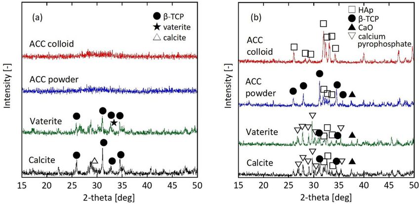
www.nature.com/scientificreports/
pH and moderate supersaturation, ACP is often the first-formed deposit, but eventually transforms into the
thermodynamically more stable HAp33,34. The role of such nanometre-sized ACP as building blocks for calcium
phosphate crystals has been debated for many years.
Based on this concept, we propose a new HAp synthesis strategy originating from ACC colloids. We dem-
onstrate that pure HAp or β-tricalcium phosphate (β-TCP; Ca3(PO4)2) is synthesised by a reaction of ACC
colloids with an orthophosphoric acid solution at room temperature and a subsequent calcination process. We
experimentally confirm that the water content or ACC concentration controls the phase purity of the calcium
phosphate compounds. Additionally, we clarify that a competitive phase transformation occurs from ACC to
ACP and a crystalline phase of calcium carbonate. We also show that the molar ratio of Ca and P plays a crucial
role on the formation of the calcium phosphate phase. Finally, we confirm that powdered ACC, vaterite, or calcite
with similar specific surface areas do not provide pure HAp. Only the ACC colloid can be used as the starting
source for pure HAp. Thus, HAp may originate from the ACC colloid.
Experimental
Materials. Calcium hydroxide (purity > 96.0%), ethanol (> 99.5%), glycerin (> 99.0%), phosphoric acid
(> 85.0%), and acetone (> 99.5%) were purchased from Kanto Chemical (Tokyo, Japan) and used as received.
ACC colloid synthesis. The preparation method for the ACC colloid dispersions is described elsewhere35.
Briefly, 25.0 g calcium hydroxide was added to 475 g solvent mixture of ethanol and glycerin (7:3 by weight)
while stirring at 400 rpm in a reaction vessel. A mixed gas of nitrogen (70 vol%) and carbon dioxide (30 vol%)
flowed at a rate of 1 L/min to start the carbonation reaction. The temperature was adjusted to 20 °C during the
carbonation reaction. The carbonation reaction was completed when the apparent pH did not change (pH ~ 8.9).
Then the unreacted coarse particles were removed by centrifugation at 3,540 g for 20 min to give an ACC colloid
dispersion.
The ACC colloid concentration was determined by thermogravimetric analysis (TG/DTA6200N, SII, Chiba,
Japan). N2 gas was introduced at a rate 80 ml/min, the rate of temperature increase was 10 °C/min, and the con-
centration was determined by the weight decrease within the temperature range of 20–500 °C. The ACC colloid
was diluted to 5 wt% or 1 wt% using the solvent mixture of ethanol and glycerin (7:3 by weight). Then it was
subjected to the phosphoric acid treatment described in “Phosphorylation” section.
ACC, vaterite, and calcite powders were prepared from the ACC colloid. The methodology and powder
characteristics are described in the supporting information.
Phosphorylation. The ACC colloid (30 g of 5 wt%) and a 5 wt% phosphoric acid solution were placed in
a container. The molar ratio of Ca and P was set to Ca/P = 1.68 (stoichiometric HAp is 1.67). Distilled water or
acetone was used as the solvent for the phosphoric acid solution. After stirring at room temperature at 800 rpm
for 180 min, the sample was centrifuged at 3540 g for 15 min, washed with ethanol, and vacuum dried for 24 h.
To identify the crystal phase of the synthetic sample, atmospheric calcination was conducted at 800 °C for 2 h
using an electric furnace (FO-200, Yamato Science, Tokyo, Japan).
The 1 wt% ACC colloid was reacted with a 5 wt% phosphoric acid aqueous solution. Subsequent operations
were the same as those for the 5 wt% ACC colloid treatment. Notably, the Ca/P ratios were set to 1.51, 1.58,
1.67, 1.71, 1.77, and 1.88.
Characterisation. The particle morphologies were observed using a scanning electron microscope (SEM,
JSM-6380A, JEOL, Tokyo, Japan). The crystal structure was analysed using an X-ray diffractometer (XRD, Mul-
tiFlex-120NP, Rigaku, Tokyo, Japan) with Cu Kα radiation (40 kV, 20 mA). The measurement range was 15°–50°
at a scan speed of 5°/min. To estimate the surface functional group on calcium compounds, Fourier-transform
infrared (FT-IR) spectra (FT/IR-460PlusK, JASCO, Tokyo, Japan) were acquired using a KBr pellet technique
with a scan range from 400 to 4000 cm−1.
Results and discussion
Competitive phase transformation of ACP and crystalline calcium carbonate. We initially
examined the phosphorylation reaction in two different systems: water and acetone. Figure 1a shows the XRD
patterns of the sample before and after calcination. For the water system (blue line), the pre-calcined sample
exhibited a clear calcite peak at 2θ = 29.4° and a broad pattern around 30°, indicating that a part of ACC under-
went a phase transformation to calcite via dissolution and recrystallisation in water23–26 prior to reacting with
the phosphate ions. In addition to ACC, the broad pattern suggests the possibility of ACP, which is an intermedi-
ate state in crystalline calcium phosphate compounds31–34. During the phosphorylation reaction, ACC partially
reacted with phosphoric acid to form ACP.
For the water system, a calcination treatment was performed to clarify the crystalline phase of the calcium
compounds. The calcined sample contained peaks of HAp, β-TCP, and calcium oxide (Fig. 1b, blue line). The
crystalline calcite in the pre-calcined sample was converted to calcium oxide during the calcination treatment.
ACP, which was generated when ACC transformed into the crystalline calcium phosphate compounds, occurred
simultaneously.
To prevent the ACC transformation into crystalline calcium carbonate, the phosphorylation reaction of the
ACC colloid was conducted in an acetone system. The pre-calcined sample was amorphous (ACP or ACC) with-
out a crystal phase (Fig. 1a, red line). Reflecting this XRD result, the main crystalline phase of the calcined sample
was HAp (Fig. 1b, red line). However, a slight peak of β-TCP and a minute calcium oxide peak were also observed.
Scientific Reports | (2021) 11:11546 | https://doi.org/10.1038/s41598-021-91064-y 2
Vol:.(1234567890)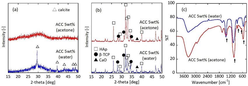
www.nature.com/scientificreports/
Figure 1. XRD patterns of (a) pre-calcined and (b) calcined particles. Peak positions in the XRD profiles
correspond to those of calcite (JCPDS no. 05-0586), hydroxyapatite (JCPDS no. 09-0432), β-TCP (JCPDS
no. 09-0169)), and calcium oxide (JCPDS no. 37-1497). (c) FTIR spectra for the pre-calcined sample.
Phosphorylation reaction is carried out in water and acetone systems using 5 wt% ACC colloid (Ca/P = 1.68).
Figure 2. Typical SEM images of the particles phosphorylated in (a,b) a water and (c,d) an acetone system. (a,c)
Pre-calcined and (b,d) calcined samples. Scale bar is 1 μm.
Figure 1c shows the FT-IR spectrum for the pre-calcined sample in the water and acetone systems. The stretch-
ing mode at 1000–1100 cm−1 and the bending mode at 550–600 cm−1 were assigned to the PO3− 4 group
36–39
. The
doubly degenerate asymmetric stretching mode at 1400–1500 cm−1, out of plane bending mode at 840–900 cm−1,
and doubly degenerate planar bending mode at 650–750 cm−1 originated from the CO2− 3 groups
40,41
. Regarding
the pre-calcined sample, the XRD results suggest the possibility of ACP in addition to the unreacted ACC. Dur-
ing the phosphorylation reaction, ACC partially reacted with phosphoric acid to form ACP. FT-IR spectrum in
Fig. 1c denotes that the modes are assigned to the CO2− 3 and PO4 groups, indicating the pre-calcined sample
3−
included ACC and ACP.
Figure 2 depicts typical SEM images of the sample before and after calcination. The images of the pre-calcine
sample (Fig. 2a,c) revealed aggregates of shapeless fine particles. In contrast, the calcined sample had nano-sized
Scientific Reports | (2021) 11:11546 | https://doi.org/10.1038/s41598-021-91064-y 3
Vol.:(0123456789)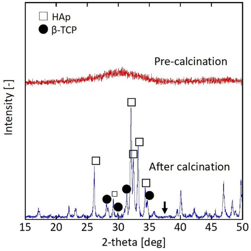
www.nature.com/scientificreports/
Figure 3. XRD patterns of pre-calcined and calcined particles phosphorylated in the water system using the
1 wt% ACC colloid (Ca/P = 1.67). Arrow denotes the peak position (2θ = 37.3°) of calcium oxide.
particles with clear edges, indicating that ACP was transformed into crystalline calcium phosphate and sustained
subsequent particle growth during the calcination treatment.
Since the aqueous system had a higher phase transformation from ACC to calcite, calcium oxide remained as
an impurity in the calcined sample. The phase transformation to crystalline polymorphs in the acetone system
was inhibited compared with the water system. Thus, the ACC reaction with phosphate ions was prioritised to
form ACP. Suppressing the phase transformation of ACC to crystalline calcium carbonate should synthesise
pure HAp.
Pure HAp synthesis. Here, we assumed the phase transformation rate of ACC (vtrs) depended on the parti-
cle concentration (C [1/m3]), which is expressed from the following Arrhenius-type equation.
Ea
vtrs = k0 exp − C (1)
RT
where Ea is the activation energy [J/mol], k0 is the frequency factor [1/s], R is the gas constant [J/(mol·K)], and T
is the absolute temperature [K]. As described in Fig. 1, the pre-calcined sample contained calcite when the start-
ing calcium source was the 5 wt% ACC colloid, and the phosphorylation reaction occurred in the water system.
According to Eq. (1), decreasing the particle concentration should inhibit the phase transformation rate. There-
fore, the starting material was the 1 wt% ACC colloid instead of the 5 wt% ACC colloid, and the phosphorylation
reaction was performed in the water system. Figure 3 summarises the XRD results before and after calcination.
The crystalline phase was not detected in the pre-calcined sample prepared from the 1 wt% ACC colloid
phosphorylated in water. Notably, the calcium oxide peak disappeared in the calcined sample, indicating that
the sample was almost ACP. The slight β-TCP peak indicated an insufficient amount of Ca (stoichiometrically
Ca/P of β-TCP is 1.50). Consequently, the 1 wt% ACC colloid is a candidate starting source to synthesise a pure
HAp phase.
Next, the molar ratio of Ca/P was adjusted in the phosphorylation reaction (see “ACC colloid synthesis” sec-
tion). Figure 4 plots the HAp/β-TCP ratio as a function of the Ca/P molar ratio. The HAp ratio was calculated
by the Reference Intensity Ratio (RIR) method. The RIR method is based on scaling all diffraction data to the
diffraction of standard reference m aterials42,43. For a Ca/P molar ratio of 1.71, the HAp ratio was 1.0, indicating
a pure phase of HAp. For Ca/P ratios of 1.51 and 1.58, which were smaller than the stoichiometric ratio for the
HAp (Ca/P = 1.67), β-TCP was the coexistence phase. Ca/P = 1.51and Ca/P > 1.71 yielded pure β-TCP and HAp,
respectively.
The ACC colloidal particle sizes in the dispersion ranged from 4 to 30 nm35. The amorphous and nano-sized
ACC is a chemically active substance, which should easily crystallise into a stable phase. These experiments
revealed the following points about the phase transformation of ACC during the phosphorylation reaction:
1. Using acetone as a solvent inhibits the dissolution and re-crystallisation of ACC to form the crystalline
calcium carbonate.
2. Decreasing the ACC concentration prevents the phase transformation and promotes the ACC reaction with
phosphoric acid to form ACP.
Scientific Reports | (2021) 11:11546 | https://doi.org/10.1038/s41598-021-91064-y 4
Vol:.(1234567890)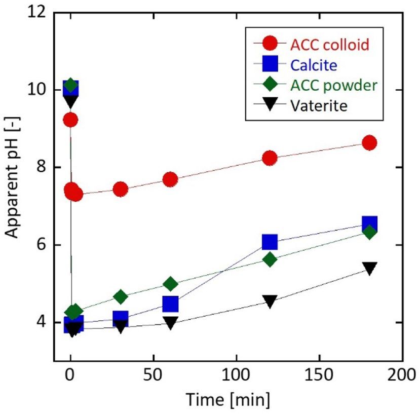
www.nature.com/scientificreports/
Figure 4. HAp/β-TCP ratio as a function of the Ca/P ratio calculated by the RIR method. Ratio = 0% denotes
the crystalline phase is β-TCP. Excess amount of Ca (Ca/P = 1.88) includes 0.6% calcium oxide due to the
unreacted ACC.
Figure 5. XRD patterns of (a) pre-calcined and (b) calcined particles. Phosphorylation reaction is performed
in a water system using a 1 wt% ACC, vaterite, or calcite powder suspension. Additionally, the reaction with a
1 wt% ACC colloid is also shown. Molar ratio of Ca/P is 1.71.
3. ACP is transformed to crystalline calcium phosphate compounds, where the main phase of calcium phos-
phate depends on the Ca/P ratio.
To confirm that the key characteristics to promote the phosphorylation reaction are "nano-particles" and
have an "amorphous" structure, phosphorylation was carried out under the conditions of Ca/P = 1.71 and a 1
wt% particle concentration.
Key factors of calcium sources for the pure HAp synthesis. We used a 1 wt% suspension of ACC,
vaterite, or calcite powder (see supporting information) as the starting source in the phosphorylation reaction.
Figure 5 shows the XRD powder patterns before and after calcination. A comparison of the ACC colloid with
a suspension of ACC powder confirmed that the pre-calcined sample was in the amorphous phase (ACP). For
the calcined samples, the 1 wt% ACC colloid produced a pure HAp peak, whereas the 1 wt% suspension of ACC
powder mainly gave a β-TCP peak.
According to the Ostwald-Freundlich equation, the solubility is substance specific when the temperature is
ecreases44 according to the following equation
constant. In contrast, the solubility increases as the particle size d
Scientific Reports | (2021) 11:11546 | https://doi.org/10.1038/s41598-021-91064-y 5
Vol.:(0123456789)www.nature.com/scientificreports/
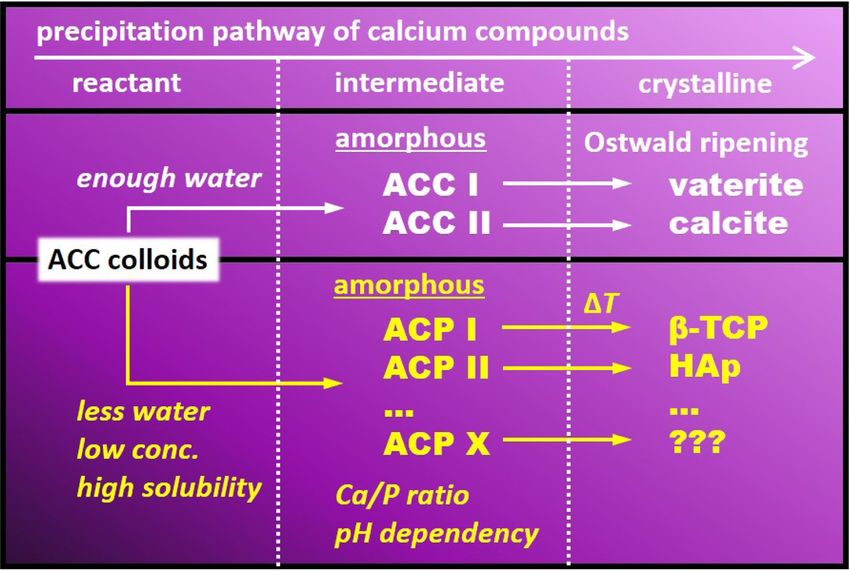
Figure 6. Schematic illustration of calcium phosphate precipitation.
xa 2γ M
ln = (2)
xa∞ RTρr
where xa [mol/L] is the solubility when the particle radius is r, xa∞ [mol/L] is the solubility when the particle
radius is infinite, γ [N/m] is the surface energy, M [g/mol] is the molar mass, R [J/(mol·K] is the gas constant, T
[K] is the absolute temperature, ρ [g/cm3] is the particle density, and r [m] is the particle radius at equilibrium.
The particle size of the ACC colloid was 4–30 nm35, whereas that of the ACC powder was 2 μm (see Fig. S1).
Because the solubility of ACC 8.4–12.4 times decreases with size, as expressed in Eq. (2), the nano-sized ACC
colloid should have a much higher reactivity with phosphoric acid than the micron-sized ACC powder. Addi-
tionally, the ACC size is a dominant factor for the final crystalline phase of calcium phosphate. Although this
study did not investigate the ACP micro-structure, the results suggested that the final crystalline phase of calcium
phosphate compounds depends on the intermediate structure of ACP.
Amorphous nano-particles were a precursor of crystalline calcium carbonate. According to Gebauer et al.29,
precursor species with different ACC phases give rise to an alternative crystallisation channel. That is, ACC I
and II are promising precursors of vaterite and calcite, r espectively29. Regarding HAp precipitation, Mahamid
et al. identified the ACP p hase45. At a neutral pH and moderate supersaturation, ACP is often the first-formed
deposit, but it eventually transforms into the thermodynamically more stable H Ap34. Thus, at least two types of
ACP, which are named ACP I and ACP II, are independently nucleated in the phosphorylation reaction (Fig. 6).
ACP I is related to an amorphous phase exhibiting a tricalcium phosphatic ordered structure, while ACP II is
related to an HAp ordered structure. Precursor species of different ACP and ACC phases give rise to alternative
crystallisation channels. When the system contains sufficient water, the ACC colloid crystallises into calcite and/
or vaterite. Notably, the thermodynamically unstable vaterite phase should transform to the stable calcite phase
via dissolution and re-crystallisation of vaterite (Ostwald ripening). In contrast, the ACC colloid transforms to
the ACP phase in a non-aqueous system (see Fig. 1). At a low ACC colloid concentration, it transforms to the
ACP phase (see Fig. 3). Ca/P (see Fig. 5) and reaction pH (see later discussion in Fig. 7) play important roles
on the ACP phase. Below the stoichiometric ratio of HAp (Ca/P < 1.67) or at acidic conditions, ACP I may be a
main phase. When the Ca/P is above 1.67 or at neutral pH, ACP II may be the main phase. ACP I and ACP II
transform to β-TCP and HAp during the calcination process, respectively. Solubility of the reactants is a funda-
mental property to decide the subsequent route (see later discussion in the last part of text).
Next, we focused on the amorphous part, which is a characteristic of ACC. The phosphorylation reaction
was performed using a 1 wt% suspension of crystalline vaterite or calcite powder. The specific surface areas of
the synthesised vaterite and calcite were similar to that for the ACC powder (Table S1). The pre-calcined sample
contained vaterite or calcite as the starting source (Fig. 5). The calcined sample contained calcium pyrophosphate
in addition to the peaks for HAp, β-TCP, and calcium oxide.
Calcium pyrophosphate was generated by dehydration of phosphoric acid. For instance, calcination of cal-
cium hydrogen phosphate dehydrate or calcium hydrogen phosphate (anhydrous salt) in air generated calcium
Scientific Reports | (2021) 11:11546 | https://doi.org/10.1038/s41598-021-91064-y 6
Vol:.(1234567890)www.nature.com/scientificreports/
Figure 7. Time dependence of the apparent pH during the phosphorylation reaction conducted in a water
system using ACC colloid, powdered ACC, vaterite, and calcite suspensions. Particle concentration is 1 wt%,
and the molar ratio of Ca/P is 1.71.
pyrophosphate. The transition from γ- to β-type, and β- to α-type occurred at 700–750 °C and 1140–1150 °C,
respectively46,47.
From this point of view, the calcination process produced calcium pyrophosphate. Therefore, to predict the
reaction of calcium carbonate with phosphoric acid, the apparent pH was measured at 0.5, 1, 3, 30, 60, 120, and
180 min. 0 min denotes the moment when the phosphoric acid aqueous solution was added. The apparent pH
at 0 min of each solution was basic (pH = 9 ~ 10), but immediately dropped to become acidic (pH ~ 4) upon the
addition of the phosphoric acid, except for the ACC colloid (Fig. 7). When the starting source was the ACC
colloid, the pH was clearly higher than those of the other sources, and the pH did not recover to pH ~ 8.5 until
180 min. However, suspensions of the powdered ACC, vaterite, and calcite remained acidic after 180 min of the
phosphorylation reaction.
HAp is stable under basic conditions, whereas calcium hydrogen phosphate and calcium hydrogen phosphate
dehydrate, which are sources of calcium pyrophosphate, are stable under acidic conditions48,49. Thus, calcium
pyrophosphate was produced in the calcined sample obtained from a vaterite or calcite suspension.
The solubility product (KSP) of calcium carbonate (KSP,V for vaterite and KSP,C for calcite) depends on the
absolute temperature (T) [K] according to the following r elationships50. In addition, the temperature-dependency
of K for ACC (KSP,ACC) is also s hown51.
3074.688
vaterite : log KSP, V = −172.1295 − 0.077993T + + 71.595 log T
T
2839.319
calcite : log KSP,C = −171.9065 − 0.077993T + + 71.595 log T
T
1247.0
ACC : logKSP,ACC = − 10.224 289 ≦ T ≦ 343
T
At 25 °C, the calculated logKSP,V and logKSP,C were – 7.91, and – 8.48, respectively, while logKSP,ACC was
– 6.04. Y. Kojima et al. reported that ACC ( CaCO3·1.5H2O) rapidly dissolves in water (20 °C), and its solubility
is 10 times higher than that of calcite after 1 min infiltration52. This corresponds to a lower Gibbs energy for
dissolution in ACC (38 kJ/mol) compared with calcite (48 kJ/mol)53. HAp was less soluble than other calcium
phosphate compounds and had a smaller solubility product of logKSP, HAp (− 56.9 to − 52.8 at 20 °C54, and − 53.51
to − 53.41 at 25 °C55).
The dissolution of calcium carbonate is a key point to precipitate HAp. The ACC colloid is an excellent source
for ACP precipitation, and subsequent calcination produces pure HAp or the β-TCP phase.
Conclusion
This study used the ACC colloid as a starting source for calcium phosphate compounds. The phosphorylation
reaction performed in the aqueous system partially confirmed the phase transformation from ACC to calcite. This
transformation was inhibited by the phosphorylation reaction in an acetone system or using a low concentration
of the ACC colloid. After calcination treatment, the main phase of the reaction was HAp. When a 1 wt% ACC
colloid was used as the starting source and the molar ratio of Ca/P was adjusted to > 1.71 or 1.51, pure HAp or
β-TCP was respectively synthesised. The precursor species of the different ACP phases gave rise to alternative
crystallisation channels: ACP I and II. ACP I and II may be a precursor for β-TCP and HAp, respectively.
Scientific Reports | (2021) 11:11546 | https://doi.org/10.1038/s41598-021-91064-y 7
Vol.:(0123456789)www.nature.com/scientificreports/
To investigate the characteristics of the ACC colloid, we conducted a phosphorylation reaction experiment
using powdered ACC, vaterite, or calcite suspensions. The following findings were obtained.
(1) Only the ACC colloid synthesised pure HAp, indicating that nano-sized particles are important due to their
higher solubility product.
(2) Phosphorylation with calcite or vaterite did not synthesise pure HAp because the solubilities of calcite and
vaterite are inferior to that of ACC.
Received: 15 April 2021; Accepted: 20 May 2021
References
1. Bystrov, V., Bystrova, A. & Dekhtyar, Y. HAP nanoparticle and substrate surface electrical potential towards bone cells adhesion:
recent results review. Adv. Colloid Interface Sci. 249, 213–219 (2017).
2. Arjama, M., Mehnath, S., Rajan, M. & Jeyaraj, M. Injectable cuttlefish HAP and macromolecular fibroin protein hydrogel for
natural bone mimicking matrix for enhancement of osteoinduction progression. React. Funct. Polym. 160, 104841 (2021).
3. Vahdat, A., Ghasemi, B. & Yousefpour, M. Mechanical properties of the hydroxyapatite and magnetic nanocomposite of
hydroxyapatite adsorbents. S. Afr. J. Chem. Eng. 33, 90 (2020).
4. Wei, X., Wang, Y., Li, X., Wu, R. & Zhao, Y. C o3O4 supported on bone-derived hydroxyapatite as potential catalysts for N 2O catalytic
decomposition. Mol. Catal. 491, 111005 (2020).
5. Das, T. K., Ganguly, S., Bhawal, P., Mondal, S. & Das, N. C. A facile green synthesis of silver nanoparticle-decorated hydroxyapatite
for efficient catalytic activity towards 4-nitrophenol reduction. Res. Chem. Intermed. 44, 1189 (2018).
6. Castro, M. A. M. et al., Synthesis of hydroxyapatite by hydrothermal and microwave irradiation methods from biogenic calcium
source varying pH and synthesis time. Bol. Soc. Esp. Ceram. Vidrio. https://doi.org/10.1016/j.bsecv.2020.06.003 (2020).
7. Phatai, P., Futalan, C. M., Kamonwannasit, S. & Khemthong, P. Structural characterization and antibacterial activity of hydroxyapa-
tite synthesized via sol-gel method using glutinous rice as a template. J. Sol-Gel Sci. Technol. 89, 764 (2019).
8. Yelten-Yilmaz, A. & Yilmaz, S. Wet chemical precipitation synthesis of hydroxyapatite (HA) powders. Ceram. Int. 44, 9703 (2018).
9. Mohd Pu’ad, N. A. S., Alipal, J., Abdullah, H. Z., Idris, M. I. & Lee, T. C. Synthesis of eggshell derived hydroxyapatite via chemical
precipitation and calcination method. Mater. Today Proc. 42, 172 (2021).
10. Batista, H. A., Silva, F. N., Lisboa, H. M. & Costa, A. C. F. M. Modeling and optimization of combustion synthesis for hydroxyapatite
production. Ceram. Int. 46, 11638 (2020).
11. Sirait, M., Sinulingga, K., Siregar, N. & Damanik, Y. F. Synthesis and characterization of hydroxyapatite from broiler eggshell. AIP
Conf. Proc. 2221, 110030 (2020).
12. Elhendawi, H., Felfel, R. M., El-Hady, B. M. A. & Reicha, F. M. Effect of synthesis temperature on the crystallization and growth
of in situ prepared nanohydroxyapatite in chitosan matrix. Int. Sch. Res. Notices 2014, 897468 (2014).
13. Javadinejad, H. R. & Ebrahimi-Kahrizsangi, R. Thermal and kinetic study of hydroxyapatite formation by solid‐state reaction. Int.
J. Chem. Kinet. 53, 583 (2021).
14. Türk, S. et al. Microwave–assisted biomimetic synthesis of hydroxyapatite using different sources of calcium. Mater. Sci. Eng. C
76, 528 (2017).
15. Chen, Y.-Y. et al. Performance and mechanism of simultaneous removal of Cd(II) and Congo red from aqueous solution by hier-
archical vaterite spherulites. Appl. Surf. Sci. 444, 224 (2018).
16. Stanić, V. et al. Synthesis, characterization and antimicrobial activity of copper and zinc-doped hydroxyapatite nanopowders. Appl.
Surf. Sci. 256, 6083 (2010).
17. Chang, M. C. Organic–inorganic interaction between hydroxyapatite and gelatin with the aging of gelatin in aqueous phosphoric
acid solution. J. Mater. Sci. Mater. Med. 19, 3411 (2008).
18. Qi, M.-L. et al. Controlled synthesis of hydroxyapatite nanomaterials regulated by different phosphorus sources. Crystals 10, 678
(2020).
19. Minha, D. P., Lyczko, N., Sebei, H., Nzihou, A. & Sharrock, P. Synthesis of calcium hydroxyapatite from calcium carbonate and
different orthophosphate sources: a comparative study. Mater. Sci. Eng. B 177, 1080 (2012).
20. da Silva, O. G., Alves, M. M., dos Santos, I. M. G., Fonseca, M. G. & Jaber, M. Mesoporous calcium phosphate using casein as a
template: application to bovine serum albumin sorption. Colloids Surf. B 158, 480 (2017).
21. Qin, J., Zhong, Z. & Ma, J. Biomimetic synthesis of hybrid hydroxyapatite nanoparticles using nanogel template for controlled
release of bovine serum albumin. Mater. Sci. Eng. C 62, 377 (2016).
22. Sheikh, L., Tripathy, S. & Nayar, S. Biomimetic matrix mediated room temperature synthesis and characterization of nano-
hydroxyapatite towards targeted drug delivery. RSC Adv. 6, 62556 (2016).
23. Addadi, L., Raz, S. & Weiner, S. Taking advantage of disorder: amorphous calcium carbonate and its roles in biomineralization.
Adv. Mater. 15, 959 (2003).
24. Faatz, M., Grohn, F. & Wegner, G. Amorphous calcium carbonate: synthesis and potential intermediate in biomineralization. Adv.
Mater. 16, 996 (2004).
25. Cölfen, H. & Mann, S. Higher-order organization by mesoscale self-assembly and transformation of hybrid nanostructures. Angew.
Chem. Int. Ed. 42, 2350 (2003).
26. Ihli, J. et al. Dehydration and crystallization of amorphous calcium carbonate in solution and in air. Nat. Commun. 5, 3169 (2014).
27. Singh, J. P., Ji, M.-J., Shim, C.-H., Kim, S. O. & Chae, K. H. Effect of precursor thermal history on the formation of amorphous and
crystalline calcium carbonate. Particuology 33, 29 (2017).
28. Pouget, E. M. et al. The initial stages of template-controlled CaCO3 formation revealed by Cryo-TEM. Science 323, 1455 (2009).
29. Gebauer, D., Völker, A. & Cölfen, H. Stable prenucleation calcium carbonate clusters. Science 322, 1819 (2008).
30. Čadež, V. et al. Amorphous calcium phosphate formation and aggregation process revealed by light scattering techniques. Crystals 8,
254 (2018).
31. Dey, A. et al. The role of prenucleation clusters in surface-induced calcium phosphate crystallization. Nat. Mater. 9, 1010 (2010).
32. Lotsari, A., Rajasekharan, A. K., Halvarsson, M. & Andersson, M. Transformation of amorphous calcium phosphate to bone-like
apatite. Nat. Commun. 9, 4170 (2018).
33. Ucar, S. et al. Formation of hydroxyapatite via transformation of amorphous calcium phosphate in the presence of alginate addi-
tives. Cryst. Growth Des. 19, 7077 (2019).
Scientific Reports | (2021) 11:11546 | https://doi.org/10.1038/s41598-021-91064-y 8
Vol:.(1234567890)www.nature.com/scientificreports/
34. Christoffersen, J., Christoffersen, M. R., Kibalczyc, W. & Andersen, F. A. A contribution to the understanding of the formation of
calcium phosphates. J. Cryst. Growth 94, 767 (1989).
35. Yamanaka, S. et al. Scalable and template-free production of mesoporous calcium carbonate and its potential to formaldehyde
adsorbent. J. Nanoparticle Res. 16, 2266 (2014).
36. Suwa, Y., Banno, H., Mizuno, M. & Saito, H. Synthesis of compositionally regulated hydroxyapatite from Ca(OH)2 and H3PO4. J.
Ceram. Soc. Jpn. 101, 659 (1993).
37. Toyama, T., Ohshima, A. & Yasue, T. Hydrothermal synthesis of hydroxyapatite whisker from amorphous calcium phosphate and
the effect of carboxylic acid. J. Ceram. Soc. Jpn. 109, 232 (2001).
38. Elliott, J. C. Structure and Chemistry of the Apatites and Other Calcium Orthophosphates (Elsevier, 1994).
39. Frost, R. L., Martens, W., Williams, P. A. & Kloprogge, J. T. Raman and infrared spectroscopic study of the vivianite-group phos-
phates vivianite, baricite and bobierrite. Mineral. Mag. 66, 1063 (2002).
40. Herzberg, C. Molecular spectra and molecular structure. In Infrared and Raman Spectra of Polyatomic Molecules, Vol. 2 (Krieger
Pub Co, 1991).
41. Adler, H. H. & Kerr, P. F. Infrared study of aragonite and calcite. Am. Mineral. 47, 700 (1962).
42. Chung, F. H. Quantitative interpretation of X-ray diffraction patterns of mixtures. I. Matrix-flushing method for quantitative
multicomponent analysis. J. Appl. Cryst. 7, 519 (1974).
43. Chung, F. H. Quantitative interpretation of X-ray diffraction patterns of mixtures. II. Adiabatic principle of X-ray diffraction
analysis of mixtures. J. Appl. Cryst. 7, 526 (1974).
44. Eslami, F. & Elliott, J. A. W. Role of precipitating solute curvature on microdrops and nanodrops during concentrating processes:
the nonideal Ostwald–Freundlich equation. J. Phys. Chem. B 118, 14675 (2014).
45. Mahamid, J., Sharir, A., Addadi, L. & Weiner, S. Amorphous calcium phosphate is a major component of the forming fin bones of
zebrafish: Indications for an amorphous precursor phase. Proc. Natl. Acad. Sci. U.S.A. 105, 12748 (2008).
46. Mclntosh, A. O. & Jablonski, W. L. X-ray diffraction powder patterns of calcium phosphates. Anal. Chem. 28, 1424 (1956).
47. Ranby, P. W., Mash, D. H. & Henderson, S. T. The investigation of new phosphors, with particular reference to the pyrophosphates.
Br. J. Appl. Phys. 6, S18 (1955).
48. Suchanek, K., Bartkowiak, A., Perzanowski, M. & Marszałek, M. From monetite plate to hydroxyapatite nanofibers by monoetha-
nolamine assisted hydrothermal approach. Sci. Rep. 8, 15408 (2018).
49. Shafie, E. R. M., Ahmad, Z. A. & Ahmad, N. Synthesis of hydroxyapatite via phase transformation of calcium hydrogen phosphate
dihydrate: effects of temperature variation on phase and morphology. Ceram. Int. 45, 21168 (2019).
50. Plummer, L. N. & Busenberg, E. The solubilities of calcite, aragonite and vaterite in C O2-H2O solutions between 0 and 90°C, and
an evaluation of the aqueous model for the system C aCO3-CO2-H2O. Geochim. Cosmochim. Acta 46, 1011 (1982).
51. Clarkson, J. R., Price, T. J. & Adams, C. J. Role of metastable phases in the spontaneous precipitation of calcium carbonate. J. Chem.
Soc. Faraday Trans. 88, 243 (1992).
52. Kojima, Y., Kawanobe, A., Yasue, T. & Arai, Y. Synthesis of amorphous calcium carbonate and its crystallization. J. Ceram. Soc.
Jpn. 101, 1145 (1993).
53. Christoffersen, M. R., Christoffersen, J. & Kibalczyc, W. Apparent solubilities of two amorphous calcium phosphates and of octa-
calcium phosphate in the temperature range 30–42°C. J. Cryst. Growth 106, 349 (1990).
54. Larsen, M. J. & Jensen, S. J. The hydroxyapatite solubility product of human dental enamel as a function of pH in the range 4.6–7.6
at 20 °C. Arch. Oral Biol. 34, 957 (1989).
55. Zhu, Y. et al. A comparative study on the dissolution and solubility of hydroxylapatite and fluorapatite at 25°C and 45°C. Chem.
Geol. 268, 89 (2009).
Acknowledgements
This work was supported by JSPS KAKENHI (Grant Number JP19K05117). We thank Dr. T. Oiso and Dr. Y.
Akimoto (Asahi Kohmatsu, Tokyo, Japan) for valuable discussions on the manuscript and for the RIR analysis.
Author contributions
S.Y. conceived the idea and directed the project; M.S. synthesized the samples and analysed the data; M.S., K. S
and S.Y. wrote the manuscript. All authors commented on the manuscript.
Competing interests
The authors declare no competing interests.
Additional information
Supplementary Information The online version contains supplementary material available at https://doi.org/
10.1038/s41598-021-91064-y.
Correspondence and requests for materials should be addressed to S.Y.
Reprints and permissions information is available at www.nature.com/reprints.
Publisher’s note Springer Nature remains neutral with regard to jurisdictional claims in published maps and
institutional affiliations.
Open Access This article is licensed under a Creative Commons Attribution 4.0 International
License, which permits use, sharing, adaptation, distribution and reproduction in any medium or
format, as long as you give appropriate credit to the original author(s) and the source, provide a link to the
Creative Commons licence, and indicate if changes were made. The images or other third party material in this
article are included in the article’s Creative Commons licence, unless indicated otherwise in a credit line to the
material. If material is not included in the article’s Creative Commons licence and your intended use is not
permitted by statutory regulation or exceeds the permitted use, you will need to obtain permission directly from
the copyright holder. To view a copy of this licence, visit http://creativecommons.org/licenses/by/4.0/.
© The Author(s) 2021
Scientific Reports | (2021) 11:11546 | https://doi.org/10.1038/s41598-021-91064-y 9
Vol.:(0123456789)You can also read


