Small Bowel Gist in a Patient with Neurofibromatosis Type 1: Revealed by Acute Abdominal Surgery: A Case Report
←
→
Page content transcription
If your browser does not render page correctly, please read the page content below
Saudi Journal of Medical and Pharmaceutical Sciences
Abbreviated Key Title: Saudi J Med Pharm Sci
ISSN 2413-4929 (Print) | ISSN 2413-4910 (Online)
Scholars Middle East Publishers, Dubai, United Arab Emirates
Journal homepage: https://saudijournals.com
Case Report Surgical Emergency
Small Bowel Gist in a Patient with Neurofibromatosis Type 1:
Revealed by
1*
Acute1 Abdominal
1
Surgery:
1
A Case Report
Dr. Maouni Iliass , M. Gridda , Y. Ouhamou , M. Elabsi
1
Surgical Emergency Department, Ibn Sina University Hospital, Rabat, Morocco
DOI: 10.36348/sjmps.2023.v09i01.007 | Received: 13.12.2022 | Accepted: 19.01.2023 | Published: 26.01.2023
*Corresponding author: Dr. Maouni Iliass
Surgical Emergency Department, Ibn Sina University Hospital, Rabat, Morocco
Abstract
We decided to report a case of gastrointestinal stromal tumor of the small intestine in a patient with neurofibromatosis
type 1 because of the increase of its incidence as shown in the literature, also, by its revelation; its occurrence in a picture
of acute peritonitis due to the rupture of the tumor. These tumors, commonly called by their English acronym GIST
(Gastro-Intestinal Stromal Tumors), found in people with neurofibromatosis type 1 generally occur in the small intestine
and are often multiple.
Keywords: GIST, small intestine, neurofibromatosis, peritonitis, tumor.
Copyright © 2023 The Author(s): This is an open-access article distributed under the terms of the Creative Commons Attribution 4.0 International
License (CC BY-NC 4.0) which permits unrestricted use, distribution, and reproduction in any medium for non-commercial use provided the original
author and source are credited.
than 10% of patients present with metastatic disease [3].
INTRODUCTION We report a case of GIST on a neurofibromatosis
Gastrointestinal stromal tumors (GISTs) occur background revealed late in an acute peritonitis picture.
mainly sporadically and represent about 1% of all
neoplasia of the gastrointestinal tract. The origin of
GISTs has a direct relationship with the interstitial cells MEDICAL OBSERVATION
of Cajal, which are part of the myenteric plexus of the The patient was 49 years old and single, with a
digestive tract and are responsible for the control of pathological history of lower limb deformity and
intestinal motility (pacemaker). They can develop from inability to walk since childhood, requiring the use of a
all segments of the digestive tract, from the esophagus wheelchair. She was referred to us with acute
to the anus, or exceptionally the mesentery and peritonitis. The interrogation with the patient on
peritoneum. They may project exophytically into the admission found a beginning of the symptomatology
lumen or less frequently they dilate through the serosa that goes back to 72h with epigastric abdominal pain
of the organ [1]. The incidence is estimated at about 15 that quickly generalized to the whole abdomen with
cases / million inhabitants/ year, the median age at vomiting. The clinical examination found a conscious
diagnosis is about 60 years, and the sex ratio of about patient, febrile at 39.5°, tachycardia at 112 beats/min,
1/1 [2]. Most mesenchymal tumors in the digestive tract blood pressure at 110/56mmhg, and polygenic at 26
have been considered as smooth muscle tumors cycles/min, the abdominal examination found a
(leiomyomas, leiomyosarcomas, etc. differential generalized contracture the rest of the examination was
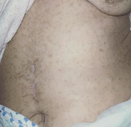
diagnosis) [3]. In 1983, Clark and Mazur introduced the characterized by the presence of skin lesions
term gastrointestinal stromal tumor (GIST) to describe a characteristic of Von Recklinghausen's disease ("Café
distinctive type of non-muscle smooth mesenchymal au lait" spots) (Fig. 1). The biological workup showed a
tumor [4]. Extensive study has shown that GISTs have hyperleukocytosis of 27500 with 88% neutrophilic
a specific etiogenesis unrelated to the gastrointestinal polynuclear, a hemoglobin level of 14.2 g/dl, a C-
smooth muscle at the expense of which, it is true, they reactive protein of 361 mg/l, the hydroelectrolytic
develop. The most frequent clinical presentation of workup showed profound hypokalemia of 2.1 mEq/l,
GIST is a digestive hemorrhage > 50% of the cases. and a correct renal function. An abdomino-pelvic
This may be chronic with anemia or massive, requiring ultrasound was performed at the beginning showed a
emergency treatment (40% of cases of hemorrhagic pelvic peritoneal effusion associated with an echogenic
GIST). The majority of cases are localized, and less collection and containing air bubbles measuring 52mm
Citation: Maouni Iliass, M. Gridda, Y. Ouhamou, M. Elabsi (2023). Small Bowel Gist in a Patient with 34
Neurofibromatosis Type 1: Revealed by Acute Abdominal Surgery: A Case Report. Saudi J Med Pharm Sci, 9(1): 34-38.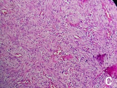
Maouni Iliass et al., Saudi J Med Pharm Sci, Jan, 2023; 9(1): 34-38
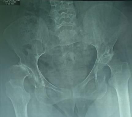
X 74 mm, then it was completed by abdomino-pelvic pneumoperitoneum bullae (Fig. 2); A standard X-ray of
CT scan with contrast injection which objectified a the pelvis showed a flattened and destroyed appearance
peri-hepatic peritoneal effusion of medium abundance, of two femoral heads (Fig. 3). This fact confirms the
peri-splenic and pelvic, and individualization of a bone involvement of his basic disease
pelvic mass of about 7 cm of great axis around which (neurofibromatosis type 1).
are agglutinated small intestines and the left colon with
Figure 1: Café au lait skin stains
Figure 2: Axial section of abdominal CT scan showing a mass in contact with the sigmoid colon
© 2023 | Published by Scholars Middle East Publishers, Dubai, United Arab Emirates 35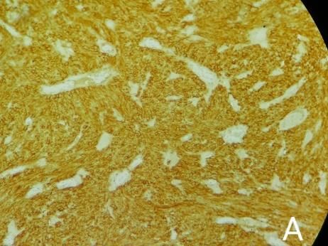
Maouni Iliass et al., Saudi J Med Pharm Sci, Jan, 2023; 9(1): 34-38
Figure 3: Bilateral femoral head necrosis
Faced with this clinical and radiological with a purulent effusion and false membranes, after
picture of peritonitis, the surgical indication was laborious liberation of the bowel, a mass was
decided after a short resuscitation and especially a discovered originating at the level of the jejunum, 20
potassium recharge by central route. The surgical cm from the duodeno-jejunal angle, about 6 cm long
exploration revealed a generalized peritonitis, neglected and invading the sigmoid colon (fig. 4).
A B
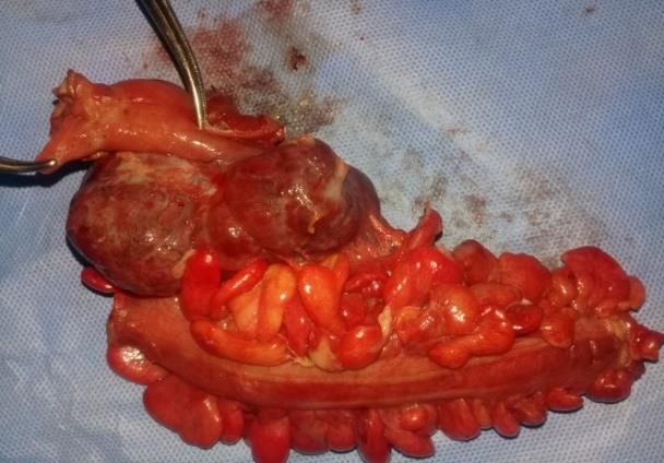
Figure 4: An Intraoperative image showing a perforated mass taking the jejunum and sigmoid colon, B: Surgical
specimen made of the sigmoid colon, jejunum and the tumor mass
This mass was perforated and made the two 6x5x5x4 cm, with spindle-shaped cells with
digestive segments communicate with the peritoneal eosinophilic cytoplasm and elongated or ovoid nuclei.
cavity; we proceeded to a resection of the mass with a The mitotic index was 1/25. These cells expressed
segment of jejunum and the sigmoid colon at a distance CD117 and DOG1 positive (Fig. 5), concluding a
from the tumor. The continuity of the jejunum was re- gastrointestinal stromal tumor (GIST) at moderate risk
established by a terminal anastomosis despite the risk of of recurrence, for which adjuvant chemotherapy was
septicemia since the resection was made in the first decided at the multidisciplinary consultation meeting
loop, while the two ends of the left colon were abutted with imatinib 400 mg per day for 4 years. We
in a double stoma to the left according to Bouilly subsequently referred the patient to a dermatologist for
Volkmann. The postoperative course was simple with management of her cutaneous Von Recklinghausen's
the patient being discharged at D+5. The disease and proposed annual surveillance by CT
anatomopathological and immunohistochemical study enterography.
of the lesion showed that it was a tumor of size
© 2023 | Published by Scholars Middle East Publishers, Dubai, United Arab Emirates 36
Maouni Iliass et al., Saudi J Med Pharm Sci, Jan, 2023; 9(1): 34-38

  Figure 5: A,B): IHC study showing positive labelling by anti CD117 antibody (C,D): Spindle cell proliferations
                                        occupying the entire grape wall

                                                                              Our patient presented with a picture of peritonitis, it is a
DISCUSSION                                                                    rare mode of discovery, seven patients (7.6%) in the
          GISTs are connective tissue tumors                                  series of Magdy A et al., had presented with rupture
characterized by the expression of the CD117 marker                           and peritonitis [10]. The stromal GIST tumor found in
(kit protein or c-kit). Only c-kit positive tumors are                        our patient is identical with the cases already reported
considered as GIST except in exceptional cases [2].                           in the course of Von Recklinghausen's disease: late
They are usually located in the stomach in 40% to 70%,                        discovery, after 49 years of age, in patients presenting
in the small intestine in 20% to 40% and less than 10%                        the characteristic cutaneous signs of the disease,
in the esophagus, colon and rectum [5, 6]. Most GISTs                         localization in the small intestine: this is the preferential
are sporadic but there are a few cases of familial                            site of occurrence of GIST associated with Von
disease. GISTs can be seen in the context of Carney's                         Recklinghausen's disease, followed by the stomach.
triad or neurofibromatosis type 1. Our case had a strong                      Complete surgical resection is the only potentially
suspicion of neurofibromatosis type 1, because of the                         curative treatment for GISTs [2]. Medical treatment is
very characteristic skin lesions and the bone defect in                       with imatinib, a pharmacological c-kit antagonist that
the pelvis. Recklinghausen's disease is an autosomal                          inhibits tyrosine kinase function, which is
dominant inherited disease that involves an abnormality                       recommended        for    advanced       GIST,      whether
on chromosome 17. It is quite frequent with an                                unresectable, metastatic or relapsed. Current data show
incidence of 1/3500 births [7]. It progresses slowly and                      that imatinib induces a 60-70% objective response rate,
is characterized by the progressive appearance of                             with 15-20% stable disease and 10-15% primary
pigment spots, skin tumors, tumors of the peripheral                          resistance. Secondary resistance (escape) is now
nerves (neurofibromas) or of the central nervous system                       reported in 10-30% of cases [2].
(gliomas) and skeletal malformations. It may be
accompanied by digestive manifestations such as
stromal tumors, lesions of the intrinsic digestive                            CONCLUSION
nervous system or endocrine tumors of the duodenum                                     The benign or malignant character of these
[7]. These stromal tumors developed in the context of                         tumors is difficult to define; several prognostic factors
neurofibromatosis type 1 do not have mutations in the                         have been proposed, but there are sometimes
KIT and PDGFRA genes. They have no morphological                              "borderline" tumors.
features but are often multiple [8]. These tumors occur
in 5% of patients with Von Recklinghausen disease [7].                                 The only potentially curative treatment is the
The diagnostic circumstances of stromal GIST tumors                           complete surgical removal of the lesion, but the use of
are variable: incidental discovery, pain, mass syndrome,                      Imatinib in recent years, a drug treatment that inhibits
anemia, hemoperitoneum, and especially digestive                              the KIT protein, has revolutionized their management.
hemorrhage which is the most frequent symptom [9].

© 2023 | Published by Scholars Middle East Publishers, Dubai, United Arab Emirates                                                     37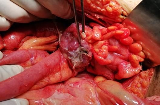
Maouni Iliass et al., Saudi J Med Pharm Sci, Jan, 2023; 9(1): 34-38
Conflict of Interest: None.                                                   6.  Chandramohan, K., Agarwal, M., Gurjar, G., Gatti,
                                                                                  R. C., Patel, M. H., Trivedi, P., & Kothari, K. C.
                                                                                  (2007). Gastrointestinal stromal tumour in
REFERENCES                                                                        Meckel's diverticulum. World Journal of Surgical
1.   Grezzana-Filho, R. J. M., Mendonça, T. B.,                                   Oncology, 5(1), 1-5.
     Golbspan, L., Kruel, C. R. P., Chedid, A. D., &                          7. Joensuu, H. (2008). Risk stratification of patients
     Kruel, C. D. P. (2009). Gists múltiplos em                                   diagnosed with gastrointestinal stromal tumor.
     neurofibromatose tipo 1: diagnóstico incidental em                           Hum Pathol, 39(10), 1411-9.
     paciente com abdome agudo. ABCD Arq Bras Cir                             8. Bensimhon, D., Soyer, P., Brouland, J. P., Boudiaf,
     Dig, 22(1), 65-8.                                                            M., Fargeaudou, Y., & Rymer, R. (2008).
2.   Zentar, A., & Alahyane Bounaim, A. (2008).                                   Gastrointestinal stromal tumors: role of computed
     Tumeur stromale multifocale diffuse de                                       tomography           before        and         after
     l'intestin. Gastroentérologie       Clinique         et                      treatment. Gastroentérologie       clinique       et
     Biologique, 32(12), 1020–22.                                                 biologique, 32(1 Pt. 1), 91-97.
3.   Bucher, P. A. R., Villiger, P., Egger, J. F., Buehler,                   9. Landi, B., Blay, J. Y., Bonvalot, S., Brasseur, M.,
     L. H., & Morel, P. (2004). Management of                                     Coindre, J. M., Emile, J. F., ... & de
     gastrointestinal stromal tumors: from diagnosis to                           Gastroentérologie, S. N. F. (2019). Gastrointestinal
     treatment. Swiss medical weekly, 134(11-12), 145-                            stromal tumours (GISTs): French Intergroup
     53.                                                                          Clinical Practice Guidelines for diagnosis,
4.   Mazur, M. T., & Clark, H. B. (1983). Gastric                                 treatments and follow-up (SNFGE, FFCD,
     stromal tumors. Reappraisal of histogenesis. Am J                            GERCOR,        UNICANCER,         SFCD,      SFED,
     Surg Pathol, 7, 507-19.                                                      SFRO). Digestive and Liver Disease, 51(9), 1223-
5.   Miettinen, M., & Lasota, J. (2006). Gastrointestinal                         1231.
     stromal tumors: review on morphology, molecular                          10. Sorour, M. A., Kassem, M. I., Ghazal, A. E. H. A.,
     pathology,       prognosis,      and       differential                      El-Riwini, M. T., & Nasr, A. A. (2014).
     diagnosis. Archives of Pathology and Laboratory                              Gastrointestinal stromal tumors (GIST) related
     Medicine, 130(10), 1466–78.                                                  emergencies. International        Journal         of
                                                                                  Surgery, 12(4), 269-280.

© 2023 | Published by Scholars Middle East Publishers, Dubai, United Arab Emirates                                                     38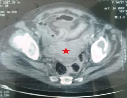
You can also read