Endometriosis in the Cecum: A Rare Clinical Entity
←
→
Page content transcription
If your browser does not render page correctly, please read the page content below
Open Access Case
                               Report                                                  DOI: 10.7759/cureus.35782

                                                Endometriosis in the Cecum: A Rare Clinical
                                                Entity
Review began 01/11/2023
                                                Ioanna Verzoviti 1 , Dimitrios Kalliouris 1 , Anastasia Boptsi 1 , Nikolaos Kiriakos 2 , Dimitrios Keramidaris 1
Review ended 02/28/2023
Published 03/05/2023                            1. Department of General Surgery, 417 Army Shared Fund Hospital, Athens, GRC 2. Department of Gastroenterology,
© Copyright 2023                                401 Army General Hospital, Athens, GRC
Verzoviti et al. This is an open access
article distributed under the terms of the      Corresponding author: Ioanna Verzoviti, jverzoviti@gmail.com
Creative Commons Attribution License CC-
BY 4.0., which permits unrestricted use,
distribution, and reproduction in any
medium, provided the original author and
source are credited.                            Abstract
                                                Cecal endometriosis is uncommon and may mimic other tumors of the colon, making it difficult to safely
                                                diagnose preoperatively. We report a case of a 50- year-old female who was found to have a cecal lesion
                                                during an endoscopic examination, which was performed for the investigation of anemia. It was also
                                                confirmed by conducting a computed tomography (CT) scan. Due to the high possibility of this mass
                                                identification as a neoplasm, the patient underwent a laparoscopic right hemicolectomy with an
                                                extracorporeal side-to-side isoperistaltic anastomosis. However, the postoperative histological diagnosis of
                                                the mass was cecal endometriosis, as the histopathology report noted endometrial tissues in the submucosa
                                                and muscolaris propria of the ileocecal region. Endometriosis of the cecum is a rare manifestation and can
                                                often be misdiagnosed as a malignant tumor. Further research is required, concerning preoperative
                                                characteristics of bowel masses in women, in order to provide optimal operative treatment and avoid
                                                unnecessary invasive procedures.

                                                Categories: Gastroenterology, General Surgery
                                                Keywords: cecum, endometriosis, cecal endometriosis, bowel endometriosis, tumor-like lesions

                                                Introduction
                                                Endometriosis, the presence of an ectopic functioning endometrial tissue outside the uterus, represents a
                                                benign condition and affects 6-10% of women in their reproductive age [1,2].

                                                It can affect almost any organ or structure; however, the pelvic cavity is the most common location for
                                                endometriotic implants. Atypical endometriosis is rare and difficult to diagnose [3].

                                                Gastrointestinal endometriosis is the most common form of extragenital endometriosis (EE). In nearly 90%
                                                of cases, it affects the rectum and sigmoid colon [4]. The small intestine follows, most commonly the ileum,
                                                in 7-12% of cases, and the appendix in 6-8% of cases [1]. The occurrence of cecal involvement is relatively
                                                rare and constitutes less than 3.6-6% of gastrointestinal endometriosis cases [1,2,5,6].

                                                The second most common form of EE is urogenital endometriosis. It affects the bladder in more than 85% of
                                                cases. The diaphragm is the most common site of thoracic endometriosis. In abdominal wall endometriosis,
                                                painful nodules arise in scars from prior abdominal surgery [4].

                                                Symptoms are nonspecific and vary according to the site of involvement, and they are usually abdominal or
                                                pelvic pain, nausea, vomiting, diarrhea, and rectal bleeding [7]. When it affects the appendix and/or ileum, it
                                                may cause clinical situations, such as acute appendicitis, perforation, or intussusception, and may be
                                                associated with menses [5,7]. Urogenital endometriosis might present with dysuria, hematuria, or irritable
                                                bladder syndrome. Endometriosis of the diaphragm may cause period-associated shoulder pain or
                                                catamenial pneumothorax. Endometriosis affecting a nerve often presents with sciatica [4]. Complete
                                                intestinal obstruction is not a common finding in patients with endometriosis [2].

                                                The differential diagnosis of intestinal endometriosis is difficult, frequently resulting in delayed diagnosis
                                                and treatment. Often it can only be confirmed postoperatively [8]. Intestinal involvement of endometriosis
                                                often causes mass-like lesions and the malignancy can only be excluded postoperatively by histopathological
                                                examination [5,6]. Laparoscopy with biopsy remains the definitive method for diagnosis [9].

                                                Roman et al. conducted a case series study concerning the surgical management of patients with deep
                                                infiltrating endometriosis of the rectum and the sigmoid colon. The cecum was involved in 6.6% of the cases
                                                [10].

                                                We present a case of a postmenopausal female patient with cecal endometriosis. In our case, endometriosis
                                                was presented as a mass during diagnostic endoscopy.

                                How to cite this article
                                Verzoviti I, Kalliouris D, Boptsi A, et al. (March 05, 2023) Endometriosis in the Cecum: A Rare Clinical Entity. Cureus 15(3): e35782. DOI
                                10.7759/cureus.35782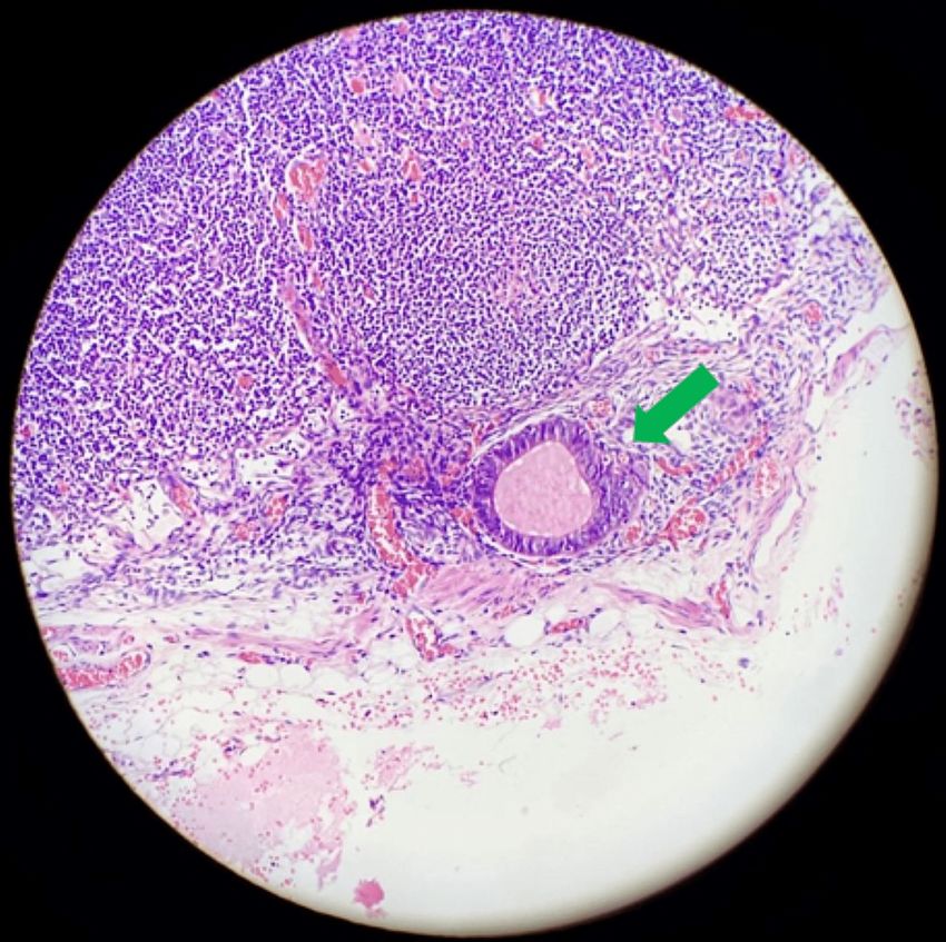
Case Presentation
A 50-year-old female, gravida 2, para 2, with a family history of colon cancer, was referred to our hospital
due to anemia (hematocrit: 29%). From her medical history, it was found that she had undergone a prior
curettage, an exploratory laparoscopy for endometriosis, 25 years ago, which was negative, and an open
appendicectomy 14 years ago. The patient was totally asymptomatic and the rest of the blood tests were
within normal limits.
An endoscopic examination of the large bowel was requested from the gastroenterology department in order
to investigate her anemia. It revealed a cecal hard mass of 2.5-3 cm, which seemed to be more of submucosal
or parietal origin, while the bowel’s mucosa seemed to be intact with no evidence of lesion. Biopsies were
taken and the histology report was indicative of normal colonic mucosa (Figure 1).
FIGURE 1: Colonoscopy demonstrating the cecal mass.
The patient was referred to our surgical department for further investigation. To clarify, a computed
tomography (CT) scan of the abdomen was performed and confirmed the presence of a mass, 2.8 x 3.2 cm,
without any lymph node pathology.
Based on all the aforementioned findings and because the patient had a family history of colon cancer, she
underwent a laparoscopic right hemicolectomy with an extracorporeal side-to-side isoperistaltic
anastomosis to remove the entire mass. Intraoperative inspection revealed no obvious implants or
abnormalities of the pelvic or the upper abdomen.
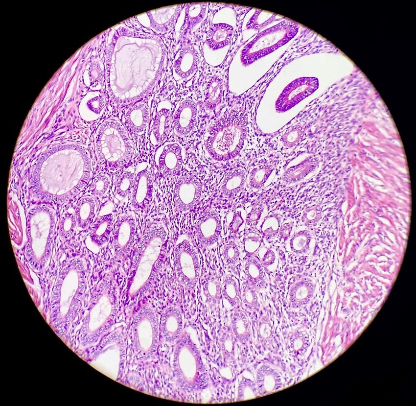
The final histological examination of the specimen revealed endometrial tissues in the colonic submucosa
and muscolaris propria of the ileo-cecal region, with negative cytology for malignant cells (Figures 2-4).
2023 Verzoviti et al. Cureus 15(3): e35782. DOI 10.7759/cureus.35782 2 of 7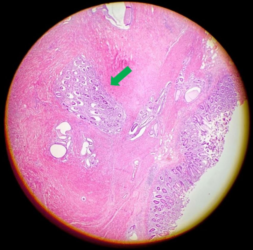
FIGURE 2: Endometrial tissue (green arrow) inside normal colonic
                                                     submucosa.

2023 Verzoviti et al. Cureus 15(3): e35782. DOI 10.7759/cureus.35782                                                    3 of 7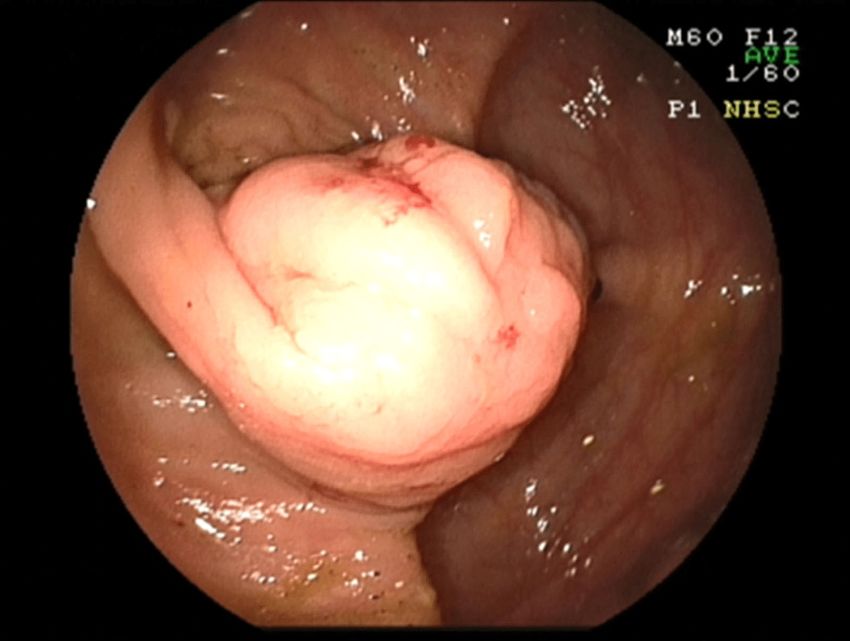
FIGURE 3: Magnification of the endometrial tissue.

2023 Verzoviti et al. Cureus 15(3): e35782. DOI 10.7759/cureus.35782                                      4 of 7
FIGURE 4: The green arrow indicates an endometrial gland inside
                                                     normal lymphatic tissue.

                                                  The postoperative period was uneventful and the patient was discharged from our clinic on the sixth
                                                  postoperative day. The patient was followed up at the first, third, sixth, and ninth months with no findings
                                                  in clinical and imaging examination.

                                                  Discussion
                                                  Women with bowel endometriosis are often diagnosed with other disorders such as irritable bowel syndrome,
                                                  inflammatory or ischemic colitis, diverticulitis, malignancy, and pelvic inflammatory disease [1]. In this
                                                  case, however, the patient was totally asymptomatic.

                                                  To date, there are no clear guidelines on the evaluation of patients with suspected bowel endometriosis.

                                                  Different imaging modalities have been proposed including transvaginal and/or transrectal ultrasonography,
                                                  magnetic resonance imaging, and double-contrast barium enema. These techniques provide useful
                                                  information regarding the presence, location, and extent of endometriosis [11].

                                                  Transvaginal sonography techniques have a sensitivity and specificity of 79% and 94%, respectively. As far
                                                  as magnetic resonance imaging (MRI) is concerned, 94% sensitivity and 77% specificity were observed. For
                                                  rectosigmoid endometriosis, pooled sensitivity and specificity of MRI were 92% and 96%, respectively [12].

                                                  The gold standard diagnostic method is laparoscopic surgery with pathological confirmation [8].

                                                  Combined hormonal contraceptives with or without nonsteroidal anti-inflammatory drugs are first-line
                                                  options for managing symptoms. Second-line treatments include gonadotropin-releasing hormone (GnRH)
                                                  receptor agonists, GnRH receptor antagonists, and danazol. Aromatase inhibitors are reserved for severe
                                                  diseases. All of these treatments seem to be effective but might cause several side effects [9].

                                                  As far as bowel endometriosis is concerned, there is no indicated treatment. Although endometriosis can be
                                                  the origin of Mullerian tumors in women, a similar situation has not been described in intestinal

2023 Verzoviti et al. Cureus 15(3): e35782. DOI 10.7759/cureus.35782                                                                                             5 of 7
endometriosis yet, due to the small number of cases of bowel endometriosis. However, it is theoretically
possible for a cecal mucinous tumor to arise concurrently from intestinal endometriosis with metaplasia [5].
The Japan Society of Obstetrics and Gynecology and the Japan Society of Endometriosis published
guidelines concerning the treatment of EE [13].
As far as rectosigmoid endometriosis is concerned, medical treatment has been reported to improve
symptoms and reduce the lesion size but did not appear to be superior to surgical treatment. For intestinal
endometriosis involving the cecum, there are no reports that assess the efficacy of medical therapy [13].
There are no guidelines specifying which lesions should be operated on and which standardized surgical
technique is recommended. For this reason, the therapeutic option should be tailored according to the
patient's symptoms. Especially in cases of bowel obstruction, surgical treatment is effective in improving
pain [11].
Fu et al. suggested that in any ileocecal lesion with mucinous epithelium or mucin extrusion, the possibility
of a cecal mucinous tumor arising from endometriosis with intestinal metaplasia must be considered [5].
Surgical treatment should also be chosen when endometriosis affects the appendix since the risk of
secondary intussusception of other bowel segments is high [14].
Laparoscopy is the ideal option as it allows precision and complete assessment of the peritoneal cavity. It is
also a well-tolerated and feasible technique [11,15].
Patients who are not candidates for surgical treatment should receive medical treatment. First-line therapy
comprises the long-term use of oral contraceptives. There are also several other medications available to
manage bowel endometriosis that actually aim to reduce circulating hormones. Generally, there is a
rationale for medical treatment before surgery to improve the patient’s symptoms, potentially negating the
need for surgical intervention [11].
In our patient, there was no suspicion of endometriosis. Malignancy was suspected due to the incidental
mass-like lesion and her family history of colon cancer. However, the final diagnosis could only be given by
the histopathological examination, which confirmed endometriosis involving the cecum without any
evidence of malignancy.
Conclusions
Endometriosis of the cecum is a rare clinical entity and presents diagnostic challenges, as limited
information is available in the literature. It is essential to include in our differential diagnosis rare diseases
that imitate symptoms of patients, who often come to the hospital with the suspicion of colon cancer.
Preoperative examination of the characteristics of bowel tumors in women could be recommended, as cecal
endometriosis might erroneously be diagnosed as a malignant tumor. To date, there are no clear guidelines
on the diagnostic and therapeutic options of cecal endometriosis. Due to the limited reported cases, optimal
management strategies are yet to be identified.
Additional Information
Disclosures
Human subjects: Consent was obtained or waived by all participants in this study. Conflicts of interest: In
compliance with the ICMJE uniform disclosure form, all authors declare the following: Payment/services
info: All authors have declared that no financial support was received from any organization for the
submitted work. Financial relationships: All authors have declared that they have no financial
relationships at present or within the previous three years with any organizations that might have an
interest in the submitted work. Other relationships: All authors have declared that there are no other
relationships or activities that could appear to have influenced the submitted work.
References
1. Lee M, Yu L: Cecal endometriosis presenting as a term intrauterine fetal demise and gastrointestinal
hemorrhage: A case report. Case Rep Womens Health. 2021, 30:e00301. 10.1016/j.crwh.2021.e00301
2. Bacalbasa N, Balescu I, Filipescu A: Ileocecal obstruction due to endometriosis - A case report and literature
review. In Vivo. 2017, 31:999-1002. 10.21873/invivo.11160
3. Chamié LP, Ribeiro DM, Tiferes DA, Macedo Neto AC, Serafini PC: Atypical sites of deeply infiltrative
endometriosis: Clinical characteristics and imaging findings. Radiographics. 2018, 38:309-28.
10.1148/rg.2018170093
4. Lukac S, Schmid M, Pfister K, Janni W, Schäffler H, Dayan D: Extragenital endometriosis in the differential
diagnosis of non- gynecological diseases. Dtsch Arztebl Int. 2022, 119:361-7. 10.3238/arztebl.m2022.0176
5. Fu X, Jiang J, Li B, Tian XY, Li Z: Cecal endometriosis with intestinal metaplasia misdiagnosed as neoplasm
2023 Verzoviti et al. Cureus 15(3): e35782. DOI 10.7759/cureus.35782 6 of 7of the cecum by intraoperative histological examination. Int J Surg Pathol. 2018, 26:24-30.
10.1177/1066896917727595
6. Iwamuro M, Tanaka T, Sugihara Y, Harada K, Hiraoka S, Kondo Y, Okada H: Two cases of endometriosis in
the cecum detected by contrast-enhanced computed tomography with air/carbon dioxide insufflation.
Intern Med. 2021, 60:1697-701. 10.2169/internalmedicine.6418-20
7. Feldhaus DJ, Harris RK, Dayal SD: Appendiceal endometriosis presenting as possible cecal mass . Am Surg.
2020, 86:1528-30. 10.1177/0003134820933606
8. Darvishzadeh A, McEachern W, Lee TK, Bhosale P, Shirkhoda A, Menias C, Lall C: Deep pelvic
endometriosis: A radiologist's guide to key imaging features with clinical and histopathologic review. Abdom
Radiol (NY). 2016, 41:2380-400. 10.1007/s00261-016-0956-8
9. Edi R, Cheng T: Endometriosis: Evaluation and treatment. Am Fam Physician. 2022, 106:397-404.
10. Roman H: A national snapshot of the surgical management of deep infiltrating endometriosis of the rectum
and colon in France in 2015: A multicenter series of 1135 cases. J Gynecol Obstet Hum Reprod. 2017, 46:159-
65. 10.1016/j.jogoh.2016.09.004
11. Habib N, Centini G, Lazzeri L, Amoruso N, El Khoury L, Zupi E, Afors K: Bowel endometriosis: Current
perspectives on diagnosis and treatment. Int J Womens Health. 2020, 12:35-47. 10.2147/IJWH.S190326
12. Bazot M, Daraï E: Diagnosis of deep endometriosis: Clinical examination, ultrasonography, magnetic
resonance imaging, and other techniques. Fertil Steril. 2017, 108:886-94. 10.1016/j.fertnstert.2017.10.026
13. Hirata T, Koga K, Kai K, et al.: Clinical practice guidelines for the treatment of extragenital endometriosis in
Japan, 2018. J Obstet Gynaecol Res. 2020, 46:2474-87. 10.1111/jog.14522
14. Costa M, Bento A, Batista H, Oliveira F: Endometriosis-induced intussusception of the caecal appendix . BMJ
Case Rep. 2014, 2014:bcr2013200098. 10.1136/bcr-2013-200098
15. Ruffo G, Rossini R: The outcomes of laparoscopic resection of bowel endometriosis . Curr Opin Obstet
Gynecol. 2013, 25:302-7. 10.1097/GCO.0b013e3283630e26
2023 Verzoviti et al. Cureus 15(3): e35782. DOI 10.7759/cureus.35782 7 of 7You can also read