Recurrent infection-induced autoimmune haemolytic anaemia complicated by pulmonary embolism: a case report and literature review
←
→
Page content transcription
If your browser does not render page correctly, please read the page content below
ACUTE MEDICAL CARE Clinical Medicine 2021 Vol 21, No 3: e306–8
Recurrent infection-induced autoimmune haemolytic
anaemia complicated by pulmonary embolism: a case
report and literature review
Authors: Shu FangA and Fan YangB
A 73-year-old woman presented with progressive dyspnoea findings revealed type 1 respiratory failure, haemoglobin (Hb)
was 71 g/L with white cell count of 23.01 × 10⁹/L, neutrophils of
ABSTRACT
up to type 1 respiratory failure. Laboratory values showed
leucocytosis, reduced haemoglobin to 71 g/L, elevated 74.2% and platelet count of 241 × 109/L; a high level of D-dimer
indirect serum bilirubin and lactic dehydrogenase. Computed (2.78 mg/L); troponin I (cTNI) was 0.091 ng/mL and brain natriuretic
tomography pulmonary angiography (CTPA) revealed peptide (BNP) was 236 pg/mL. A 12-lead electrocardiography
peripheral pulmonary embolism (PE). Echocardiography (ECG) showed no abnormalities in ST segment and T wave (Fig 1).
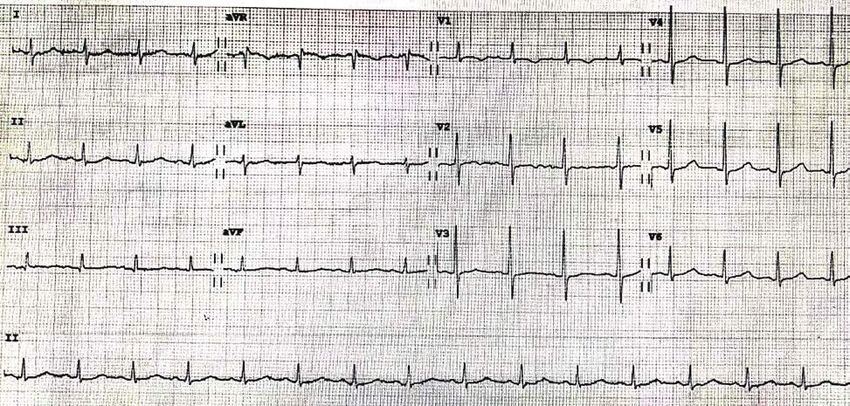
showed enlarged right ventricle, elevated estimated Emergent computed tomography pulmonary angiography (CTPA)
pulmonary arterial systolic pressure (57.2 mmHg) and normal showed bilateral subsegmental and segmental pulmonary emboli
left ventricular ejection fraction. The patient was diagnosed (Fig 2a).
with autoimmune haemolytic anaemia (AIHA), which was
induced by recurrent infections without standard treatment Initial management
in the past year. AIHA is the cause of PE due to the absence of
common predisposing factors and other thrombophilia. The The patient was sent to the cardiac care unit (CCU).
patient became better after administration of glucocorticoids, Echocardiography showed enlarged right ventricle and elevated
intravenous immunoglobulin and rivaroxaban. estimated pulmonary arterial systolic pressure (57.2 mmHg)
and normal left ventricular ejection fraction (81.2%). Dynamic
KEYWORDS: autoimmune haemolytic anaemia, pulmonary changes of laboratory results are summarised in supplementary
embolism, infection material S1. We exclude common predisposing factors and
other thrombophilia for pulmonary embolism (PE) by a detailed
DOI: 10.7861/clinmed.2021-0112 history and laboratory test (cardiolipin anti-antibody, anti-β2-
glycoprotein 1 antibody, lupus anticoagulant, protein C or S
were normal). The patient suffered from haemolytic anaemia
Case presentation as judged by elevated reticulocyte and serum bilirubin. Further
tests showed peripheral blood smears, paroxysmal nocturnal
A 73-year-old woman was referred to the emergency department haemoglobinuria (PNH) clone, haemoglobin electrophoresis,
(ED) with progressive dyspnoea, weakness and dark urine for erythrocyte penetration fragility test, G6PD activity, complement
about 10 days. She complained of recurrent fever for 1 year due H factor concentration, complement H factor antibody and
to pneumonia or urinary tract infection (UTI). She presented to ADAMTS13 were all normal. Coombs test was positive, and
local hospital and was diagnosed with suspicious infection due to resulted in the diagnosis of autoimmune haemolytic anaemia
leucocytosis. Although treating with moxifloxacin, her symptoms
failed to relieve. Past medical history included reflux oesophagitis
for many years and was treated with omeprazole. She has no
history of illicit substance use.
Vital signs were blood pressure at 116/62 mmHg, heart rate
of 90 beats/min, respiratory rate of 26 breaths/min and oxygen
saturation of 85% on room air. Physical examination showed high-
pitched P2, no crackles and rhonchi in both lungs, no peripheral
oedema, no erythema and no palpable cords. The initial significant
Authors: Aresident physician, Peking University First Hospital, Beijing,
China; Battending physician, Peking University First Hospital, Beijing,
China Fig 1. Electrocardiography on admission.
e306 © Royal College of Physicians 2021. All rights reserved.
Pulmonary embolism and autoimmune haemolytic anaemia

                                                                       follow up, reinspection of CTPA revealed disappearance of
                                                                       thrombosis (Fig 2b). At the recent follow-up, 12 months after the
                                                                       initial presentation, she required 5 mg od of prednisolone and
                                                                       20 mg od of rivaroxaban to keep her haemoglobin (Hb) over
                                                                       10 g/dL and had no thromboembolic episodes.

                                                                       Discussion
                                                                       Autoimmune haemolytic anaemia (AIHA) is a rare and
                                                                       heterogeneous disorders characterised by the destruction of red
                                                                       blood cells through warm or cold autoantibodies.2 The overall
                                                                       estimated incidence in adults is 0.8–3 per 100,000/year, a
                                                                       prevalence of 17:100,000 and a mortality rate of 3–4%.3,4 AIHA is
                                                                       the higher risk for thromboembolism due to hypercoagulability.2
                                                                       AIHA occur in systemic lupus erythematosus (SLE), chronic
                                                                       lymphocytic leukaemia (CLL), infection, drug exposure and other
                                                                       mixed disorders.2,5,6 As early as 1967, Allgood et al reported
                                                                       that pulmonary embolism (PE) was the most common cause of
                                                                       death in AIHA, and revealed the association between PE and
                                                                       AIHA.7 Patients with AIHA have 2.6-fold higher risk of venous
                                                                       thromboembolism (VTE) compared with non-AIHA.8 In two large
                                                                       studies, the prevalence of thromboembolism in AIHA patients was
                                                                       recorded up to 15%.4,9 Anticoagulant prophylaxis can effectively
                                                                       reduce the occurrence of a thromboembolic episode in acute
                                                                       exacerbation of AIHA (4.7% vs 33.3%).10 The reasons for patients
                                                                       with AIHA were at an increased risk of VTE remain unclear, which
                                                                       may include the increased expression of phosphatidylserine,
                                                                       microparticles and cytokine-induced monocytes or endothelial
                                                                       tissue factors.8,11 Thromboembolic episodes are relatively common
                                                                       during active phases of the disease, but also occur during disease
                                                                       maintenance therapy.12 High leucocyte count and severe anaemia
Fig 2. Comparison of computed tomography pulmonary angiography.        were associated with VTE.9 PE is a life-threatening complication
a) Image on admission showing bilateral subsegmental and segmental
pulmonary embolism. b) Image at 3 months after discharge showing no    of AIHA. Clinical manifestations of PE include sudden dyspnoea,
pulmonary embolism.                                                    hypoxaemia, and chest pain, which can easily be masked by
                                                                       severe anaemia caused by AIHA, leading to delayed diagnosis.13
                                                                       Therefore, close attention as well as timely treatment plays a
                                                                       crucial role.
                                                                          The causes of AIHA and PE are complex and varied, which
(AIHA). The works for secondary AIHA included other autoimmune         need comprehensive evaluation in patients with PE at the
diseases (antinuclear antibody, anti-dsDNA were negative), serum       first occurrence.1,2 In our case, considering that the patient
immunofixation electrophoresis (no monoclonal band) and a              had suffered recurrent infection without sufficiently effective
computed tomography to exclude lymphoma and solid tumours.             treatment and haemoglobin gradually decreased in the past year,
In addition, T-SPOT.TB, the immunoglobulin M of mycoplasma,            which strongly suggested that the prodromal infection induced
Epstein–Barr virus (EBV), cytomegalovirus (CMV), influenza virus,      AIHA. Previous literature reported that infection associated AIHA
rubella virus, measles, adenovirus and parvovirus B19, hepatitis B     is not rare, accounting for 14% of all patients.11 Drug-related AIHA
surface antigen (HBsAg) and HIV antibodies were all negative.          was excluded because the anaemia did not worsen after taking
Finally, PE attributed to AIHA was considered due to no common         drugs such as moxifloxacin.
aetiology of PE.                                                          The first-line therapy for AIHA is glucocorticoids and
  According to guidelines, the patient was stratified as Pulmonary     approximately 80% of patients would respond to the therapy.
Embolism Severity Index (PESI) class IV, simplified PESI (sPESI)       There are no studies in comparing tapering regimens, where long
≥1, the risk of early death is intermediate–high.1 She received        duration and slow reduction are recommended. No response
low-molecular weight heparin (LMWH) and discontinued other             to steroids after 21 days or relapse during/after a steroid wean
suspicious drugs. During hospitalisation, she once received            is an indication for the second-line therapy.2,3 This patient was
intravenous methylprednisolone sodium succinate 40 mg once             administered with rivaroxaban sequent to LMWH, although there
daily (od)) and intravenous immunoglobulin 10 g od for 7 days. The     was insufficient evidence-based practice in the alternative of
patient didn’t receive anti-infective nor blood transfusion therapy.   novel oral anticoagulants to vitamin K antagonists. Direct oral
                                                                       anticoagulants (DOACs) are not recommended in antiphospholipid
                                                                       syndrome (APS)-induced VTE.14 Notably, numbers of AIHA
Case progression and outcome
                                                                       patients complicated with thromboembolism were reported with
The patient was discharged after 3 weeks with oral prednisolone        positive antiphospholipid antibodies.11 The thrombotic risk is
40 mg qd and rivaroxaban 15 mg twice daily (bid). At 3-month           reported to not correlate with traditional risk factors during AIHA.13

© Royal College of Physicians 2021. All rights reserved.                                                                              e307
Shu Fang and Fan Yang

Vitamin K antagonists reduce functional coagulation factors             4 Barcellini W, Zaninoni A, Fattizzo B et al. Predictors of refractoriness
in the extrinsic and intrinsic pathways of coagulation, which             to therapy and healthcare resource utilization in 378 patients with
showed a stronger anticoagulant effect.15 Thus, it is uncertain that      primary autoimmune hemolytic anemia from eight Italian refer-
DOACs may be a perfect substitute to warfarin in VTE, especially          ence centers. Am J Hematol 2018;93:e243–6.
                                                                        5 Newman K, Owlia MB, El-Hemaidi I, Akhtari M. Management of
attributed to thrombophilia or autoimmune diseases. In our case,
                                                                          immune cytopenias in patients with systemic lupus erythematosus
DOACs are safe and effective for this patient, which providing a
                                                                          - Old and new. Autoimmunity Reviews 2013;12:784–91.
clinical evidence for anticoagulant therapy of AIHA-induced PE.         6 Hodgson K, Ferrer G, Pereira A, Moreno C, Montserrat E.
Given the higher risk of thrombosis, long-term anticoagulation and        Autoimmune cytopenia in chronic lymphocytic leukaemia: diag-
follow-up should be recommended.12                                        nosis and treatment. Br J Haematol 2011;154:14–22.
                                                                        7 Allgood JW, Chaplin H, Jr. Idiopathic acquired autoimmune hemo-
Key points                                                                lytic anemia. A review of forty-seven cases treated from 1955
                                                                          through 1965. Am J Med 1967;43:254–73.
>> AIHA is a hypercoagulable disorder, requiring vigilance against      8 Ungprasert P, Tanratana P, Srivali N. Autoimmune hemolytic anemia
   the complication of deep vein thrombosis.                              and venous thromboembolism: A systematic review and meta-
>> Consider a wide range of differentials when patients present           analysis. Thromb Res 2015;136:1013–7.
   with PE attributed to AIHA.                                          9 Barcellini W, Fattizzo B, Zaninoni A et al. Clinical heterogeneity and
                                                                          predictors of outcome in primary autoimmune hemolytic anemia:
>> No guidelines to recommend anticoagulant therapy for AIHA
                                                                          a GIMEMA study of 308 patients. Blood 2014;124:2930–6.
   induced PE. DOACs may be effective and safe in APS-negative
                                                                       10 Hendrick AM. Auto-immune haemolytic anaemia--a high-risk dis-
   AIHA. ■                                                                order for thromboembolism? Hematology 2003;8:53–6.
                                                                       11 Hoffman PC. Immune hemolytic anemia--selected topics.
Supplementary material                                                    Hematology Am Soc Hematol Educ Program 2009:80–6.
                                                                       12 Audia S, Bach B, Samson M et al. Venous thromboembolic
Additional supplementary material may be found in the online              events during warm autoimmune hemolytic anemia. PLoS One
version of this article at www.rcpjournals.org/clinmedicine:              2018;13:e0207218.
S1 – Dynamic changes of laboratory results and process of              13 Lecouffe-Desprets M, Néel A, Graveleau J et al. Venous throm-
diagnosis and treatment from the initial infection to follow-up.          boembolism related to warm autoimmune hemolytic anemia: a
                                                                          case-control study. Autoimmunity Reviews 2015;14:1023–8.
                                                                       14 Pengo V, Denas G, Zoppellaro G et al. Rivaroxaban vs warfarin
Acknowledgements                                                          in high-risk patients with antiphospholipid syndrome. Blood
Written informed consent was obtained from the patient to                 2018;132:1365–71.
publish the clinical details and images in this article.               15 Cohen H, Hunt BJ, Efthymiou M et al. Rivaroxaban versus warfarin
                                                                          to treat patients with thrombotic antiphospholipid syndrome, with
                                                                          or without systemic lupus erythematosus (RAPS): a randomised,
References                                                                controlled, open-label, phase 2/3, non-inferiority trial. Lancet
                                                                          Haematology 2016;3:e426–36.
1 Konstantinides SV, Meyer G, Becattini C et al. 2019 ESC Guidelines
  for the diagnosis and management of acute pulmonary embolism
  developed in collaboration with the European Respiratory Society
  (ERS). Eur Heart J 2020;41:543–603.
2 Jäger U, Barcellini W, Broome CM et al. Diagnosis and treatment
  of autoimmune hemolytic anemia in adults: Recommendations
  from the First International Consensus Meeting. Blood Rev 2020;
  41:100648.                                                           Address for correspondence: Dr Fan Yang, Department of
3 Hill QA, Stamps R, Massey E et al. The diagnosis and management      Cardiology, Peking University First Hospital, No.8 Xishiku
  of primary autoimmune haemolytic anaemia. Br J Haematol              Street, Xicheng District, Beijing 100034, China.
  2017;176:395–411.                                                    Email: dryangf@126.com

e308                                                                                           © Royal College of Physicians 2021. All rights reserved.
You can also read