Case Report Giant Abdominal Lipoma in Adult
←
→
Page content transcription
If your browser does not render page correctly, please read the page content below
Hindawi
Case Reports in Surgery
Volume 2021, Article ID 6610533, 4 pages
https://doi.org/10.1155/2021/6610533

Case Report
Giant Abdominal Lipoma in Adult

          Nihal Cinar Ozcan , Akay Edizsoy , and Tahsin Colak
          Department of General Surgery, Medical Faculty, Mersin University, 33110 Mersin, Turkey

          Correspondence should be addressed to Nihal Cinar Ozcan; nihal@drnihalcinarozcan.com

          Received 30 December 2020; Revised 15 February 2021; Accepted 20 February 2021; Published 3 March 2021

          Academic Editor: Beth A. Schrope

          Copyright © 2021 Nihal Cinar Ozcan et al. This is an open access article distributed under the Creative Commons Attribution
          License, which permits unrestricted use, distribution, and reproduction in any medium, provided the original work is
          properly cited.

          Lipomas arising from the omentum are extremely rare in adults. Omental lipomas are typically asymptomatic, but very large ones
          may cause nonspecific abdominal symptoms and discomfort. Rarely they can cause omental torsion and present with an acute
          abdomen. We report a 41-year-old female patient with a giant lipoma (40 × 26 × 8 cm and 11,520 g) who presented with mild
          abdominal discomfort. Workup included abdominal ultrasound (USG) and magnetic resonance imaging (MRI). Surgical
          resection was performed without complication. No recurrence was observed during 4-year follow-up.

1. Introduction                                                       nontender, smooth solid mass occupying almost the entire
                                                                      abdomen. There was no evidence of mechanical bowel
Although lipomas are the most common encapsulated                     obstruction. There was no history of any abnormalities of
benign mesenchymal tumors of soft tissue, omental lipomas             the digestive system, liver or renal failure, gynecological malig-
are extremely rare. We found no case series published in              nancy, or any other reason to cause abdominal ascites. All
the global literature. Only a few case reports have been              hematological and biochemical parameters were within
reported in the literature, and most of these are in childhood        normal range. Also, tumor markers (please be specific) were
[1]. Furthermore, giant omental lipomas in adults are                 normal. A solid, well-defined mass lesion measuring approxi-
extremely rare. Less than 10 cases have been published so             mately 40 cm in greatest diameter was reported on ultrasound
far in the literature [2–10]. Most of the cases were asymptom-        (USG). On magnetic resonance imaging (MRI), T1-weighted
atic and diagnosed incidentally [2]. Some cases presenting            axial and coronal MR images showed a hyperintense giant
with acute abdomen due to torsion or compression to other             mass filling most of the abdomen and displacing the bowels.
organs were reported [11, 12]. This report presents a patient         The lesion was also compressing the liver. In fat-saturated
with a giant lipoma which occupied nearly the entire abdom-           T1-weighted MR images, the signal of the mass was sup-
inal cavity yet caused minimal ambiguous nonspecific symp-             pressed similarly to fat. The mass did not enhance with con-
toms and discomfort.                                                  trast. The lesion measured approximately 34 cm × 26 cm in
                                                                      size (Figures 1–3). Given the symptomatic nature of the mass,
2. Case Presentation                                                  surgical resection was recommended. A laparotomy was per-
                                                                      formed by midline abdominal incision. Operative exploration
A 41-year-old woman was admitted to the hospital with mild            revealed a giant mass with a smooth surface, encapsulated and
abdominal pain, discomfort, and bloating on the abdomen. In           lobulated that was originating from the greater omentum.
recent years, the patient had mildly suffered from these symp-             It was extending along the falciform ligament to the poste-
toms. The patient did not complain of weight loss, nausea,            rior liver and filled the entire abdomen and pelvis caudad
vomiting, weakness, or anorexia, but she was worried about            without strong adhesion to adjoining tissues and organs. The
increasing waist circumference and some dyspeptic symp-               mass displaced the intestines posteriorly but did not cause
toms. Physical examination revealed a big, relatively mobile,         obstruction. Also, the entire colon was intact. The mass was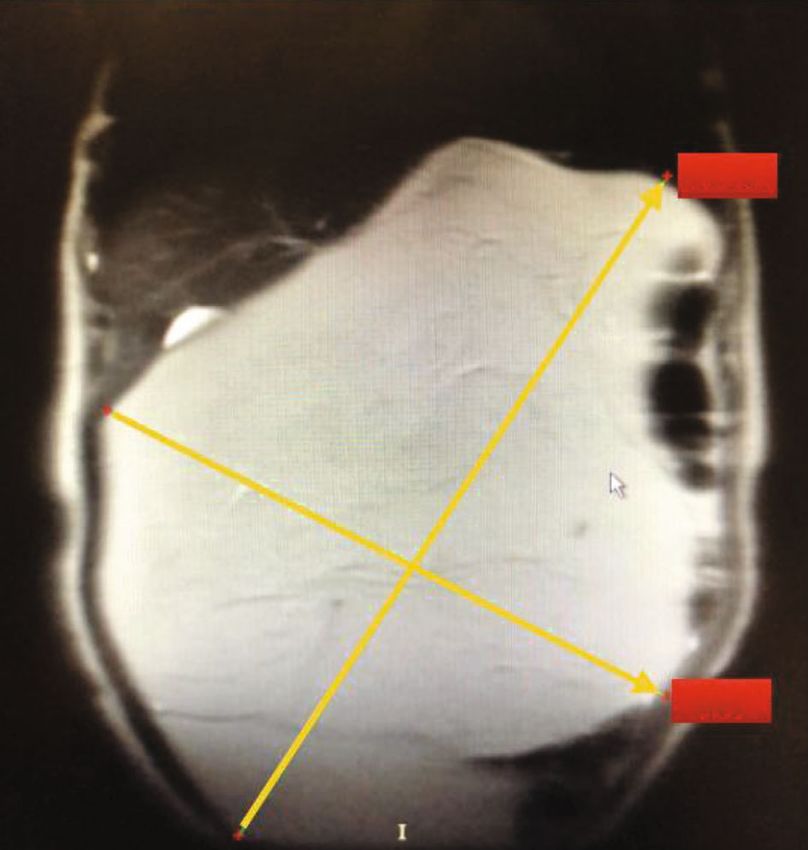
2 Case Reports in Surgery
26.66 cm
12.35 cm
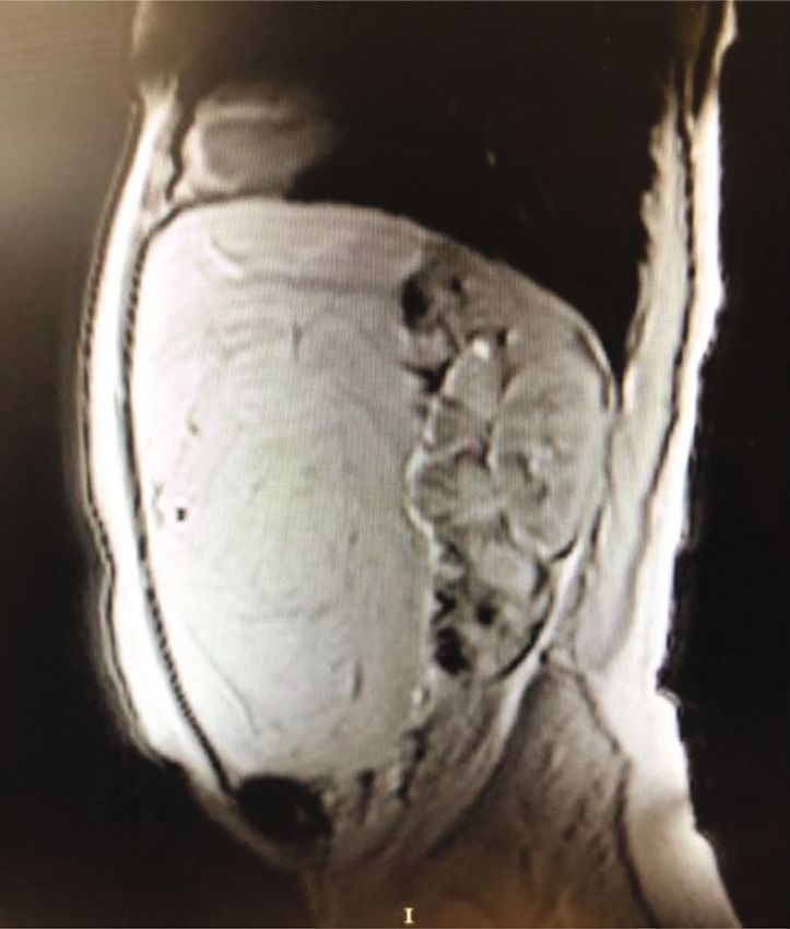
Figure 1: Axial view of magnetic resonance imaging shows that the
lesion is encapsulated and lobulated.
34.04 cm
Figure 3: Sagittal view of magnetic resonance imaging shows that
the lesion caused the intestines to move towards the posterior.
40.00 cm
26.72 cm
Figure 2: The lesion measured as approximately 34 cm × 26 cm in
size on coronal view of magnetic resonance imaging. Figure 4: It was reported that the long diameter of the pathology
specimen was measured as 40 cm.
completely extracted without any violation of lipoma capsule.
The final specimen measured 40 × 26 × 8 cm and weighed
11,520 g (Figure 4). Macroscopically, the specimen was a soft, are rarely seen, with omental lipomas being exceedingly
encapsulated yellow fatty mass, and the cut surface consisted rare [8]. Only about 20 cases that considered all ages have
of homogenous fatty tissue. Pathological examination with been reported [3], and less than 10 cases arising from the
hematoxylin-eosin staining revealed fine-encapsulated mature greater omentum in adults have been reported. A case was
adipose tissue without atypia or malignant degeneration or published as the largest ever reported omental lipoma
necrotic tissue, consistent with a mature benign lipoma. In weighing 6900 g in an adult [3]. The present case is one
postoperative follow-up, liquid food was tolerated on the first of the biggest omental lipomas reported with 11,520 g in
postoperative day and solid food on the second day. She was weight. Early satiety, intermittent vomiting, abdominal dis-
discharged without any complications on postoperative day comfort due to displacement of intestines, and pressure
3. No recurrence was observed during the 4-year follow-up. effect of surrounding structures were frequently reported
as the symptoms in the huge lipoma cases. Also, gradual
3. Discussion abdominal distention and/or expansion in waist circumfer-
ence were often seen in noncomplicated giant lipomas.
Although lipomas are the most common mesenchymal The presented case had abdominal distention mildly, waist
tumors on any part of the body, intra-abdominal lipomas expansion without weight gain, and mild dyspeptic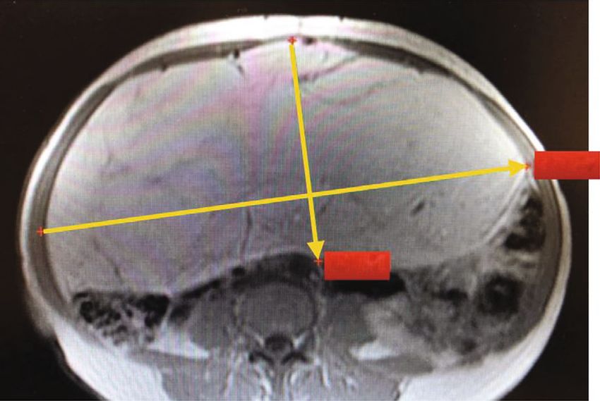
Case Reports in Surgery                                                                                                              3

symptoms caused minimal discomfort. As in this case,               differentiated liposarcomas should be included in the differ-
most of the patients with omental lipoma were diagnosed            ential diagnosis, and reasonable effort should be made to
after symptoms of abdominal discomfort were reported,              discriminate these masses before the operation. Liposarco-
or incidentally when the patients presented for different           mas generally occur in the gluteal region, but they can rarely
reasons [2].                                                       occur in the abdomen with similar characteristics of lipomas
     USG and abdominal computerized tomography (CT) are            in other locations, such as an encapsulated and smooth
the preferred imaging studies for abdominal lipomas. On            surface without invasion of neighboring organs. Elevated
USG, the lipomas appear as homogenous echogenic well-              serum amylase levels have also been reported [18].
bounded masses. Because USG is largely user-dependent,                 Surgery is the treatment of choice for omental lipomas.
most surgeons need further diagnostic imaging study                Because lipomas are encapsulated and seldom infiltrate the
including CT or MRI. CT shows the specific characteristics          surrounding structures, the excision technique is generally
of fat content and aids in distinguishing from the other           uncomplicated. However, every effort should be made for
intra-abdominal masses including leiomyosarcoma, lipo-             complete excision with an intact capsule to avoid recurrence.
sarcoma, fibroma, fibrosarcoma, or hemangiopericytoma                The rate of recurrence after excision is reported to be less
[8]. CT also determines the size of the tumor and its rela-        than 5% [19]. Preoperative imaging is important for surgical
tionship to neighboring structures [13]. It has also been          planning. In the light of the literature, omental lipomas may
reported that abdominal CT is useful for understanding             create nonspecific signs or symptoms or be asymptomatic
the arterial supply of the tumor and its relationship with         and may rarely lead to emergent surgery. Also, omental lipo-
other organs [1]. MRI also provides excellent tissue charac-       mas are surgical entities that need to be made a differential
terization, with lipomas appearing identical to subcutaneous       diagnosis from malignancies and can reach large sizes.
fat on all pulse sequences and any fibrous septa within exhi-
biting low signal intensity on T1- and T2-weighted images.         4. Conclusion
The fatty nature of the tissue can be confirmed on chemical
shift imaging and frequency selective fat suppression tech-        This case report revealed that omental lipomas can remain
niques [13]. In T1- and T2-weighted MR images, homoge-             silent until they reach a giant size. In these cases, surgery is
neous low signal intensity should be detected. In distinction      the definitive treatment option, and preoperative cross-
to liposarcoma, which may show thick, irregular, or nodular        sectional imaging is critical for surgical planning.
septations larger than 2 millimeters in both CT and MRI, fat-
free nodular foci increased septal contrast [14]. In the current   Conflicts of Interest
case, MRI was used to distinguish soft tissues, and the Dopp-
ler USG was used for vascularity. No findings suggesting            The authors declare that they have no conflict of interests and
other lesions with predominantly macroscopic fat include           no financial support.
myelolipoma, angiomyolipoma, liposarcoma, teratoma, epi-
ploic appendagitis, fat infarction, and mesenteric panniculitis    References
were detected. Pereira et al. reported that MRI imaging tech-
niques were often powerful tools in the noninvasive evalua-         [1] H. Shiroshita, Y. Komori, M. Tajima et al., “Laparoscopic
tion of these lesions. Knowledge of clinical, anatomic, and             examination and resection for giant lipoma of the omentum,”
imaging features is important in formulating an appropriate             Surgical Laparoscopy, Endoscopy & Percutaneous Techniques,
differential diagnosis and guiding patient care, often obviat-           vol. 19, no. 5, pp. e217–e220, 2009.
ing invasive diagnostic procedures [13].                            [2] M. R. Sanchez, F. M. Golomb, J. A. Moy, and J. R. Potozkin,
     In this patient, the lipoma was arising from the greater           “Giant lipoma: case report and review of the literature,” Jour-
omentum and associated with the falciform ligament. It                  nal of the American Academy of Dermatology, vol. 28, no. 2,
                                                                        pp. 266–268, 1993.
caused minimal discomfort and abdominal distention. But
different unexpected clinical scenarios or acute abdominal           [3] D. Sen, R. Chakrabarti, M. Ranjith, and D. Gulati, “Giant
                                                                        omental lipoma in an elderly female patient,” Medical Journal
syndrome occurred in some of the other cases. Hishiki et al.
                                                                        Armed Forces India, vol. 74, no. 4, pp. 377–379, 2018.
reported a case arising from the lesser omentum, presenting
                                                                    [4] W. J. Tan and W. H. Chan, “Giant omental lipoma,” Singapore
as an acute abdomen due to torsion [11]. Other reports                  Medical Journal, vol. 53, no. 6, pp. e131–e132, 2012.
include patients with omental lipomas in an inguinal hernia
                                                                    [5] V. Chaudhary, M. K. Narula, R. Anand, I. Gupta, G. Kaur, and
sac were noted [12, 15]. An omental lipoma case that caused             K. Kalra, “Giant omental lipoma in a child,” Iranian Journal of
amenorrhea and abdominal distention was reported; there-                Radiology, vol. 8, no. 3, pp. 167–169, 2011.
fore, it was considered pregnancy [10]. Another patient             [6] G. C. Beattie and S. T. Irwin, “Torsion of an omental lipoma
who presented with abdominal pain developed an infarct in               presenting as an emergency,” International Journal of Clinical
the lipoma originating from the falciform ligament [16].                Practice, vol. 59, pp. 130-131, 2005.
The lipoma was excised from this patient who was operated           [7] X. Luo, W. Gao, and J. Zhan, “Giant omental lipoma in
on urgently due to the acute abdomen. A case with chronic               children,” Journal of Pediatric Surgery, vol. 40, no. 4,
diseases and bleeding spontaneously and disrupting the                  pp. 734–736, 2005.
patient’s hemodynamic was observed [17]. Transarterial              [8] V. Barauskas, D. Malcius, and V. Jazdauskiene, “Lipoma of the
embolization was applied to this patient, who was inoperable.           greater omentum in a child,” Medicina, vol. 40, no. 9, pp. 860–
In the presentation of large abdominal masses, well-                    863, 2004.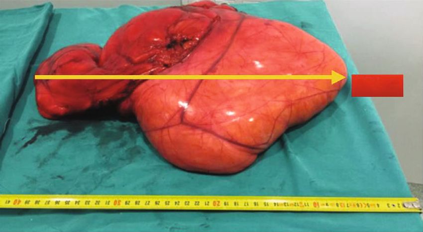
4                                                                       Case Reports in Surgery

 [9] V. E. Grankin, “Torsion of lipoma of the greater omentum,”
     Vestnik Khirurgii Imeni I. I. Grekova, vol. 104, no. 2, p. 132,
     1970.
[10] H. Chandra, “Omental lipoma,” Indian Medical Gazette,
     vol. 76, no. 7, pp. 420-421, 1941.
[11] S. Hishiki, H. Fukushima, T. Nishimura, J. Horiguchi,
     T. Shibayama, and J. M. Chong, “A case of lipoma of the lesser
     omentum with torsion of the pedicle in a twenty-eight-year-
     old woman,” Nihon Shokakibyo Gakkai Zasshi, vol. 107,
     no. 9, pp. 1450–1455, 2010.
[12] T. W. Yang, Y. W. Tsuei, C. C. Kao, W. H. Kuo, Y. L. Chen,
     and Y. Y. Lin, “Torsion of a giant antimesenteric lipoma of
     the ileum: a rare cause of acute abdominal pain,” American
     Journal of Case Reports, vol. 18, pp. 589–592, 2017.
[13] J. M. Pereira, C. B. Sirlin, P. S. Pinto, and G. Casola, “CT and
     MR imaging of extrahepatic fatty masses of the abdomen and
     pelvis: techniques, diagnosis, differential diagnosis, and pit-
     falls,” Radiographics, vol. 25, no. 1, pp. 69–85, 2005.
[14] S. Zama Ali and S. Srinivasan, “Comment on: giant omental
     lipoma,” Singapore Medical Journal, vol. 53, no. 10, pp. 697-
     698, 2012.
[15] E. Turk, E. Karagulle, H. Oguz, and E. Toprak, “Indirect
     hernial sac containing the uterus, ovary, and fallopian tube
     in association with a giant intraabdominal lipoma: report
     of a case,” Hernia, vol. 16, no. 5, pp. 593–595, 2012.
[16] B. Coulier, V. Cloots, and A. Ramboux, “US and CT diagnosis
     of a twisted lipomatous appendage of the falciform ligament,”
     European Radiology, vol. 11, no. 2, pp. 213–215, 2001.
[17] P. Tirukonda, S. Wu, J. Brar, K. S. Ng, and S. Mirsadraee,
     “Trans arterial embolization of spontaneous intra-abdominal
     haemorrhage from omental lipoma,” Case Reports in Radiol-
     ogy, vol. 2018, Article ID 2926143, 5 pages, 2018.
[18] S. Hashimoto, J. Arai, M. Nishimuta et al., “Resection of lipo-
     sarcoma of the greater omentum: a case report and literature
     review,” International Journal of Surgery Case Reports,
     vol. 61, pp. 20–25, 2019.
[19] A. I. Squillaro, M. D. Chow, F. Arias, E. T. Sadimin, and Y. H.
     A. Lee, “A giant childhood mesenteric lipoblastoma with
     extensive maturation,” Frontiers in Pediatrics, vol. 8, p. 404,
     2020.
You can also read