Case Report A Patient with Complex Gout with an Autoinflammatory Syndrome and a Sternoclavicular Joint Arthritis as Presenting Symptoms
←
→
Page content transcription
If your browser does not render page correctly, please read the page content below

Hindawi
Case Reports in Rheumatology
Volume 2020, Article ID 5026490, 4 pages
https://doi.org/10.1155/2020/5026490

Case Report
A Patient with Complex Gout with an Autoinflammatory
Syndrome and a Sternoclavicular Joint Arthritis as
Presenting Symptoms

          M. M. Fedeli ,1 M. Vecchi,2 and P. Rodoni Cassis2,3,4
          1
            Department of Internal Medicine, Ospedale La Carità, Locarno, Switzerland
          2
            Centro Medico, Chiasso, Switzerland
          3
            Department of Radiology, Clinica Sant’Anna, Lugano-Sorengo, Switzerland
          4
            Department of Radiology, Clinica Ars Medica, Gravesano, Switzerland

          Correspondence should be addressed to M. M. Fedeli; marcofedeli89@gmail.com

          Received 30 September 2019; Revised 22 December 2019; Accepted 13 January 2020; Published 31 January 2020

          Academic Editor: James V. Dunne

          Copyright © 2020 M. M. Fedeli et al. This is an open access article distributed under the Creative Commons Attribution License,
          which permits unrestricted use, distribution, and reproduction in any medium, provided the original work is properly cited.
          A 50-year-old man presented to the emergency department with widespread pain, especially at the chest level, fever, and night
          sweats. Physical examinations revealed a swelling with localized pain in the left sternoclavicular joint. Laboratory tests showed a
          CPR of 134 mg/l and an ESR of 70 mm/h. The patient’s anamnesis is, for a chronic gouty arthritis, poorly controlled type 2 diabetes
          and a lumbosacral radicular syndrome. Home therapy includes metformin, sitagliptin, gliclazide, naproxen with partial benefit on
          pain, and febuxostat. Differential diagnoses of sternoclavicular swelling include infection, crystal or psoriatic arthropathy, tumor
          pathology, SAPHO syndrome, and osteoarthritis. An ultrasound scan performed at the thoracic level showed the presence of
          effusion in the sternoclavicular joint. A thoracoabdominal CT scan, performed in doubt of neoplasias, shows no masses but
          osteostructural nonspecific alterations of the sternoclavicular joint. We performed a dual energy CT (DECT) which reports a
          gouty arthropathy at the sternoclavicular joints (in the literature, only three similar cases are proved). Because of the poor
          therapeutic effects using febuxostat and systemic corticosteroids, the patient was treated with anakinra, an interleukin 1 receptor
          antagonist, which led, 6 months after the event, to a total remission.

1. Case Report                                                          compartment of the right hand because of severe pain and
                                                                        losing range of motion in the affected joint.
R. is a 50-year-old patient who has, since a month, a state of              He also had high uric acid levels (Figure 1) as he con-
general malaise characterized by widespread pain and in-                sumed febuxostat (allergic to allopurinol).
termittent episodes of fever and night sweats. He refers fever              He is also known for type 2 diabetes (diagnosis of 2011)
with stakes up to 39°C in two days prior to hospitalization at          in pharmacological treatment with metformin, sitagliptin,
the Department of Rheumatology. The pains are especially at             and gliclazide poorly controlled because of wrong eating
night to the chest level. Despite a home drug therapy with              habits without a regular physical activity. In fact, in phys-
NSAID (nonsteroidal anti-inflammatory drug), symptoms                    iological anamnesis, the patient declares he drinks 2–3 liters
do not regress.                                                         of carbonated soft drinks a day.
    The patient’s anamnesis is for a chronic gouty arthritis                He has a class 1 obesity (BMI 31.5 Kg/m2) that is why he
characterized by the presence of tophi and frequent arthritis;          underwent gastric banding surgery in 2004 with mediocre
the first gouty attack was in 2011 on the ankles treated with            success. Another cardiovascular risk factor is a chronic renal
steroids. In 2014, he has undergone surgical operation with             failure (Figure 2) G3a (KDIGO, 2012) [2] due to diabetic
the removal of tophaceus masses [1] at the sixth                        nephropathy (albuminuria grade A2) [3].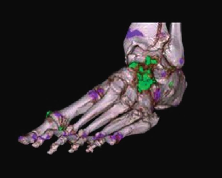
2                                                                                                        Case Reports in Rheumatology

                                                            Uric acid levels (mmol/l)
                800
                700
                600
                500
                400
                300
                200
                100
                  0
                      10/02/2000
                      24/06/2000
                      09/01/2001
                      07/08/2001
                      26/10/2001
                      27/12/2001
                      28/07/2003
                      17/05/2011
                      04/06/2012
                      20/08/2012
                      15/10/2012
                      22/05/2013
                      25/03/2014
                      10/05/2014
                      11/11/2014
                      26/11/2014
                      15/12/2014
                      21/01/2015
                      01/04/2015
                      13/07/2015
                      15/09/2015
                      13/10/2015
                      13/11/2015
                      26/01/2016
                      22/03/2016
                      18/05/2016
                      23/06/2016
                      25/10/2016
                      07/02/2017
                      13/06/2017
                      16/11/2017
                      08/02/2018
                      14/08/2018
                      19/02/2018
                      01/04/2018
                      09/05/2019
                      04/09/2019
                                         Figure 1: Monitoring uric acid levels over the years.

                                                            Creatinine (µmol/l)
             160
             140
             120
             100
              80
              60
              40
              20
               0
            06/07/2009   18/11/2010   01/04/2012   14/08/2013   27/12/2014    10/05/2016    22/09/2017   04/02/2019   18/06/2020
                                       Figure 2: Monitoring renal function levels over the years.

    Lastly, he suffered from lumbosacral radicular syn-
drome, irritative L3 on the left side.
    On examination, the patient is in good general condi-
tion, 102 kg weight, 180 cm height, with eupnea, normal
heart rate, normal heart rhythm, and normal cardiac
rhythm. Clinically, there was a florid inflammation at the
level of the first metatarsophalangeal joint of the left foot
with a swelling of the left ankle without pain on pressure or
mobilization. The knees appear bilaterally swollen, warm to
thermotouch, aching with the presence of a joint effusion
mostly expressed on the left. We deduce inflammation to the
metacarpophalangeal joints (II, III, and IV) of the left hand
where, in the past, tophi had been removed surgically. The
movement in extension of the left elbow is deficient. Lumbar                       Figure 3: Functional ultrasound scan of the knee.
spine mobility is reduced by 1/3 in the lateral-bilateral
flexion and in forward flexion with soreness at the lum-
bosacral level. At chest level, there is a swelling with localized           (iv) Psoriatic arthropathy
pain in the left sternoclavicular joint. Valid heart sounds, no               (v) SAPHO syndrome (synovitis, acne, pustulosis, hy-
murmurs were observed. At lung auscultation, there was                            perostosis, and osteitis)
vesicular murmur spreading ubiquitously. The abdomen is
                                                                             (vi) Osteoarthritis
treatable and painless on palpation. No evidence of asso-
ciated neurological deficits.                                                Proceeding by elimination, we have eradicated from
    The electrocardiogram revealed no significant alterations.           our list psoriatic arthritis in a patient who showed no skin
Laboratory tests show a CRP of 134 mg/l and an ESR of                   changes, and with silent family history for this disease. A
70 mm/h. Uric acid is within the normal limit (263 mmol/l).             thoracoabdominal CT scan, performed in doubt of neo-
    The differential diagnosis of sternoclavicular swelling:             plasias, shows no masses but osteostructural nonspecific
                                                                        alterations of the sternoclavicular joint. Procalcitonin is
      (i) Infectious arthropathy
                                                                        negative reducing the chance of bacterial infection.
     (ii) Crystals arthropathy (uric acid or calcium pyro-              Trauma was not reported in the recent period. We have
          phosphate crystals)                                           also excluded osteoarthritis because of the absence of the
    (iii) Tumor pathology                                               classical radiological signs (narrowing of joint space,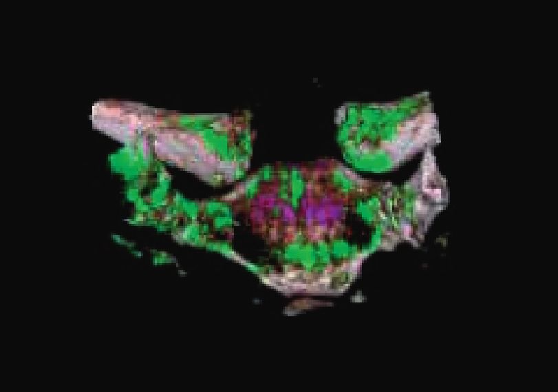
Case Reports in Rheumatology                                                                                                   3

                            Figure 4: Sternoclavicular details with Dual Energy CT 3D reconstruction.

                                                                      A chest ultrasound showed the presence of effusion in
                                                                  the sternoclavicular joint and thickening of the synovial
                                                                  capsule.
                                                                      Because of the lack of joint fluid, we could not carry out
                                                                  arthrocentesis of sternoclavicular joint. We performed a CT
                                                                  with the DUAL ENERGY (DECT) method, which con-
                                                                  firmed the suspicion of gout-originated arthropathy at the
                                                                  sternoclavicular joints, explaining the patient’s chest
                                                                  symptoms. Specifically, we show the 3D reconstruction of
                                                                  the dual energy CT of the sternoclavicular joints and the foot
                                                                  (Figures 4 and 5) [1]. The uric acid deposits appear in green,
                                                                  while the bone calcium appears in purple.
                                                                      Our patient has a systemic disease with destructive
  Figure 5: Left foot with Dual energy CT 3D-reconstruction.
                                                                  deforming arthritis. Indeed, it has some tophaceous lesions
                                                                  (Figure 6) [1].

                                                                  2. Discussion
                                                                  A gouty arthritis with a sternoclavicular localization is a
                                                                  very rare finding (in the literature, only three similar cases
                                                                  are proved [6]). Classically, imagining a classic case of gout,
                                                                  we thought more frequently to the metatarsophalangeal
                                                                  joint (podagra), to proximal and distal interphalangeal, to
                                                                  elbow and knee [1, 5, 7] Our patient had an intermediate
                                                                  probability of having gout (7.5 points) [8]. In this case, the
                                                                  gold standard for the diagnosis of gout remains arthro-
                                                                  centesis with the detection of uric acid crystals [8]. The
                                                                  diagnostic alternatives include ultrasound, with the classic
                                                                  double contour of the cartilage surface typical of ar-
                                                                  thropathies with crystal deposits, and the Dual Energy CT
                                                                  [1, 7, 9]. With this method [9], the images are acquired
                                                                  simultaneously, using two different energy levels. Com-
                                                                  paring the specific attenuation at 80 and 140 kVp, it is
                                                                  possible to differentiate the chemical composition of tissues
Figure 6: Erosion and intra-articular swelling with tophaceous    subjected to CT. Uric acid crystals are thus differentiated
masses (CT of the left foot).
                                                                  from the bone or from calcium-based dystrophic calcifi-
                                                                  cation. Recent studies have shown that CT Dual Energy a
osteophytes, subchondral bony sclerosis, and subchondral          sensitivity of 75 to 90% and a specificity between 83 and
cysts) [4, 5].                                                    93%, respectively [9]. This type of examination, therefore,
    Therefore, we continued with a functional ultrasound          represents an excellent alternative for the diagnosis of gout
scan to the knee that showed synovitis with joint effusion         and in particular, in the case of clinical doubts or incon-
and the characteristic double contour that means the              clusive microscopic analysis.
presence of deposits of urate at the femoral condyles car-            The abundant consumption of drinks, given the high-
tilage level (Figure 3) [1].                                      fructose content, may have helped to maintain the hyper-
    The synovial fluid, drawn at the knee level, confirmed          uricemia and played a role in causing acute attacks through
this hypothesis given the presence of uric acid crystals.         urea fluctuations [10, 11].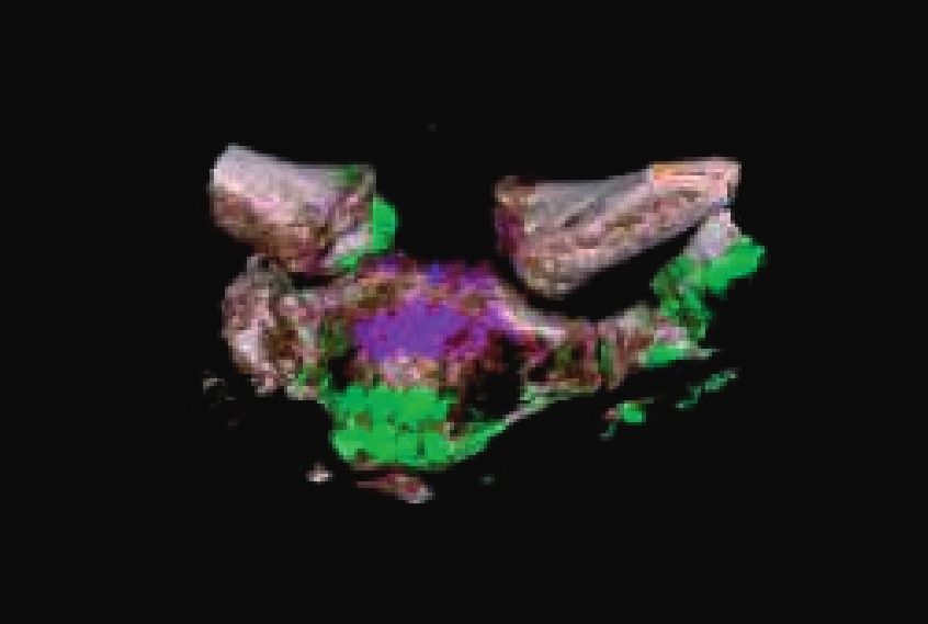
4                                                                                                      Case Reports in Rheumatology

    Another useful starting point in clinical practice is the           [9] T. R. C. Johnson, C. Fink, S. O. Schönberg, and M. F. Rei-ser,
uric acid levels were normal; this indicates how important                  Dual Energy CT in Clinical Practice, Springer, Berlin, Germany,
are the uric acid fluctuations in blood rather than the peak in              2011.
parallel with acute attacks of gout [7].                               [10] H. K. Choi and G. Curhan, “Soft drinks, fructose con-
    At last, a brief mention of therapy. Regarding the acute                sumption, and the risk of gout in men: prospective cohort
                                                                            study,” BMJ, vol. 336, no. 7639, pp. 309–312, 2008.
attack, you should first use AINS or colchicine and, in case of
                                                                       [11] J. W. J. Choi, E. S. Ford, X. Gao, and H. K. Choi, “Sugar-
monoarticular attack, also corticosteroid infiltrations. Be-                 sweetened soft drinks, diet soft drinks, and serum uric acid
cause of the failure benefit of AINS, the failure tolerance of               level: the third national health and nutrition examination
colchicine and polyarticular localization, we have resorted to              survey,” Arthritis & Rheumatism, vol. 59, no. 1, pp. 109–116,
prednisone starting with a dosage of 20 mg to scale gradually               2008.
down. Febuxostat treatment has been kept, and because of
the poor therapeutic effects, we decide to use anakinra, an
inhibitor of interleukin 1, which led to total remission. The
total duration of the gout flare was 6 months.

3. Take Home Message
Summarizing, the key points are:
      (i) Gout can occur in a generalized and systematic way
          and affect any joint [1]
     (ii) Dual-Energy CT or ultrasound scan are the best
          functional tests chosen in cases of doubt or for
          special localizations of gout [5, 7]
    (iii) The abuse of carbonated soft drinks, rich in fructose,
          is a risk factor comparable with beer for gout
          [10, 11]
    (iv) Normal uric acid levels do not exclude a gouty
          attack [5, 7]

Conflicts of Interest
The authors declare that they have no conflicts of interest.

References
 [1] P. Towiwat, A. Chhana, and N. Dalbeth, “The anatomical
     pathology of gout: a systematic literature review,” BMC
     Musculoskeletal Disorders, vol. 20, no. 1, p. 140, 2019.
 [2] Kidney Disease: Improving Global Outcomes (KDIGO),
     “KDIGO clinical practice guide-line for the evaluation and
     management of chronic kidney disease,” Kidney International
     Supplements, vol. 3, pp. 1–150, 2013.
 [3] F. Persson and P. Rossing, “Diagnosis of diabetic kidney
     disease: state of the art and future perspective,” Kidney In-
     ternational Supplements, vol. 8, no. 1, pp. 2–7, 2018.
 [4] D. J. Hunter and D. T. Felson, “Osteoarthritis,” BMJ, vol. 332,
     no. 7542, pp. 639–642, 2006.
 [5] M. C. Hochberg, E. M. Gravallese, A. J. Silman, J. S. Smolen,
     M. E. Weinblatt, and M. H. Weisman, Rheumatology, Elsevier,
     Amsterdam, Netherlands, 2018.
 [6] http://journals.plos.org/plosmedicine/article.
 [7] P. Richette, M. Doherty, E. Pascual et al., “2018 updated
     European League against Rheumatism evidence-based rec-
     ommendations for the diagnosis of gout,” Annals of the
     Rheumatic Diseases, vol. 79, no. 1, pp. 31–38, 2019.
 [8] H. M. Janssens, J. Fransen, E. H. van de Lisdonk,
     P. M. van Riel, C. van Weel, and M. Janssen, “A diagnostic rule
     for acute gouty Arthritis in primary care without joint fluid
     analy-sis,” Archives of Internal Medicine, vol. 170, no. 13,
     pp. 1120–1126, 2010.
You can also read