A Versatile and Affordable Plunge Freezing Instrument for Preparing Frozen Hydrated Specimens for Cryo Transmission Electron Microscopy (CryoEM)
←
→
Page content transcription
If your browser does not render page correctly, please read the page content below
Downloaded from https://www.cambridge.org/core. IP address: 46.4.80.155, on 14 Oct 2021 at 03:51:14, subject to the Cambridge Core terms of use, available at https://www.cambridge.org/core/terms. https://doi.org/10.1017/S1551929500054432
           A Versatile and Affordable Plunge Freezing
            Instrument for Preparing Frozen Hydrated
           Specimens for Cryo Transmission Electron
                      Microscopy (CryoEM)
                               Linda Melanson
                            Gatan, Inc., Pleasanton, CA
                               lmelanson@gatan.com
             CryoEM is a powerful tool in the arsenal of structural biolo-
        gists and soft polymer chemists. Hydrated specimens require
        a preservation method that will counteract the effects of the
        electron beam and the high vacuum environment of the electron
        microscope. Classical specimen preparation techniques using
        chemical fixatives are not able to capture the native structure
        of the once hydrated specimen perfectly. In contrast to classi-
        cal methods for preserving specimens for electron microscopy,
        rapid freezing of radiation-sensitive specimens such as dispersed
        biological macromolecular assemblies, 2D crystals, and colloids
        allows the structural details of the specimen to be captured in
        their essentially native state to near atomic resolution.
             Key to the entire process of cryoEM is the preparation of
        the specimen. A successful cryo preparation requires that the
        specimen is rapidly frozen so that all motion and metabolic
        activities are instantaneously arrested and that the fluid within
        or surrounding the specimen freezes without the formation of
        ice crystals.
             There are three main steps for preparing samples for cryoEM:         Figure 1: Cryoplunge™3 showing support column (A), humidity
        • A small aliquot (3 µL) of a fluid suspension containing the         chamber (B), safety shield (C), and cryogenic workstation (D).
           sample is applied to the surface of a supporting substrate
           such as a fenestrated carbon film (Quantifoil® or C-flat™) or      requires a minimum of space on a standard laboratory bench.
           a continuous carbon film that is attached to the surface of a      The support column of a Cp3 houses all of the pneumatic logic
           standard TEM specimen grid.                                        and electrical components including the ethane temperature con-
        • The droplet is blotted with filter paper until only a thin layer    troller, the blotting timer and the temperature/humidity meter.
           (approximately 100 nm thick) is left on the support substrate.          The humidity chamber safeguards the specimen from drying
        • The thin fluid layer is rapidly immersed into a suitable cryo-      during the blotting process. A simply designed humidity wand
           gen of high heat capacity, which gives instantaneous and
           contaminant-free freezing.
             A suitable cryogen must be capable of achieving a freezing
                                  6
        rate of approximately 10 Kelvin per second. At room tempera-
        ture and pressure, the heat capacity of liquid nitrogen (boiling
        point -196 °C) is too low to properly freeze the specimen; a speci-
        men placed within the liquid nitrogen will cause rapid boiling
        in the area around the specimen forming an insulating layer of
        nitrogen gas. The cooling rate of liquid nitrogen can be improved
        provided that it is maintained at a temperature near its melting
        point of -210 °C by applying a low vacuum for several seconds
        just prior to freezing the specimen. Alternatively, very rapid
        cooling for plunge freezing specimens can be obtained using
        liquid ethane maintained close to its melting point (-181.76 °C)
        using liquid nitrogen as the primary coolant.†
             Cryoplunge™3 (also known as Cp3) is Gatan’s new, semi-au-
                                                                                     Figure 2: Cryoplunge™3 workstation (shown at room temperature with the
        tomatic plunge freezing instrument specifically designed for the        workstation covers removed): ethane pot (e), cryo grid box transfer pot (t) that
        preparation of frozen hydrated specimens for cryoEM, figure 1.          accepts round or square cryo grid boxes, liquid nitrogen remote fill funnel(f),
             Everything is included with the Cp3 for fast, easy set-up.         workstation overflow port (o), cryo grid box staging area (s), filter paper clip
                                                                                (c) for blotting excess ethane from frozen hydrated grid prior to storage or
        Weighing only 12.5 kg, the Cp3 is portable, and its small footprint     viewing on the TEM.

        14 n MICROSCOPY TODAY March 2009

ABMelanson.indd 1                                                                                                                                      02/27/2009 12:28:02 PM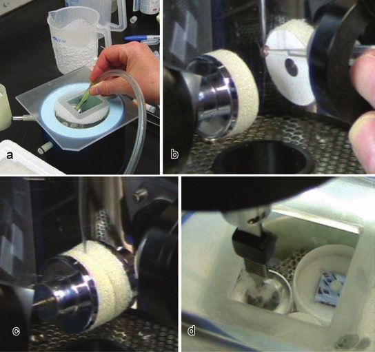
Downloaded from https://www.cambridge.org/core. IP address: 46.4.80.155, on 14 Oct 2021 at 03:51:14, subject to the Cambridge Core terms of use, available at https://www.cambridge.org/core/terms. https://doi.org/10.1017/S1551929500054432

                                                                                                                                                                                                                                                02/27/2009 12:32:41 PM
                                                                                                                                                                                                                                                MT Full Page Ad Master.indd 1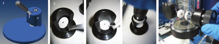
Downloaded from https://www.cambridge.org/core. IP address: 46.4.80.155, on 14 Oct 2021 at 03:51:14, subject to the Cambridge Core terms of use, available at https://www.cambridge.org/core/terms. https://doi.org/10.1017/S1551929500054432
                 Figure 3: Cp3’s filter paper die cutter (1) lets one select the filter paper best suited to the experiment. Mounting the blotting filter paper with the easy-to-use filter
            paper loading jig: loading the filter paper disk onto the inverted filter paper securing pin (2), loading the protective backer (3), which prevents contamination of the
            blotter’s foam pad during the blot cycle, attaching the filter paper and protective backer onto a blot assembly (4), and inserting the blotter into the humidity chamber (5).

        with a replaceable cellulose sponge provides high humidity to the                            specimen grid. One press of the quick-disconnect-pushbutton on
        chamber approximately 15 minutes after insertion. A humidity/                                the plunge rod allows the operator to quickly and easily discon-
        temperature sensor monitors the atmospheric conditions within                                nect the plunging tweezers and safely transfer the frozen hydrated
        the chamber. The blot assemblies, humidity wand, and the sen-                                grid to a pre-cooled cryo grid storage box located within the
        sor are incorporated into the humidity chamber and all of these                              cryo grid box transfer pot. The frozen hydrated grids are fully
        components, including the humidity chamber itself, can be easily                             protected as the liquid nitrogen filled transfer pot is removed
        removed for cleaning.                                                                        from the workstation and transferred to a liquid nitrogen storage
             There are two safety interlocks on Cp3. The safety interlock                            dewar or to the pre-cooled workstation of a cryo transfer holder
        on the safety shield protects the user during the blotting and                               for low electron dose imaging on the TEM.
        plunging cycle. The safety interlock on the cryogenic worksta-                                    A typical protocol for preparing a frozen hydrated speci-
        tion ensures that the workstation is perfectly aligned with the                              men using Cp3 might involve the following steps, illustrated in
        plunge piston, which will not release unless this safety connec-                             figure 4.
        tion is made.                                                                                • Pre-cool the workstation and fill the ethane pot of Cp3.
             The specially designed, removable cryogenic workstation,                                • Adjust the ethane temperature so that the ethane is maintained
        figure 2, maintains the cryogen (ethane) at a temperature just                                  at a temperature just above its melting point.
        above its melting point to ensure consistent freezing of the speci-                          • Insert a pre-treated, carbon support grid (C-flat™ and Quan-
        men. As liquid nitrogen is added to the workstation through the                                 tifoil® are commonly used) into the Cp3 plunging tweezers.
        attached funnel, a blanket of cold, dry nitrogen gas fills the work-                         • Attach the tweezers to the plunge post and raise the plunge
        station chamber. This minimizes condensation of atmospheric                                     post to position the tweezer and grid within the humidity
        oxygen and water vapor onto the surface of the ethane; it also                                  chamber of Cp3.
        generates a protective cryo interface for transferring the frozen
        hydrated grid within the workstation.
             The adjustable pneumatic blotting action for Cp3 is triggered
        by an electronic timer that is accurate to 0.1 seconds. The speci-
        men grid can be blotted one time or multiple times.  Cp3 comes
        with two interchangeable blot assemblies for two sided blotting
        and one blanking plug that provides the capability for one-sided
        front or back side blotting of the specimen grid.  By setting the
        blot timer to zero, one can override the automatic blotting action
        altogether to facilitate other methods for applying the specimen,
        which may not require blotting.
             The main specimen loading port is in the front of the humid-
        ity chamber but the specimen can also be applied to the TEM
        grid from either the left- or right-hand side of the chamber by
        removing one of the blotters. A specially designed die cutter
        allows one to cut the filter paper of their choice for blotting the
        specimen, Figure 3. The easy to clean filter paper loading jig
        ensures a virtually contamination free means for loading the
        blotting filter paper disks onto the blotters.
             The pneumatically assisted, multi-positional plunge piston
                                                                                                              Figure 4: Condensing ethane into the pre-cooled ethane pot within the
        fires the freshly blotted grid into a temperature monitored ethane                               liquid nitrogen workstation (a), applying the 3 µL sample aliquot to the
        vessel at 1.7 m/sec. The workstation covers are positioned to al-                                surface of a pre-treated support film (b), 2-sided blotting of the specimen
        low easy access for removal and transport of the frozen hydrated                                 grid to produce the thin film (c), plunging the blotted grid into the ethane
                                                                                                         to freeze the sample (d).

        16 n MICROSCOPY TODAY March 2009

ABMelanson.indd 2                                                                                                                                                                  02/27/2009 12:28:03 PM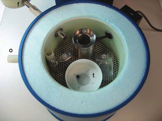
Downloaded from https://www.cambridge.org/core. IP address: 46.4.80.155, on 14 Oct 2021 at 03:51:14, subject to the Cambridge Core terms of use, available at https://www.cambridge.org/core/terms. https://doi.org/10.1017/S1551929500054432
Figure 5: The three images above are an example of the high quality of frozen hydrated preparations produced with Cryoplunge™3. (1) Image
of a single grid square; TEM magnification ~900X, electron dose 0.01 e-/Ǻ2. (2) Higher magnification image of a portion of the grid square; 4700X,
0.1 e-/Ǻ2. (3) Image of part of one hole; 59KX, 20 e-/Ǻ2. The frozen-hydrated microtubule specimen was prepared on Quantifoil® R1.2/1.3 macro
machined holey carbon grids which were plasma cleaned using the Gatan Solarus® 950 Advanced Plasma Cleaning System for 15 seconds at 50
Watts using hydrogen and oxygen plasma. All images were recorded on an FEI Tecnai F30 TEM with a Gatan 626 70° single tilt liquid nitrogen
cryo transfer holder and a Gatan Ultrascan® 4000. Images courtesy of Dr. Chen Xu, Rosenstiel Basic Medical Sciences Research Center, Brandeis
University, Waltham, MA.
• Insert the blotters pre-loaded with the filter paper of choice Bald WB (1984) The relative efficiency of cryogenic fluids
that will be used for blotting the specimen grid. used in the rapid quench-cooling of cryogenic samples. Journal
• Apply 3 µL of the fluid suspension that contains the specimen of Microscopy, 134, 261-270
to the surface of the grid. Bald WB (1987) Quantitative Cryofixation. Adam Hilger,
• Set the blot timer for the desired blotting time. Bristol and Philadelphia
• Press the start button on the Cp3; the grid is blotted to pro- Battersby BJ, Sharp JCW, Webb RI, Barnes GT (1994) Vitri-
duce a thin fluid film and immediately plunged into the liquid fication of aqueous suspensions from a controlled environment
ethane where the specimen is frozen. for electron microsocopy: an improved plunge-cooling device.
• Transfer the frozen hydrated grid to a cryo grid box for stor- Journal of Microscopy, 176, 110-120
age. Dubochet, J., Groom, M. and Mueller-Neuteboom, S. (1982),
• From this point forward the frozen hydrated specimen must Mounting of macromolecules for electron microscopy with
be maintained near liquid nitrogen temperature either by particular reference to surface phenomena and treatment of
storage in a liquid nitrogen dewar or by transferring it into a support films by glow discharge. Advances in optical and electron
purpose-built cryo TEM using a cryo transfer holder main- microscopy, Barrer, R. and Cosslett, V. E. (eds.), Academic Press,
tained at liquid nitrogen temperature. London, New York. 107-135.
• Images of the specimen are recorded under low electron dose Dubochet J, Adrian M, Chang J-J, Homo J-C, Lepault J, Mc-
conditions (typically 10 to 25 e-/Å2). Dowall AW, Schulz P (1988) Cryo-electron microscopy of vitrified
The Cryoplunge3 is easy to use and provides clean, consis- specimens, Quarterly Review of Biophysics 21, 129-228
tent results for the preparation of frozen hydrated specimens for Echlin P (1992) Low Temperature Microscopy and Analysis.
cryoEM, figure 5. The Cp3 incorporates many of the features Plenum Publishing Corporation, New York
found in more expensive plunge freezing instruments at a frac-
Fukami A, Adachi K. (1965) A new method of preparation of
tion of the cost while providing a versatile platform to facilitate
a self-perforated micro plastic grid and its application. J Electron
a variety of specimen preparation protocols.
Microscopy (Japan). 14(2):112-118.
Acknowledgements
Glaser, R, Downing, K, DeRosier, D, Chiu, W, Fran, J. (2007)
The author thanks Dr. Chen Xu of Brandeis University for Electron Crystallography of Biological Macromolecules. Oxford
outstanding technical assistance and helpful discussions. University Press. 150-166.
† Short quotes from Wikipedia contributors. Cryofixation Melanson, L. “Cryoplunge™3 and Solarus® 950: a perfect duet
[Internet]. Wikipedia, The Free Encyclopedia; 2008 Sep 12, 11:54 for consistent, high quality frozen hydrated specimen prepara-
UTC [cited 2009 Feb 1]. Available from: http://en.wikipedia. tions for cryo transmission electron microscopy (cryoEM).” [On-
org/w/index.php?title=Cryofixation&oldid=237916959. appear line]. Available: http://www.gatan.com/resources/Answers-10.
in the introductory paragraphs. php. (Nov 7, 2008).
General References Steinbrecht, RA, Zierold, K. (1987) Cryotechniques in biologi-
Allison DP, Daw CS and Rorvik MC (1987) The construction cal electron microscopy. Berlin: Springer-Verlag. 47-54.
and operation of a simple and inexpensive slam freezing device
for electron microscopy. Journal of Microscopy, 147, 103-108
MICROSCOPY TODAY March 2009 n 17
ABMelanson.indd 3 02/27/2009 12:28:03 PM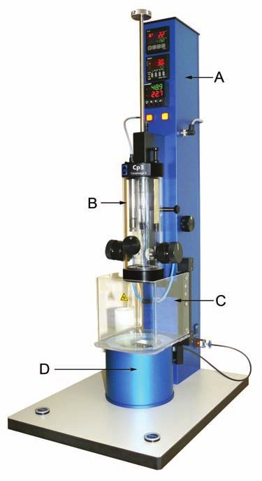
You can also read