Is it Lupus or Not? A Clinical Approach to Pediatric SLE - By Angela Migowa, MBCHB-UON, MMed (Pediatrics and Child Health)-AKUHN Pediatric ...
←
→
Page content transcription
If your browser does not render page correctly, please read the page content below
Is it Lupus or Not?
   A Clinical Approach to Pediatric SLE

By Angela Migowa, MBCHB-UON, MMed (Pediatrics and Child Health)-AKUHN
                     Pediatric Rheumatology Fellow
       Montreal Children’s Hospital, McGill University Health Centre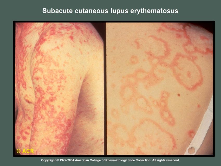
Mahatma Gandhi

• “….Live as if you were to
  die tomorrow. Learn as
  if you were to live
  forever…”
Objectives

• Recognize the presentation of Pediatric
  Systemic Lupus Erythematosus (pSLE).

• Develop a clinical approach to the initial
  work up and management of pSLE.
Introduction

• A central concept to the pathogenesis of
  autoimmune diseases is that “Tolerance to self
  is violated.”

                     Pascual et al. Annu Rev Immunol. 2010 April 23; 28: 535–571.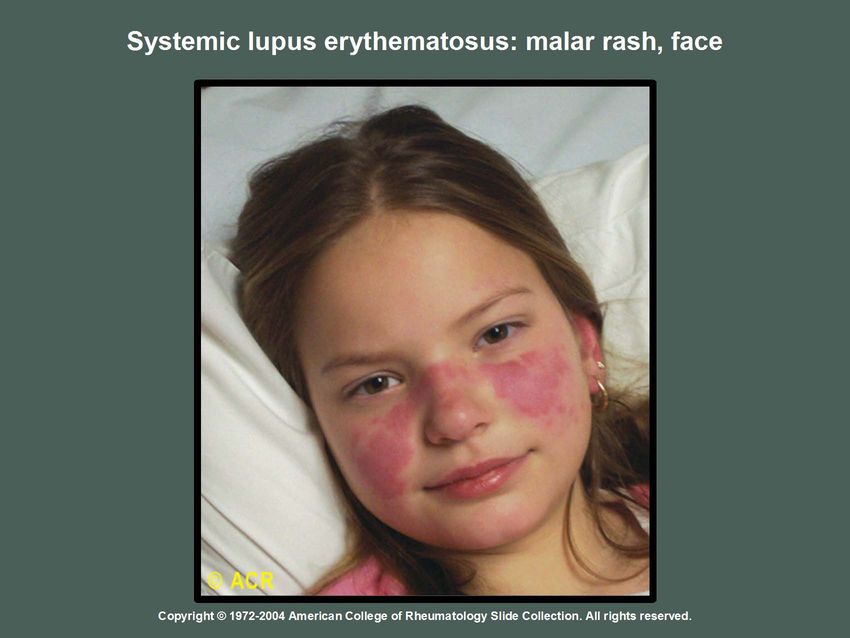
Definition

• SLE has been classically described as a prototypic
  autoimmune disease with a wide array of clinical
  manifestations and characterized by the production
  of auto-antibodies to components of the cell
  nucleus.

                             Gilbert et al. Pediatric Rheumatology 2014, 12:16
Epidemiology
• pSLE makes up 20% of all cases.

• Incidence in North America is 0.3 – 2.5 cases per 100,000.
• Frequently in African-Americans, Asians and Hispanics.

• More common in adolescent age group.
• Average age at onset 12 yrs; Rare before 5 yrs.
• Female: Male = 9:1; however before puberty, it is 3:1.

                                           Pineles et al.Lupus 2011;20:1187-1192.
                                    Weiss JE. Pediatrics in Review 2012;33:62-73.
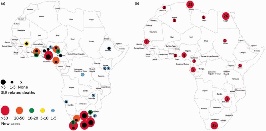
Lupus in Africa - under reported?
• Incidence is unknown

• Early reports (1960s-1970s): lupus rare
   – (Uganda, Nigeria, South Africa)

• Recent (1980s-1990s): increased reports
   – (Uganda, Zimbabwe, South Africa)

• “Current” (2000’s): case series
   – 2001 (Molokhia: West Africa prevalence)
   – 2005 (Faller: South Africa pediatrics)
   – 2008 (Abdou: Senegal nephritis)
   – 2009 (Adelowo, Oguntona: Nigeria)

                          Slide courtesy of Dr C.Hitchon
New lupus cases/deaths

                          New cases/ deaths                                Case series Publications

N Tiffin et al. Lupus 2014;23:102-111         Slide courtesy of Dr C. Hitchon
Copyright © by SAGE Publications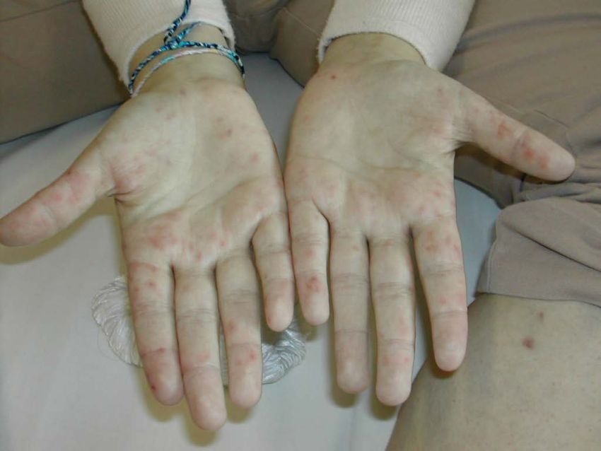
Pathophysiology
Joints
         Tsokos NEJM Dec 2011
photosensitivity
Serositis
Arthritis
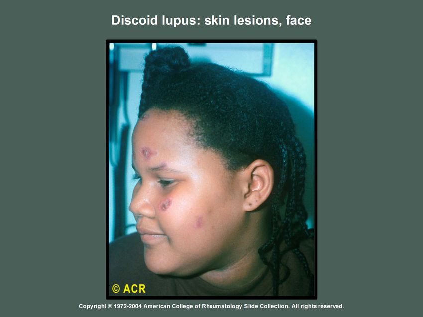
Discoid rash
Malar Rash
Neurologic
Infection
Renal
Presentation of lupus in Africa: survey
Slide courtesy of Dr C. Hitchon
N Tiffin et al. Lupus 2014;23:102-111SLE - Diagnostic Criteria
• Malar Rash • CNS involvement
• Discoid Rash – seizures
• Photosensitivity – psychosis
• Oral or nasal ulcers • Positive ANA
• Nonerosive arthritis • Positive Immunoserology
• Pleuritis or pericarditis – Ab to dsDNA
• Cytopenias – Ab to Sm
• Nephritis – Positive anti-phospholipid
Ab: anti-cardiolipin Ab,
– proteinuria > 0.5 g/day
lupus anti-coagulant, or
– cellular casts false + VDRL x 6 mos
Hochberg MC et al. Arthritis Rheum 1997; 40:1725.
4 out of 11 criteria Tan EM et al. Arthritis Rheum 1982; 25:1271-77.SLICC Criteria
Wangari wa Maathai
It’s the little things
   citizens do…..That’s
   what will make the
   difference. My little
   thing is planting trees.
Amelia 14 yr old teenager
History & Examination                       Work up
•   Ref for management of incision site     •   WBC 1.7, N 0.8, L 0.8, RBC 3.72,Hb
    abscess mid Sept 2014 foll.                 112 g/l, Plt 137, CRP 3.4mg/l, ESR
    Appendicectomy.                             44mm/hr
                                            •   Peripheral blood film- 1 myelocyte
•   2 months prior, hair loss, 1 month hx   •   Bone Marrow Aspirate-No
    of appetite loss and papular skin           malignancy
    rash.
                                            •   U/A-blood, Urine prot:creat 1.85 g/g
•   HR86 T40.5 RR 18 BP109/66               •   Renal biopsy-?
•   On exam malar rash, vasculitic rash,    •   ANA+ve (1:160), C3 0.56[N],C4 0.1
    palatal ulcer, cervical                 •   Anti ds DNA>800, anti La +ve, anti
    lymphadenopathy and abscess at              RNP +ve
    incision site.
Palatal ulcer in patient with Systemic Lupus Erythematosus Courtesy of Dr R Scuccimarri
Systemic Lupus Erythematosus Malar Rash
         Courtesy of Dr C. Hlela
pSLE – Renal disease
• Present to some degree in all children.
• More significant disease occurs in at least 75% and is often more
severe in children than in adults.
• Can present with either asymptomatic proteinuria or hematuria
(or both); or with hypertension; nephrotic syndrome; or renal
failure.
• The degree of urinary abnormalities does not always predict the
renal lesion thus biopsy is recommended if evidence of renal
involvement.
Klein-Gitelman M et al. Rheum Dis Clin N Am 2002; 28:561-577.
Benseler SM et al. Pediatr Clin N Am 2005: 52:443-467.pSLE – Renal evaluation • Urinalysis/microscopy: proteinuria, RBCs, casts. • Urine protein/creatinine. • Serum creatinine, electrolytes, albumin, total protein. • May need renal biopsy.
pSLE - Clinical Findings
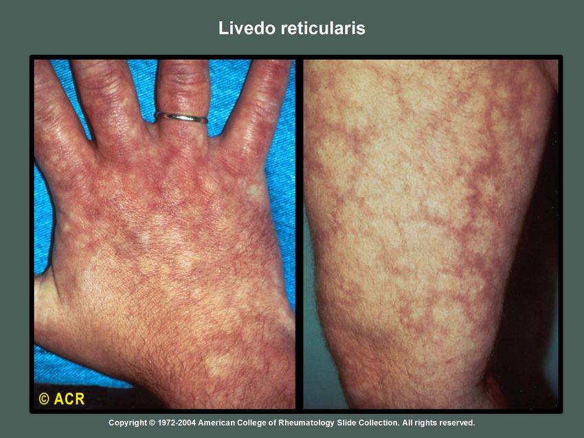
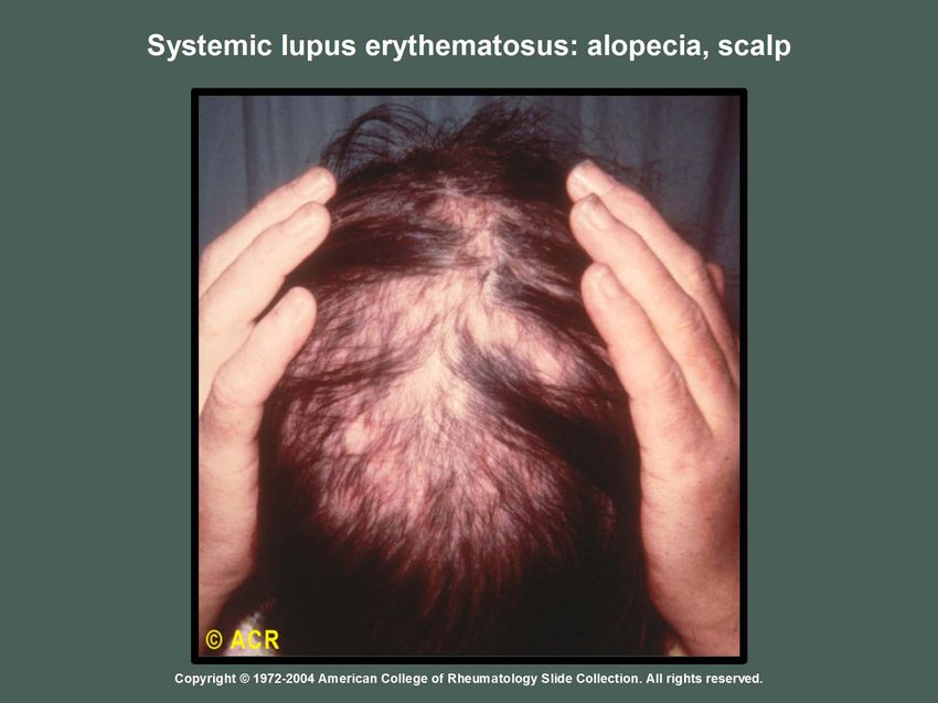
Other rashes
• Photosensitivity • Lupus profundus
• Periungal erythema (panniculitis)
• Nailfold infarcts • Recurrent urticaria
• Raynaud’s phenomenon • Erythema multiforme
• Livedo reticularis • Bullae
• Ulcerating lesions • Alopecia
• Erythema nodosum • Hypo/hyperpigmentationMax 3 ½ yr old boy
History and Examination Work up
• 2 yr history of multiple joint pain assoc • HR 106 RR24 T 36.7
with night awakening
• Early Oct 2014 3 day hx of fever ranging
38-38.5 C • WBC 6, N 2.8,L 2.6, RBC 4.47,Hb 122
g/l,Plt 204
• Loss of appetite and weight loss
• Known for adenotonsillar hypertrophy • Anti dsDNA+ve, ANA +ve 1;160, C3 1.13g/l
[N], C4 0.19g/l [N]
• Family history of Psoriasis
• Normal systemic examination. Referral • Urinalysis normal, Urine prot:creat normal
note ANA +ve, antiDNA +vepSLE - Laboratory Findings Immunoserology • Antinuclear Antibodies (ANA) – always positive (>95%) – usually of high titre – BUT not diagnostic / not specific – + ANA does NOT confirm the dx of SLE
Prevalence of Normal Individuals with Positive ANA

                                     Adams BB et al. Int J Dermatol. 2000;39(12):887-91.
Slide courtesy of Dr R Scuccimarri
pSLE - Laboratory Findings
Immunoserology
• Antibodies to anti-dsDNA
– seen in 60-70% of SLE pts
– specific for SLE (but not sensitive)
– seen in high titres with active nephritis
• Extractable Nuclear Antigens (ENA)
i. Anti-sm (Smith)
– seen in 40-50% of SLE pts
– specific for SLE
ii. Anti-ro; anti-la; anti-RNP can be seen; not specific
Benseler SM et al.Pediatr Clin N Am 2005: 52:443-467.Julia 14 yr old teenager
History and Examination Work up
• 1 month history of fatigue and rash on palms • WBC 2.63, N 1.39, L 0.9,Hb 113 g/l,Plt
and soles initially diagnosed as eczema. 1 day 130,CRP 0.7 mg/l, ESR 55 mm/hr, Uric acid
history of fever. 201, LDH 251 U/L
• Weight loss, ongoing poor concentration • C3 0.51[L], C4 0.1[L]
since July 2015.
• Urinalysis-normal, prot:creat-ve
• 2 years prior white then red discolouration of
the fingers and toes worse in the winter. • Bone marrow-No malignancy
• On exam T 37.1, RR 12, HR 76, O2 99% • Hand and feet Xrays-no bony changes or
erosions, soft tissue swelling
• No lymphadenopathy, vasculitic rash, malar • ANA +ve (1:160 ), anti DNA +ve (473),anti
erythema, polyarthritis,tender along both Sm+ve, anti RNP+ve
shins. • Lupus anticoagulant+veVasculitic rash in patient with Systemic Lupus Erythematosus Courtesy of Dr R.Scuccimarri
pSLE - Central Nervous System
             Disease
• Ranks 2nd after nephritis as a cause of
  morbidity and mortality in children with SLE.

• Occurs in 20-30% of patients.

• Most common presentation is headache,
  seizures and psychosis.
                                   Weiss JE. Pediatrics in Review 2012;33:62.
               Klein-Gitelman M et al.Rheum Dis Clin N Am 2002; 28:561-577.
pSLE – Hematological Findings
• Anemia
– of chronic disease.
– 30% are coombs positive without overt
hemolysis.
– < 10% have hemolytic anemia.
Benseler SM et al. Pediatr Clin N Am 2005: 52:443-467.pSLE – Haematological Findings
• Thrombocytopenia (< 100 000)
– Can be due to peripheral platelet destruction or
anti-platelet Ab.
– Some children with ITP or Evan’s syndrome can progress to
SLE; usually occurs within first year.
– Splenectomy should be avoided.
• Leukopenia (< 4)
– occurs in up to 40% of pts.
– lymphopenia (pSLE – Hematological Findings
• Coagulopathy
– A prolonged PTT is the most common coagulation defect.
– Due to circulating anticoagulant that blocks activation of
the prothrombin activation complex in vitro.
– The antiphospholipid Ab often cross-reacts with
anticardiolipin Ab and is responsible for the false positive
VDRL.pSLE – Hematological Findings
• Coagulopathy continued
– increased risk of thromboembolic events.
– can have recurrent venous or arterial thromboses,
emboli, stroke, chorea, livedo reticularis, hypertension,
thrombocytopenia, spontaneous abortions, and fetal
loss.
– PT/PTT/INR, Lupus anticoagulant, Anti-cardiolipin Ab.Maria 12 year old girl
History and Examination Work up
• 1 month history of recurrent fever (38C- • WBC 5.1, N 3.3, L1.3,Hb 114g/l, Platelet
40.8C). 200
• Treated initially for strep throat infection. • Urinalysis-normal, urine prot:creat
• At presentation had a rash, and 0.01g/mmol
complained of muscle pain, difficulty • C3 1.19 g/l [N], C4 0.24 g/l [N]
swallowing and weight loss.
• 12th Jan-CK 8089 U/L,17th -3421, 20th-
• On examination HR 100 RR24 BP 118/70 2590,27th -3408, 2nd Feb-5247
T36.7C
• Erythema nodosum on arms and shins. • Anti Sm, RNP, anti DNA, ANA +ve
• Weak neck flexors and proximal muscles. • Lupus anticoagulant-ve
No gottron’s, heliotrope rash or cuticular
hypertrophy.pSLE – Musculoskeletal System • Arthralgias and myalgias are common. • Non-erosive arthritis – usually polyarticular involvement. – pain is often severe even with minimal findings. • Myositis – proximal muscle involvement.
pSLE – Cardiac Disease

• Pericarditis / Pericardial effusion
   – Most common cardiac feature.
   – Patients may be a/symptomatic.

• Myocarditis

• Accelerated atherosclerosis
                    Benseler SM et al. Pediatr Clin N Am 2005: 52:443-467.
pSLE – Pleuropulmonary Disease • Pleural effusions or pleuritis • Pneumonitis • Pulmonary hemorrhage • Restrictive lung disease • Pulmonary embolus • Shrinking lung – loss of lung volume due to diaphragmatic dysfunction.
Differential Diagnosis
• Infection
   –   Parvovirus B19 - rash, arthritis, fever, cytopenia.
   –   CMV - fatigue, cytopenia, abdominal pain, LFT abnormality.
   –   HIV - fever, weight loss, lymphadenopathy, oral ulcer.
   –   Hepatitis B - arthritis, antibodies.
   –   Post-streptococcal disease – arthritis, fever, rash.
   –   Tuberculosis – fever, weight loss, lymphadenopathy, arthritis.

• Malignancy
   – Lymphoma; leukemia: fever, weight loss, arthralgias, cytopenia,
     lymphadenopathy, rash, ANA.

• Other autoimmune disease
   – JIA, MCTD, systemic vasculitis.

• Drug induced lupus
   – Minocycline, hydralazine, procainamide, isoniazid, anti-seizure.
pSLE - Treatment
Depends on extent and severity of the disease:
• Hydroxychloroquine/Chloroquine alone.
• Hydroxychloroquine + NSAID.
• Hydroxychloroquine + NSAID + Methotrexate.
• Hydroxychloroquine + low dose prednisone.
• Hydroxychloroquine + high dose prednisone.
• Hydroxychloroquine + high dose prednisone + cytotoxic
agent.
• Cytotoxic agents- azathioprine, mycophenolate mofetil,
cyclophophamide.
• Treatment of specific organ system complications.
• Sun protection; Bone Health; Immunizations.
Slide courtesy of Dr R ScuccimarriWallace, D. J. et al. Nat. Rev. Rheumatol.
Anti-malarials in pSLE

• Risk of retinopathy

   – Reaches 1% after 5-7
     years of use or
     cumulative dose
     of >1000 gm.

   – Need baseline ocular
     exam and yearly exam in
     children.

                            Retinopathy: Early and Late Bull’s Eye lesion
                                 Brunner HI et al. Nature Reviews 2011; 7:225-233.
                                Marmor MF et al. Ophthalmology 2011;118:415-22.
Course and Prognosis
• Characterized by exacerbations and remissions.
• Chronic disease that varies in severity.
• Western world: 5 year survival 94-100%
10 year survival 81-92%
• Delayed diagnosis; access to care and treatment are significant
challenges in some countries.
• End stage renal disease a significant cause of morbidity.
• Morbidity is related to disease and treatment.
• Infection is the leading cause of death.Summary • Diagnosis requires a combination of clinical features and relevant investigations. • Skin, MSK and renal are the most common organ involvement in children; renal and CNS are the most severe. • Management requires tailoring treatment to lupus specific features and severity of organ involvement.
Acknowledgments

• Thanks to our patients who inspire us to
  improve and be better healthcare providers.
• Thanks to all my mentors and teachers in my
  professional journey.
• Thanks to Dr. R. Scuccimarri for her guidance
  and support in preparation for the scientific
  talks and lectures.
• Thanks to Dr. C. Hlela for her support in
  providing pictures for this talk.
Mwalimu Julius Nyerere

•   "...Intellectuals have a special
    contribution to make to the
    development of our nation, and to
    Africa. And I am asking that their
    knowledge, and the greater
    understanding that they should
    possess, should be used for the
    benefit of the society of which we are
    all members."

Julius Kambarage Nyerere, from his book Uhuru na
     Maendeleo (Freedom and Development), 1973.
     -
Asante! Thank you! Merci!
NAIROBI CITYYou can also read