Systematic Lymph Node Dissection May Be Abolished in Patients With Apparent Early-Stage Low-Grade Mucinous and Endometrioid Epithelial Ovarian Cancer
←
→
Page content transcription
If your browser does not render page correctly, please read the page content below
ORIGINAL RESEARCH
                                                                                                                                      published: 06 September 2021
                                                                                                                                     doi: 10.3389/fonc.2021.705720

                                              Systematic Lymph Node Dissection
                                              May Be Abolished in Patients With
                                              Apparent Early-Stage Low-Grade
                                              Mucinous and Endometrioid
                                              Epithelial Ovarian Cancer
                                              Jiayu Chen , Jie Yin , Yan Li , Yu Gu , Wei Wang , Ying Shan , Yong-Xue Wang , Meng Qin ,
                                              Yan Cai , Ying Jin * and Lingya Pan *
                           Edited by:
                       Fabio Martinelli,      Department of Obstetrics and Gynecology, Peking Union Medical College Hospital, Chinese Academy of Medical Sciences
Istituto Nazionale dei Tumori (IRCCS),        and Peking Union Medical College, Beijing, China
                                  Italy

                        Reviewed by:          Objective: To investigate whether systematic lymph node dissection can confer clinical
                        Nicolò Bizzarri,
 Università Cattolica del Sacro Cuore,
                                              benefits in patients with apparent early-stage low-grade epithelial ovarian cancer.
                                   Italy      Methods: Patients with apparent early-stage low-grade epithelial ovarian cancer seen at
                          Lucas Minig,
  Universidad CEU Cardenal Herrera,           Peking Union Medical College Hospital from January 1, 2005, to December 31, 2015,
                                 Spain        were retrospectively enrolled. Patients with other histological types and those who did not
                  *Correspondence:            receive necessary adjuvant chemotherapy were excluded. Data collection and long-term
                           Lingya Pan
                                              follow-up were performed. According to the removed lymph node number, three groups
                      panly@pumch.cn
                              Ying Jin        based on surgical methods were used: abnormal lymph node resection, pelvic
                    jinying@pumch.cn          lymphadenectomy, and systematic lymph node dissection to control surgical quality.
                                              Their effects on prognosis were analyzed in pathological subgroups.
                   Specialty section:
         This article was submitted to        Results: A total of 196 patients were enrolled; 30.1% of patients had serous, 42.3% of
             Gynecological Oncology,
               a section of the journal
                                              patients had mucinous, and 27.6% of patients had endometrioid carcinoma, of which 51
                 Frontiers in Oncology        (26.0%), 96 (49.0), and 49 (25.0%) patients were treated with the above surgical methods,
            Received: 06 May 2021             respectively. The occult lymph node metastasis rate was 14 (7.1%), and only five (2.6%) of
        Accepted: 16 August 2021              apparent early-stage patients were upstaged due to lymph node metastasis alone.
     Published: 06 September 2021
                                              Systematic lymph node dissection did not benefit progression-free survival or disease-
                            Citation:
           Chen J, Yin J, Li Y, Gu Y,         specific overall survival of apparent early-stage low-grade mucinous and endometrioid
       Wang W, Shan Y, Wang Y-X,              epithelial ovarian cancer but prolonged progression-free survival of apparent early-stage
Qin M, Cai Y, Jin Y and Pan L (2021)
 Systematic Lymph Node Dissection
                                              low-grade serous patients (OR, 0.231, 95% CI, 0.080, 0.668, p = 0.007).
  May Be Abolished in Patients With           Conclusions: Systematic lymph node dissection may be abolished in patients with
   Apparent Early-Stage Low-Grade
        Mucinous and Endometrioid
                                              apparent early-stage low-grade mucinous and endometrioid epithelial ovarian cancer but
           Epithelial Ovarian Cancer.         may be considered for apparent early-stage low-grade serous patients.
           Front. Oncol. 11:705720.
    doi: 10.3389/fonc.2021.705720             Keywords: epithelial ovarian cancer, low grade, lymph nodes, metastasis, lymph node dissection

Frontiers in Oncology | www.frontiersin.org                                        1                                  September 2021 | Volume 11 | Article 705720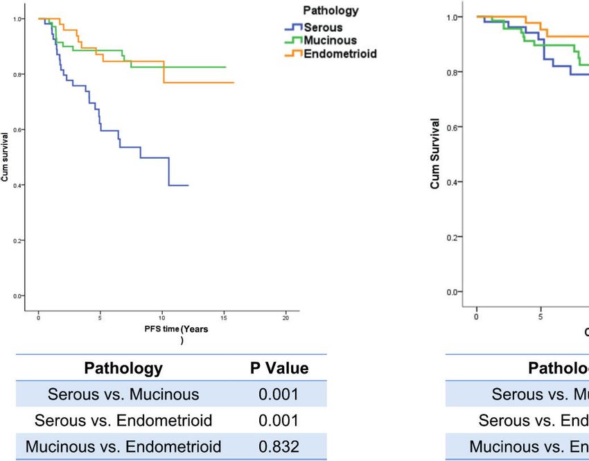
Chen et al. SLND for Apparent Early-Stage LGEOC
INTRODUCTION stage disease; and 3) underwent staging surgery. Literature
reported that all International Federation of Gynaecology and
Ovarian cancer is the most lethal tumor of all gynecological Obstetrics (FIGO) grade 1 and some FIGO grade 2 patients
malignancies, approximately 90% of which are epithelial ovarian might belong to low grade according to the two-tier grading
cancer (EOC) (1). Complete staging surgery and necessary criteria (12), so two independent pathologists reclassified the
adjuvant chemotherapy are the standard treatments for EOC primary lesion pathological sections of those patients into low
patients according to the National Comprehensive Cancer and high grades in terms of the two-tier grading criteria
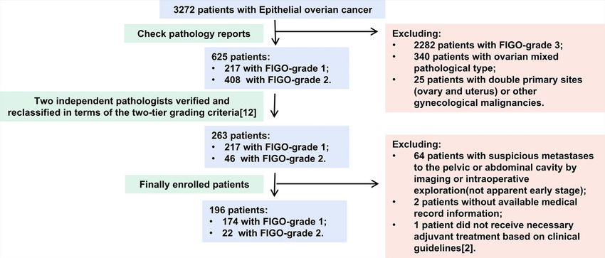
Network (NCCN) guidelines (2). Systematic lymph node (Figure 1). An apparent early stage was defined as a tumor
dissection (SLND) is an essential procedure that has been a localized to the bilateral adnexa (ovaries and fallopian tubes) and
part of complete staging procedures since 1988, including pelvic uterus on preoperative imaging and intraoperative exploration,
and para-aortic lymphadenectomy (2, 3). In early-stage EOC, similar to FIGO I–IIA stage (13). The exclusion criteria were as
SLND helps doctors acquire a sufficient number of lymph nodes follows: 1) ovarian mixed pathology, double primary sites
(LNs) to identify occult LN metastases and guide adjuvant (ovary and uterus), and other gynecological malignancies;
chemotherapy decisions by accurate staging (4, 5). 2) no available medical record information; and 3) did
However, the low LN metastatic rate and upstaging rate in not receive necessary adjuvant treatment based on clinical
apparent early-stage low-grade EOC (LGEOC) reported in few guidelines (2).
studies challenge the necessity of SLND (6, 7). Nevertheless, those
studies had intrinsic limitations: uncontrolled surgery quality, non- Clinical Data Collection and Organization
parallel prognostic factors, and partially missing clinical and This retrospective single-center study was conducted at the
prognostic data. As a result, the role of SLND in apparent early- Department of Obstetrics and Gynecology, Peking Union
stage LGEOC is still unclear. Low incidence increases the difficulty Medical College Hospital, between January 1, 2005, and
of studying LGEOC, but its unique features compared with high- December 31, 2015, and approved by the Institutional Review
grade EOC (HGEOC) have increased the urgency and necessity of Board. Medical history, surgical and pathological data, and
studying its clinical characteristics and establishing an postoperative treatment and follow-up data were collected
individualized treatment (8–11). continuously once a patient met the inclusion criteria and
This study aims to determine the LN metastatic patterns of lacked the exclusion criteria. The general physical condition
apparent early-stage LGEOC patients, including patients with low- was assessed with the American Society of Anesthesiologists
grade serous ovarian cancer (LG-SOC), low-grade mucinous ovarian (ASA) classification (14). Pathologically explicit FIGO stages I to
cancer (LG-MOC), and low-grade endometrioid ovarian cancer (LG- IIa were defined as early stage. The LN dissection methods were
EOC), and to explore the survival benefit of SLND on them. The classified into three categories to control quality (5, 7, 15–19):
primary endpoint is disease-specific overall survival (OS), and the
secondary endpoint is progression-free survival (PFS). 1. Group 1: no LN dissection or LN sampling—removal of none
or a few LNs (less than ten pelvic LNs)
2. Group 2: pelvic lymphadenectomy—removal of more than 10
MATERIALS AND METHODS pelvic LNs
3. Group 3: SLND—removal of more than 10 pelvic LNs and
Patients five para-aortic LNs
The inclusion criteria were as follows: 1) diagnosed with LGEOC
—LG-SOC, LG-MOC, or LG-EOC; 2) presented apparent early- All LN excision numbers were confirmed by pathology.
FIGURE 1 | The flowchart of patients’ inclusion and exclusion.
Frontiers in Oncology | www.frontiersin.org 2 September 2021 | Volume 11 | Article 705720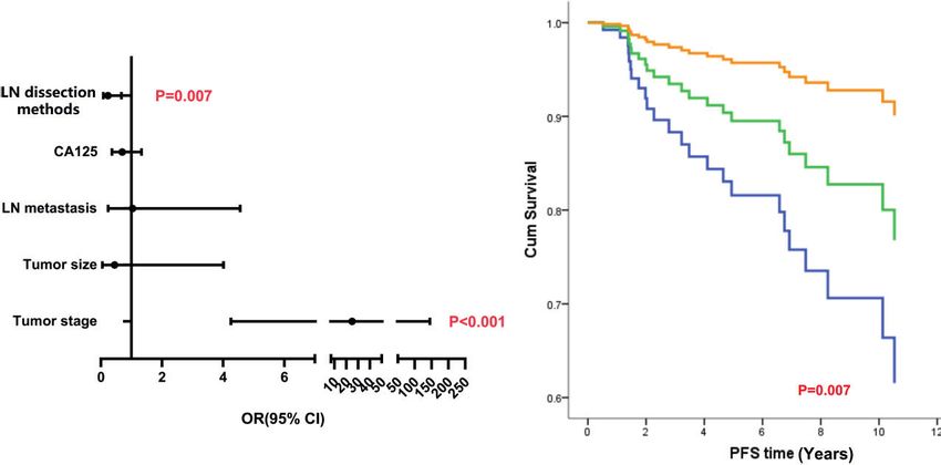
Chen et al.                                                                                                                      SLND for Apparent Early-Stage LGEOC

Follow-Up                                                                                disease). One hundred ninety-six patients were eventually
PFS was defined as the time between the date of diagnosis and                             included in the study (Figure 1), of which 59 (30.1%) had LG-
the date of the first recurrence, the last follow-up, or death,                           SOC, 83 (42.3%) had LG-MOC, and 54 (27.6%) had LG-EOC.
whichever occurred first; while OS was the interval period from                           Their clinical features are depicted in Table 1: more than half of
the date of diagnosis to the date of disease-specific death or last                       patients were younger than 40 years at diagnosis and had a
follow-up. Follow-up was conducted by consulting clinic records                          history of borderline ovarian tumor (BOT). The CA125 level
or telephone contact, and the cutoff date was between December                           varied remarkably, ranging from 0.32 to 65,065 U/ml.
2020 and January 2021.                                                                   Recurrence occurred in 24.6% of patients, and disease-specific
                                                                                         death occurred in 14.3% of patients. The 5-year survival rate was
Statistical Analysis                                                                     88.0% (95% CI, 82.1%, 93.9%), and the 10-year survival rate was
The measurement data were analyzed by ANOVA or a non-                                    74% (95% CI, 62.2%, 85.8%). Notably, 33 apparent early patients
parametric test (Mann–Whitney U test), and the chi-square test                           were classified as advanced patients due to postoperative pathology.
was used to analyze hierarchical data. Patients lost to follow-up were
excluded from the survival analysis. The reverse Kaplan–Meier                            Different Lymph Node Dissection Modes
method was used to calculate the median follow-up time; and 5- or                        and Lymph Node Metastasis Status
10-year PFS rates and OS rates were estimated according to the                           The three groups recruited 51, 96, and 49 patients, and all
Kaplan–Meier curves. The log-rank test and Kaplan–Meier test                             indexes but pathological type were balanced among them
were adopted as univariate analysis methods for identifying risk                         (Table 1). The number of LNs removed by different surgical
factors for PFS and OS, and those variables with p-values less than                      methods and the LN metastatic status are described in Table 2.
0.2 were enrolled in the multivariate Cox regression analysis to                         Fourteen patients (7.1%) had occult LN metastasis, including
identify independent risk factors. All p-values were two-sided, and                      contralateral metastasis, bilateral metastasis, and skip metastasis
differences were considered statistically significant with p ≤ 0.05 and                   that only had para-aortic LN metastasis and no pelvic LN
when the 95% confidence interval (CI) did not cross 1. All statistical                    metastasis. The most common metastatic site was iliac LNs
analyses were conducted with IBM SPSS Statistics 20 (IBM,                                (13/14, 92.9%), followed by para-aortic LNs (4/14, 28.6%),
Armonk, NY, USA).                                                                        while only one patient had common iliac LN metastases (p <
                                                                                         0.001). LG-SOC had a significantly higher LN involvement rate
                                                                                         than LG-MOC and LG-EOC (18.6% vs. 1.2% and 3.7%,
                                                                                         p < 0.001).
RESULTS
                                                                                         The Effect of Lymph Node Dissection
The Clinical Features of Low-Grade                                                       Mode on the Lymph Node Metastasis
Epithelial Ovarian Cancer                                                                Detection and Upstaging Rates
In over 3,272 EOC patients, 263 (8.04%) were diagnosed with                              Although a significantly higher number of pelvic and para-aortic
LGEOC (217 had FIGO-G1 disease and 46 had FIGO-G2                                        LNs were removed from SLND than from other patients, no

TABLE 1 | Clinical information of apparent early-stage patients with different LN resection methods.

Mode of lymph node resection                               Total                       1                          2                        3                 p-Value
                                                          n = 196                    n = 51                     n = 96                   n = 49

Age (years)                  ≤40                      103 (52.6%)                  28 (54.9%)                 51 (53.1%)               24 (49.0%)             0.356
                             40–60                     76 (38.8%)                  16 (31.4%)                 37 (38.6%)               23 (46.9%)
                             >60                        17 (8.6%)                   7 (13.7%)                  8 (8.3%)                 2 (4.1%)
Menopause                    No                       146 (74.5%)                  35 (68.6%)                 73 (76.0%)               38 (77.6%)             0.567
                             Yes                       50 (25.5%)                  16 (31.4%)                 23 (24.0%)               11 (22.4%)
BMI                                                   22.88 ± 3.76                22.67 ± 7.34               22.77 ± 3.72             22.31 ± 3.94            0.482
BOT history                  No                        97 (51.3%)                  20 (51.3%)                 47 (51.1%)               30 (62.5%)             0.129
                             Yes                       92 (48.6%)                  19 (48.7%)                 45 (48.9%)               18 (37.5%)
ASA classification            I                        110 (57.0%)                  28 (56.0%)                 58 (61.1%)               24 (50.0%)             0.159
                             II                        77 (39.9%)                  18 (36.0%)                 36 (37.9%)               23 (48.0%)
                             III                        6 (3.1%)                    4 (8.0%)                   1 (1.0%)                 1 (2.0%)
CA125 level (U/ml)                                   66.3 (23.9, 227)            77.7 (37.3, 116)          49.1 (20.3, 228.5)        76.7 (28.8, 410)         0.243
Tumor size (cm)                                         10 (7, 15)                10 (7.75, 10)                10 (6, 15)               10 (7, 13)            0.752
Pathology                    Serous                    59 (30.1%)                  22 (43.1%)                 25 (26.0%)               12 (24.4%)             0.011
                             mucinous                  83 (42.3%)                  18 (35.3%)                 49 (51.0%)               16 (32.7%)
                             Endometrioid              54 (27.6%)                  11 (21.6%)                 22 (23.0%)               21 (42.9%)
Tumor stage                  Early                    163 (83.2%)                  38 (74.5%)                 84 (87.5%)               41 (83.7%)             0.135
                             Late                      33 (16.8%)                  13 (24.5%)                 12 (12.5%)                8 (16.3%)

BMI, body mass index; BOT, borderline tumor; ASA, American Society of Anesthesiologists; LN, lymph node.

Frontiers in Oncology | www.frontiersin.org                                          3                                    September 2021 | Volume 11 | Article 705720
Chen et al.                                                                                                                    SLND for Apparent Early-Stage LGEOC

TABLE 2 | LN removed number, LN+ detection rate, and upstaging only due to LN metastasis among three LN dissection groups in all subgroups and pathological subgroups.

Mode of lymph node resection                                                                    1                  2                       3                  p-Value

All N = 196                  Number   of pelvic LNs removed                                 0 (0, 2.5)         20 (16, 28)        25.5 (19.25, 30.75)
Chen et al.                                                                                                                    SLND for Apparent Early-Stage LGEOC

                             A                                                    B

 FIGURE 3 | The analysis of independent risk factors on PFS of LG-SOC. (A) The forest figure of Cox multiple regression for PFS of LG-SOC, including items with
 p-values less than 0.3 in univariate analysis. Any item in which a p-value was less than 0.05 and the 95% CI for OR did not cross 1 was considered statistically
 significant. The p-value of the multivariate regression model was less than 0.001. (B) The survival curves of LN dissection methods on PFS after controlling other
 variables by the Cox test. OR, odds ratio; 95% CI, 95% confidence interval of OR; PFS, progression-free survival; LG-SOC, low-grade serous ovarian cancer; LN,
 lymph node.

OS and PFS (Appendix 2). LN dissection methods did not affect                         patients, a low LN metastasis rate, and a favorable prognosis (5,
survival (PFS: OR, 0.530, 95% CI, 0.155, 1.811, p = 0.311; OS: OR,                    7–11, 18, 20–25).
0.684, 95% CI, 0.173, 2.694, p = 0.587), while tumor stage was the                       Since neither preoperative imaging nor intraoperative LN
only risk factor affecting both PFS and OS (Appendix 4).                              observation can predict LN metastasis precisely, 20%–30% of
    In LG-EOC, not enough items could be considered in the Cox                        apparent early-stage EOC patients have LN metastasis (26–29).
regression analysis of PFS; the p-value of the log-rank test for LN                   As a result, the aim of SLND in apparent early-stage
resection mode was 0.059 (Appendix 3). Age, mode of LN                                EOC is to find occult LN metastasis and guide surgical–
resection, and tumor size were considered in the Cox                                  pathological staging (4, 5). The patients who experience
regression analysis of OS, but we failed to find any significant                        upstaging receive adjuvant chemotherapy, which may benefit
risk factors (Appendixes 3, 4).                                                       the prognosis.
                                                                                         However, the significantly lower incidences of LN metastasis
The Effect of Lymph Node Dissection                                                   and upstaging in LGEOC than in HGEOC challenge the
Mode on Operation-Related Complications                                               necessity of SLND in apparent early-stage patients. The LN
The operative time, blood loss, perioperative complication                            involvement rate of LGEOC was 2.9% (7). Similarly, Nasioudis
incidence, and incidence of lymphocysts significantly increased                        et al. (25) recognized that the LN metastasis rates of LG-SOC,
as the number of LNs removed increased (Table 3).                                     LG-MOC, and LG-EOC patients were significantly lower than
                                                                                      those of high-grade patients (9.0% vs. 14.4% and 1.7% vs. 5.1%
                                                                                      and 1.7% vs. 8.6%, respectively). Moreover, Minig et al. (6)
DISCUSSION                                                                            observed that only 2.4% of apparent stage I LGEOC was
                                                                                      upstaged by LN involvement alone. A meta-analysis of
As a rare form of ovarian cancer, LGEOC has a low incidence,                          retrospective studies reported that the proportion was only
accounting for approximately 6%–8% of EOC cases (8–11), and                           2.9% (7). In our study, the LN involvement rate was only 7.1%,
has unique clinical features as compared with HGEOC: low onset                        and only 2.6% of apparent early-stage LGEOC patients were
age, a history of BOT, an increased proportion of early-stage                         upstaged by LN involvement alone. Although SLND significantly

TABLE 3 | Comparison of operative time, blood loss, blood transfusion, and perioperative complications among different lymph node resection methods.

Mode of lymph node resection                               1                                   2                                 3                            p-Value
                                                         N = 51                              N = 96                            N = 49

Operative time (min)                                 190.3 ± 84.9                         213.1 ± 53.5                       251.1 ± 38.2                      0.001
Blood loss (ml)                                     300 (137.5, 600)                     300 (200, 400)                     400 (300, 500)                     0.001
Blood transfusion                                      8 (15.7%)                           15 (15.6%)                         5 (10.2%)                        0.671
Perioperative complication                             7 (13.7%)                           19 (19.8%)                         17 (34.7%)                       0.031
Lymphatic cyst                                         1 (2.0%)                            16 (16.7%)                         13 (26.5%)                       0.002

Frontiers in Oncology | www.frontiersin.org                                       5                                    September 2021 | Volume 11 | Article 705720
Chen et al. SLND for Apparent Early-Stage LGEOC
increased the LN involvement rate among early stage EOC in one improve patient staging or prognosis or increase surgery risk.
randomized controlled trial (RCT) research (18), the LN Patients with apparent LG-SOC may still need SLND,
dissection methods did not affect the LN+ detection rate or considering its prolongation of PFS.
upstaging rate in this study. This may be due to the low rate of
LN metastasis in LGEOC, considering more than half of patients
included in Maggioni’s study were FIGO stage 3, and 60 patients
were diagnosed as clear-cell, undifferentiated, and other pathology
types. Given these findings, we believe that upstaging should not DATA AVAILABILITY STATEMENT
be the reason for performing SLND in apparent early-stage
The original contributions presented in the study are included in
LGEOC patients. Notably, LG-SOC had a higher LN+ rate and
the article/Supplementary Material. Further inquiries can be
upstaging rate than the other two pathological types.
directed to the corresponding authors.
Prolonging survival is the other reason for SLND, based on
the hypothesis that dissection of chemotherapy-resistant
metastatic LNs could improve patient prognosis (referred to as
the chemotherapeutic drug sanctuary hypothesis) (30). In a
multicenter retrospective study including 639 patients with ETHICS STATEMENT
apparent early-stage EOC, researchers found that pelvic and
para-aortic lymphadenectomy improves disease-free survival but Written informed consent was obtained from the individual(s)
not OS (31). However, proof of a survival benefit of SLND in for the publication of any potentially identifiable images or data
apparent early-stage LGEOC patients is still lacking. In this included in this article.
paper, SLND did not prolong PFS or OS among LG-EOC and
LG-MOC patients, but it did significantly prolong PFS in LG-
SOC patients. LGEOC patients diagnosed at younger age have
longer survival and may experience multiple recurrences, so a
shorter PFS means those patients may need to undergo multiline AUTHOR CONTRIBUTIONS
treatment in a longer time, resulting in a significant decrease in
quality of life and an increase in financial burden. Although the All authors contributed to the study conception and design and
European Society for Medical Oncology–European Society of material preparation. The first draft of the manuscript was
Gynaecological Oncology (ESMO–ESGO) consensus conference written by JC, and all authors commented on previous versions
recommends that SLND may be questioned in some of the manuscript. All authors contributed to the article and
histological subtypes (LG-SOC or mucinous carcinoma of expansile approved the submitted version.
subtype) due to a low prevalence of LN metastases (33), we insist that
LG-SOC patients may still need SLND in terms of PFS benefit.
Another concern of performing SLND in LGEOC patients is
that SLND is a complicated surgery, so even experienced
gynecological oncologists encounter various complications (7). FUNDING
A study reported that 26.9% of patients with SLND experienced This project was supported by CAMS Innovation Fund for
perioperative complications, and 54.7% had postoperative Medical Sciences (CIFMS-2017-I2M-1-002) and The Fund of
complications (32). Therefore, it is necessary to balance the The National Key R&D Program of China 2016YFC1303700
benefits with the risks. We observed that the operative time, (Affiliated project 2016YFC1303701).
blood loss, perioperative complications, and lymphocyst count
were significantly increased with an increase in the LN
removal scope.
This retrospective study has inherent limitations. We could not
control or include all prognostic factors. In addition, the ACKNOWLEDGMENTS
information collection had some deficiencies, such as insufficient
details of LN metastatic sites, possible omissions regarding surgical We are thankful for the help from the Pathology Department
complications, and incomplete Immunohistochemistry (IHC) colleagues of PUMCH, especially Professor Yan You.
information, making it impossible to analyze correlated issues.
SUPPLEMENTARY MATERIAL
CONCLUSION
The Supplementary Material for this article can be found online at:
In conclusion, SLND may be abolished in patients with apparent https://www.frontiersin.org/articles/10.3389/fonc.2021.705720/
early-stage LG-MOC and LG-EOC since it did not significantly full#supplementary-material
Frontiers in Oncology | www.frontiersin.org 6 September 2021 | Volume 11 | Article 705720Chen et al. SLND for Apparent Early-Stage LGEOC
REFERENCES of the Arbeitsgemeinschaft Gynäkologische Onkologie AGO Study Group”
[Eur J Cancer 49 (2013) 1905-1914]. Eur J Cancer (2016) 65:192–3. doi:
1. Siegel RL, Miller KD, Jemal A. Cancer Statistics, 2019. CA: A Cancer J Clin 10.1016/j.ejca.2016.06.014
(2019) 69:7–34. doi: 10.3322/caac.21551 22. Wong KK, Gershenson D. The Continuum of Serous Tumors of Low
2. National Comprehensive Cancer Network. (NCCN) Clinical Practice Malignant Potential and Low-Grade Serous Carcinomas of the Ovary. Dis
Guidelines in Oncology. Ovarian Cancer, Version 1 (2020). Available at: Markers (2007) 23(5-6):377–87. doi: 10.1155/2007/204715
https://www.nccn.org/professionals/physician_gls/f_guidelines.asp 23. Nasioudis D, Chapman-Davis E, Witkin SS, Holcomb K. Prognostic Significance
(Accessed 11 March 2020). of Lymphadenectomy and Prevalence of Lymph Node Metastasis in Clinically-
3. Di Re F, Fontanelli R, Raspagliesi F, Di Re E. Pelvic and Para-Aortic Apparent Stage I Endometrioid and Mucinous Ovarian Carcinoma. Gynecol Oncol
Lymphadenectomy in Cancer of the Ovary. Baillieres Clin Obstet Gynaecol (2017) 144(2):414–9. doi: 10.1016/j.ygyno.2016.11.038
(1989) 3(1):131–42. doi: 10.1016/s0950-3552(89)80048-3 24. Wafa M, Braicu EI, Muallem MZ, Richter R, Taube E, Sehouli J, et al.
4. Trimbos JB. Lymphadenectomy in Ovarian Cancer: Standard of Care or Incidence and Pattern of Spread of Lymph Node Metastasis in Patients With
Unnecessary Risk. Curr Opin Oncol (2011) 23(5):507–11. doi: 10.1097/ Low-Grade Serous Ovarian Cancer. Anticancer Res (2019) 39(10):5617–21.
CCO.0b013e32834847e7 doi: 10.21873/anticanres.13757
5. Mikami M. Role of Lymphadenectomy for Ovarian Cancer. J Gynecol Oncol 25. Nasioudis D, Mastroyannis SA, Ko EM, Latif NA. Does Tumor Grade Influence
(2014) 25(4):279–81. doi: 10.3802/jgo.2014.25.4.279 the Rate of Lymph Node Metastasis in Apparent Early Stage Ovarian Cancer? .
6. Minig L, Heitz F, Cibula D, Bakkum-Gamez JN, Germanova A, Dowdy SC, Arch Gynecol Obstet (2018) 298(1):179–82. doi: 10.1007/s00404-018-4789-2
et al. Patterns of Lymph Node Metastases in Apparent Stage I Low-Grade 26. Paik ES, Shim M, Choi HJ, Lee YY, Kim TJ, Lee JW, et al. Impact of
Epithelial Ovarian Cancer: A Multicenter Study. Ann Surg Oncol (2017) 24 Lymphadenectomy on Survival After Recurrence in Patients With
(9):2720–6. doi: 10.1245/s10434-017-5919-y Advanced Ovarian Cancer Without Suspected Lymph Node Metastasis.
7. Lago V, Minig L, Fotopoulou C. Incidence of Lymph Node Metastases in Gynecol Oncol (2016) 143(2):252–7. doi: 10.1016/j.ygyno.2016.08.321
Apparent Early-Stage Low-Grade Epithelial Ovarian Cancer: A 27. Harter P, Gnauert K, Hils R, Lehmann TG, Fisseler-Eckhoff A, Traut A, et al.
Comprehensive Review. Int J Gynecol Cancer (2016) 26(8):1407–14. doi: Pattern and Clinical Predictors of Lymph Node Metastases in Epithelial
10.1097/IGC.0000000000000787 Ovarian Cancer. Int J Gynecol Cancer (2007) 17(6):1238–44. doi: 10.1111/
8. Hacker KE, Uppal S, Johnston C. Principles of Treatment for Borderline, j.1525-1438.2007.00931.x
Micropapillary Serous, and Low-Grade Ovarian Cancer. J Natl Compr Canc 28. Ditto A, Martinelli F, Reato C, Kusamura S, Solima E, Fontanelli R, et al.
Netw (2016) 14(9):1175–82. doi: 10.6004/jnccn.2016.0124 Systematic Para-Aortic and Pelvic Lymphadenectomy in Early Stage Epithelial
9. Kaldawy A, Segev Y, Lavie O, Auslender R, Sopik V, Narod SA. Low-Grade Ovarian Cancer: A Prospective Study. Ann Surg Oncol (2012) 19:3849–55. doi:
Serous Ovarian Cancer: A Review. Gynecol Oncol (2016) 143(2):433–8. doi: 10.1245/s10434-012-2439-7
10.1016/j.ygyno.2016.08.320 29. Powless CA, Aletti GD, Bakkum-Gamez JN, Cliby WA. Risk Factors for
10. Slomovitz B, Gourley C, Carey MS, Malpica A, Shih IM, Huntsman D, et al. Lymph Node Metastasis in Apparent Early-Stage Epithelial Ovarian Cancer:
Low-Grade Serous Ovarian Cancer: State of the Science. Gynecol Oncol (2020) Implications for Surgical Staging. Gynecol Oncol (2011) 122:536–40. doi:
156(3):715–25. doi: 10.1016/j.ygyno.2019.12.033 10.1016/j.ygyno.2011.05.001
11. Pauly N, Ehmann S, Ricciardi E, Ataseven B, Bommert M, Heitz F, et al. Low- 30. Morice P, Joulie F, Rey A, Atallah D, Camatte S, Pautier P, et al. Are Nodal
Grade Serous Tumors: Are We Making Progress? Curr Oncol Rep (2020) 22 Metastases in Ovarian Cancer Chemo Resistant Lesions? Analysis of Nodal
(1):8. doi: 10.1007/s11912-020-0872-5 Involvement in 105 Patients Treated With Preoperative Chemotherapy. Eur J
12. Malpica A, Deavers MT, Lu K, Bodurka DC, Atkinson EN, Gershenson DM, Gynaecol Oncol (2004) 25:169–74.
et al. Grading Ovarian Serous Carcinoma Using a Two-Tier System. Am J Surg 31. Bizzarri N, du Bois A, Fruscio R, De Felice F, De Iaco P, Casarin J, et al. Is
Pathol (2004) 28:496–504. doi: 10.1097/00000478-200404000-00009 There Any Therapeutic Role of Pelvic and Para-Aortic Lymphadenectomy in
13. Prat J. FIGO Committee on Gynecologic Oncology. Staging Classification for Apparent Early Stage Epithelial Ovarian Cancer? . Gynecol Oncol (2021) 160
Cancer of the Ovary, Fallopian Tube, and Peritoneum. Int J Gynaecol Obstet (1):56–63. doi: 10.1016/j.ygyno.2020.10.028
(2014) 124(1):1–5. doi: 10.1016/j.ijgo.2013.10.001 32. Simon V, Ngo C, Pujade-Lauraine E, Ferron G, Pomel C, Leblanc E, et al. Should
14. Irlbeck T, Zwißler B, Bauer A. ASA-Klassifikation: Wandel Im Laufe Der Zeit We Abandon Systematic Pelvic and Paraaortic Lymphadenectomy in Low-Grade
Und Darstellung in Der Literatur [ASA Classification: Transition in the Serous Ovarian Cancer? . Ann Surg Oncol (2020) 27(10):3882–90. doi: 10.1245/
Course of Time and Depiction in the Literature]. Anaesthesist (2017) 66 s10434-020-08361-5
(1):5–10. doi: 10.1007/s00101-016-0246-4 33. Colombo N, Sessa C, du Bois A, Ledermann J, McCluggage WG, McNeish I,
15. Carnino F, Fuda G, Ciccone G, Iskra L, Guercio E, Dadone D, et al. et al. ESMO-ESGO Consensus Conference Recommendations on Ovarian
Significance of Lymph Node Sampling in Epithelial Carcinoma of the Cancer: Pathology and Molecular Biology, Early and Advanced Stages,
Ovary. Gynecol Oncol (1997) 65:467–72. doi: 10.1006/gyno.1997.4633 Borderline Tumours and Recurrent Disease†. Ann Oncol (2019) 30(5):672–
16. Pereira A, Irishina N, Pe´rez-Medina T, Magrina JF, Magtibay PM, Kovaleva A, 705. doi: 10.1093/annonc/mdz062
et al. Defining the Optimal Lymphadenectomy Cut-Off Value in Epithelial Ovarian
Cancer Staging Surgery Utilizing a Mathematical Model of Validation. Eur J Surg Conflict of Interest: The authors declare that the research was conducted in the
Oncol (2013) 39:290–6. doi: 10.1016/j.ejso.2012.12.006 absence of any commercial or financial relationships that could be construed as a
17. Ataseven B, Grimm C, Harter P, Prader S, Traut A, Heitz F, et al. Prognostic Value potential conflict of interest.
of Lymph Node Ratio in Patients With Advanced Epithelial Ovarian Cancer.
Gynecol Oncol (2014) 135(3):435–40. doi: 10.1016/j.ygyno.2014.10.003 Publisher’s Note: All claims expressed in this article are solely those of the authors
18. Maggioni A, Benedetti Panici P, Dell’Anna T, Landoni F, Lissoni A, Pellegrino and do not necessarily represent those of their affiliated organizations, or those of
A, et al. Randomised Study of Systematic Lymphadenectomy in Patients With the publisher, the editors and the reviewers. Any product that may be evaluated in
Epithelial Ovarian Cancer Macroscopically Confined to the Pelvis. Br J Cancer this article, or claim that may be made by its manufacturer, is not guaranteed or
(2006) 95(6):699–704. doi: 10.1038/sj.bjc.6603323 endorsed by the publisher.
19. Plaxe SC. Epidemiology of Low-Grade Serous Ovarian Cancer. Am J Obstet
Gynecol (2008) 198(4):e1–459.e4.59E19:459. doi: 10.1016/j.ajog.2008.01.035 Copyright © 2021 Chen, Yin, Li, Gu, Wang, Shan, Wang, Qin, Cai, Jin and Pan. This
20. Gockley A, Melamed A, Bregar AJ, Clemmer JT, Birrer M, Schorge JO, et al. is an open-access article distributed under the terms of the Creative Commons
Outcomes of Women With High-Grade and Low-Grade Advanced-Stage Attribution License (CC BY). The use, distribution or reproduction in other forums is
Serous Epithelial Ovarian Cancer. Obstet Gynecol (2017) 129(3):439–47. doi: permitted, provided the original author(s) and the copyright owner(s) are credited and
10.1097/AOG.0000000000001867 that the original publication in this journal is cited, in accordance with accepted
21. du Bois A, Ewald-Riegler N, de Gregorio N, Reuss A, Mahner S, Fotopoulou academic practice. No use, distribution or reproduction is permitted which does not
C, et al. Corrigendum to “Borderline Tumours of the Ovary: A Cohort Study comply with these terms.
Frontiers in Oncology | www.frontiersin.org 7 September 2021 | Volume 11 | Article 705720You can also read