Testing telediagnostic obstetric ultrasound in Peru: a new horizon in expanding access to prenatal ultrasound - BMC ...
←
→
Page content transcription
If your browser does not render page correctly, please read the page content below
Toscano et al. BMC Pregnancy and Childbirth             (2021) 21:328
https://doi.org/10.1186/s12884-021-03720-w

 RESEARCH ARTICLE                                                                                                                                   Open Access

Testing telediagnostic obstetric ultrasound
in Peru: a new horizon in expanding access
to prenatal ultrasound
Marika Toscano1*†, Thomas J. Marini2†, Kathryn Drennan1, Timothy M. Baran2, Jonah Kan3, Brian Garra4,
Ann M. Dozier5, Rafael L. Ortega3, Rosemary A. Quinn3, Yu T. Zhao3, Miguel S. Egoavil6, Lorena Tamayo6,
Claudia Carlotto6 and Benjamin Castaneda7

  Abstract
  Background: Ninety-four percent of all maternal deaths occur in low- and middle-income countries, and the majority
  are preventable. Access to quality Obstetric ultrasound can identify some complications leading to maternal and
  neonatal/perinatal mortality or morbidity and may allow timely referral to higher-resource centers. However, there are
  significant global inequalities in access to imaging and many challenges to deploying ultrasound to rural areas. In this
  study, we tested a novel, innovative Obstetric telediagnostic ultrasound system in which the imaging acquisitions are
  obtained by an operator without prior ultrasound experience using simple scan protocols based only on external body
  landmarks and uploaded using low-bandwidth internet for asynchronous remote interpretation by an off-site specialist.
  Methods: This is a single-center pilot study. A nurse and care technician underwent 8 h of training on the
  telediagnostic system. Subsequently, 126 patients (68 second trimester and 58 third trimester) were recruited
  at a health center in Lima, Peru and scanned by these ultrasound-naïve operators. The imaging acquisitions
  were uploaded by the telemedicine platform and interpreted remotely in the United States. Comparison of
  telediagnostic imaging was made to a concurrently performed standard of care ultrasound obtained and
  interpreted by an experienced attending radiologist. Cohen’s Kappa was used to test agreement between
  categorical variables. Intraclass correlation and Bland-Altman plots were used to test agreement between
  continuous variables.
  (Continued on next page)

* Correspondence: Marika_toscano@urmc.rochester.edu
An oral presentation of the current study was presented at the Society for
Maternal Fetal Medicine 41st Annual Pregnancy Meeting, January 25th-30th,
2021.
†
 Marika Toscano and Thomas J. Marini are co-first authors
1
 Department of Obstetrics & Gynecology, Division of Maternal/Fetal
Medicine, University of Rochester Medical Center, 601 Elmwood Ave, Box
668, Rochester, NY 14642, USA
Full list of author information is available at the end of the article

                                        © The Author(s). 2021 Open Access This article is licensed under a Creative Commons Attribution 4.0 International License,
                                        which permits use, sharing, adaptation, distribution and reproduction in any medium or format, as long as you give
                                        appropriate credit to the original author(s) and the source, provide a link to the Creative Commons licence, and indicate if
                                        changes were made. The images or other third party material in this article are included in the article's Creative Commons
                                        licence, unless indicated otherwise in a credit line to the material. If material is not included in the article's Creative Commons
                                        licence and your intended use is not permitted by statutory regulation or exceeds the permitted use, you will need to obtain
                                        permission directly from the copyright holder. To view a copy of this licence, visit http://creativecommons.org/licenses/by/4.0/.
                                        The Creative Commons Public Domain Dedication waiver (http://creativecommons.org/publicdomain/zero/1.0/) applies to the
                                        data made available in this article, unless otherwise stated in a credit line to the data.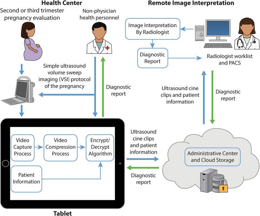
Toscano et al. BMC Pregnancy and Childbirth      (2021) 21:328                                                      Page 2 of 13

 (Continued from previous page)
 Results: Obstetric ultrasound telediagnosis showed excellent agreement with standard of care ultrasound allowing
 the identification of number of fetuses (100% agreement), fetal presentation (95.8% agreement, κ =0.78 (p < 0.0001)),
 placental location (85.6% agreement, κ =0.74 (p < 0.0001)), and assessment of normal/abnormal amniotic fluid volume
 (99.2% agreement) with sensitivity and specificity > 95% for all variables. Intraclass correlation was good or excellent for
 all fetal biometric measurements (0.81–0.95). The majority (88.5%) of second trimester ultrasound exam biometry
 measurements produced dating within 14 days of standard of care ultrasound.
 Conclusion: This Obstetric ultrasound telediagnostic system is a promising means to increase access to diagnostic
 Obstetric ultrasound in low-resource settings. The telediagnostic system demonstrated excellent agreement with
 standard of care ultrasound. Fetal biometric measurements were acceptable for use in the detection of gross
 discrepancies in fetal size requiring further follow up.
 Keywords: Global health, Handheld ultrasound, Low- and middle-income countries, Low-resource setting, Perinatal
 morbidity and mortality, Point-of-care ultrasound, Portable ultrasound, rural medicine, Telemedicine, Volume sweep imaging

Background                                                       with the highest maternal morbidity and mortality [10,
Globally, more than 800 women die each day from com-             11]. Increased use of Obstetric teleultrasound in rural
plications of pregnancy or childbirth, and for each death,       areas could contribute towards improving these outcomes,
20 women suffer serious morbidity [1]. The majority of           and several reports have appeared in the recent literature
these maternal deaths occur in low- and middle-income            demonstrating this [6, 12–24].
countries (LMICs) and are mostly preventable [2, 3]. Ac-           Deploying ultrasound to rural areas is limited by many
cess to quality Obstetric ultrasound can identify some           factors including lack of trained sonographers and sonolo-
complications leading to maternal mortality or morbidity         gists, lack of appropriate equipment, and lack of infrastruc-
and may allow timely referral to higher-resource centers         ture [8, 11, 13, 15, 25]. To overcome these challenges, we
[4–9]. Unfortunately, Obstetric ultrasound remains un-           developed a novel telediagnostic ultrasound system which
available in two-thirds of the world, particularly in regions    acquires images in the absence of a specialist (Fig. 1) [15].

 Fig. 1 Overview of the Obstetric Telediagnostic System
Toscano et al. BMC Pregnancy and Childbirth (2021) 21:328 Page 3 of 13
Operators without prior ultrasound experience obtain cine sweeps and arcs the transducer over the gravid abdomen
clips of the target region using scan protocols that rely to obtain cine clips containing a full volumetric acquisi-
only on external body landmarks for proper transducer tion of the target region. The ideal imaging speed is 1–2
placement to obtain adequate imaging of anatomy and cm/s, and an acquisition rate of 10–15 frames per sec-
pathology. Acquisitions are sent using a telemedicine plat- ond yields an image every 1–2 mm [15]. No specialized
form requiring low internet bandwidth for asynchronous anatomical or ultrasound knowledge is necessary to per-
remote interpretation. form the VSI protocol, therefore any individual may
A previous pilot study of this system for Obstetric teleul- serve as an operator. The probe motions are simple, and
trasound in the rural rainforest of Peru demonstrated en- ultrasound presets obviate the need for image adjust-
couraging results but was limited by small sample size and ments. All image interpretation is done remotely by a
lack of standard imaging for comparison [15]. Therefore, the specialist.
goal of this study was to test the system in comparison to The 8-step Obstetric VSI protocol (Fig. 2) was devel-
standard imaging in a larger sample of Peruvian women. The oped for simplicity, image redundancy, and optimal
primary endpoint was to obtain sufficient data to test agree- acoustic windows. Training an operator on the protocol
ment in descriptive variables between the telediagnostic sys- can be accomplished effectively in a short time (in our
tem (interpreted remotely) and standard of care (SOC) experience, 8 h is sufficient to demonstrate mastery of
ultrasound (interpreted locally). Evaluation of image quality the scan protocol and the telemedicine platform). Use of
and calculation of intraclass correlation coefficients (ICC) for the protocol is currently limited to second or third tri-
fetal biometry were also studied. We hypothesized that the mester examinations as the sweeps are solely transab-
system would yield diagnostic interpretations in agreement dominal, and first trimester examinations may require
with SOC ultrasound and successfully identify critical fea- an additional transvaginal examination for optimal
tures of the pregnancy. visualization. A video of the protocol, the training pos-
ter, and sample sweeps are available as supplemental
Methods material (Supplemental Materials 1, 2, 3, 4). In total, a
Obstetric volume sweep imaging (VSI) VSI examination takes about 10–20 min to perform, less
Obstetric volume sweep imaging (VSI) is a simple ultra- than 5 min to upload with standard internet, and 5–10
sound examination technique in which the operator min to interpret.
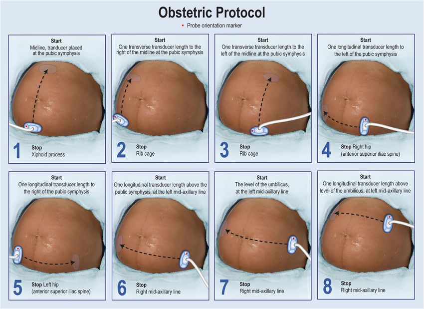
Fig. 2 Illustration of the Obstetrics VSI Protocol. The Obstetrics protocol involves 8 sweeps each beginning and ending with an arc (fan) of the
probe that has not been illustrated for simplicity. The arcing of the probe allows maximal visualization. Steps 1-3 involve transverse sweeps of the
probe from the pelvis to the upper abdomen. Steps 4 and 5 are sagittal sweeps at the base of the pelvis. Steps 6-8 are sagittal sweeps on the
upper gravid abdomenToscano et al. BMC Pregnancy and Childbirth (2021) 21:328 Page 4 of 13
Telediagnostic system with a 4.5 mHz curvilinear transducer. For VSI exams,
The technical specifications of the “Medical for this was attached to a Windows 10 tablet containing the
Ultrasound” (Med4US) telemedicine platform used in telemedicine software. A low- to moderate-speed, 3G
this study were previously published [26]. Briefly, internet connection in the health clinic allowed upload
this comprehensive telemedicine platform is installed times within 5 min.
on a tablet capable of sending imaging acquisitions
over very low bandwidths, roughly equivalent to Image interpretation
dial-up internet, with reasonable transmission times A single Peruvian radiologist acquired the SOC images,
[26]. The software prompts the operator to input the interpreted them, and generated a report per standard
patient’s clinical information, guides them in per- guidelines [27, 28]. The radiologist did not view the VSI
forming each of the 8 sweeps, then compresses, en- scans at any point. VSI images were interpreted remotely
crypts, and uploads the imaging data to the cloud. by a Maternal-Fetal Medicine fellow in the United States
In the absence of an internet connection, images are who was blinded to the results of SOC ultrasound. VSI
saved locally and can later be uploaded. Remote in- images were evaluated for ability to confirm a live fetus
terpretation by a specialist can subsequently be per- (based on cardiac activity), fetal number, fetal presenta-
formed asynchronously. Clips are not viewed as tion, placental location, and amniotic fluid volume.
video but rather as scrollable image stacks. The Image quality was assigned using 3-point Likert scale (1-
structured diagnostic report generated can then be “excellent,” 2-“acceptable,” or 3-“poor”) as was reader
sent back to the tablet and shared with the provider confidence in imaging findings (1-“confident,” 2-“inter-
and patient. mediate,” 3-“not confident”).
This version of the Obstetric VSI protocol was not de-
Testing the obstetric telediagnostic system signed for estimation of fetal biometry because the
This study was conducted in accordance with the proper imaging planes needed for accurate and reprodu-
Declaration of Helsinki and approved by the Institu- cible biometric measurements are not always present in
tional Review Board (IRB) at the Hospital Nacional the cine clips. However, as an exploratory outcome, we
Docente Madre Nino San Bartolome on 2/6/2018. investigated three methods of estimating fetal gestational
Two operators, a nurse and a care technician, with age using the protocol to identify gross discrepancies in
no prior ultrasound experience, underwent an 8 fetal size requiring further follow up. These included 1)
h training on the protocol and use of the telemedi- a gross visual estimation of fetal gestational age (second
cine software. Training involved approximately 4 h of versus third trimester), 2) fetal biometry using strict cri-
didactic sessions, containing visual and video aids to teria, and 3) fetal biometry using relaxed criteria. A strict
teach the operators the external body landmarks of measurement was an optimal biometric measurement in
interest, the pattern of ultrasound sweeps across the the correct plane closely or perfectly fulfilling current
abdomen, and the techniques for scanning. This was guidelines [29]. A relaxed measurement was a close esti-
followed by approximately 4 h of hands-on training to mate when the measurement of interest was not in the
practice and reinforce the protocol until the operators optimal plane or orientation.
could comfortably and accurately reproduce it After initial analysis, cases that were discrepant be-
independently. tween VSI ultrasound and SOC ultrasound underwent
Second or third trimester patients over age 18 who adjudication by an experienced Maternal-Fetal Medicine
presented to the Conde de la Vega Health Center in physician to determine the most accurate interpretation.
Lima, Peru were recruited from 6/2018–3/2019. All
patients participated in the informed consent process Statistical analysis
and then underwent a SOC ultrasound exam per- Agreement on categorical variables was assessed by
formed by a radiologist followed by VSI ultrasound overall agreement between VSI and SOC ultrasound and
using telediagnostic system performed by one of the Cohen’s kappa. Resultant kappa values were compared
two previously ultrasound-naïve operators who had to a theoretical mean of zero using a one-sample t-test
undergone training. The acquired VSI clips were to determine whether the kappa value was significantly
uploaded to the cloud for remote interpretation at better than zero. Biometry was compared between VSI
the time of their acquisition but were not immedi- and SOC ultrasound using paired t-tests, calculation of
ately accessed and reviewed during the recruitment ICC, and Bland-Altman analysis. ICC values were calcu-
phase of the study, and no feedback was provided to lated using a two-way mixed effects model for absolute
the ultrasound-naïve operators. agreement. ICC was classified as “excellent” with values
All exams were performed using a portable Mindray greater than 0.9, “good” with values between 0.75 and
DP-10 (Mindray, China), set to the Obstetrics preset, 0.9, “moderate” with values between 0.5 and 0.75, andToscano et al. BMC Pregnancy and Childbirth (2021) 21:328 Page 5 of 13
“poor” with values less than 0.5 [30]. Both ICC values visualized secondary to fast sweep speed or early second
and Bland-Altman bias were independently compared to trimester fetus.
a theoretical mean of zero using a one-sample t-test. The vast majority of examinations were interpreted as
These analyses were repeated separately by trimester normal with high agreement between VSI and SOC
and by the number of biometry measurements achiev- ultrasound (95.2% agreement, κ =0.55 (p < 0.0001)). For
able on VSI. Sweep length was compared between cases abnormal cases, there was 100% agreement (4/4 cases)
with “acceptable” versus “excellent” image quality using for non-cephalic presentation in the third trimester with
unpaired t-tests. Differences in sweep length based on high confidence by VSI reader for all assessments. VSI
the number of biometry measurements were assessed identified 5 cases (4 after adjudication) of possible low
with ordinary one-way ANOVA with Tukey’s test used lying/placenta previa which were not reported on SOC
for pairwise comparisons. All statistical analysis was per- ultrasound. All were second trimester fetuses with the
formed using MATLAB (v2019b, The Mathworks, Inc., potential for re-imaging later in pregnancy for confirm-
Natick, MA) and SPSS (v26, IBM Corporation, Armonk, ation. VSI identified 1 case of suspected oligohydramnios
NY). in a third trimester gestation (verified on adjudication)
which was not reported on SOC ultrasound.
Results Gross visual estimation of fetal gestational age by tri-
One hundred twenty-six patients (inclusive of 68 sec- mester (with fetuses straddling the trimester definition
ond trimester and 58 third trimester pregnancies) (26–30 weeks) counted as agreement if reported as ei-
with a mean age of 25.7 years participated in the ther second or third) yielded only 5 disagreements. Dis-
study. All images were successfully uploaded using agreements differed by only a few days outside of
the health clinic’s low- to moderate-bandwidth inter- defined ranges for each trimester. After adjudication, all
net connection and were accessed in the United 5 discrepant cases were grouped into the correct trimes-
States for remote interpretation. Only one VSI im- ter yielding 100% agreement.
aging study was rated of “poor” imaging quality ICC between VSI and SOC measurements was good-
(visualization of an early second trimester fetus was excellent for individual biometric measurements (ICC =
limited by maternal body habitus). The remaining im- 0.81–0.93) and excellent for overall estimated gestational
aging clips (> 99%) provided a diagnostic exam of “ac- age (EGA) (ICC = 0.95) when examining all exams
ceptable” (38.1%) or “excellent” (61.1%) image quality. (Table 2). For subgroups, second trimester demonstrated
The overwhelming majority of sweeps were performed moderate correlation for individual measurements
correctly (suboptimal sweep position was only identi- (ICC = 0.67–0.84) and excellent correlation for overall
fied in two exams). Sample images and clips obtained EGA (ICC = 0.94), and third trimester demonstrated
from the VSI protocol and uploaded by the telediag- poor - moderate correlation for individual measurements
nostic system are seen in Figs. 3, 4 and Supplemental (ICC = 0.28–0.68) and moderate correlation for overall
Materials 3, 4. The average file size of uncompressed EGA (ICC = 0.64). VSI slightly over-estimated all bio-
clips was 12.5 megabytes and average sweep time was metric measurements except femur length and slightly
12.6 seconds. Sweeps 3 (p = 0.0006) and 7 (p = 0.01) overestimated EGA in all exams (Table 2). The magni-
of the protocol were significantly longer for cases tude of this difference is not likely to be clinically signifi-
with excellent image quality. No differences in sweep cant for the intended application of teleultrasound.
length based on the total number of attained biomet- Bland-Altman plots were constructed to identify any sys-
ric measurements were found. tematic differences between VSI and SOC ultrasound,
Results for overall agreement between VSI and SOC and these demonstrated low-level fixed bias but also
ultrasound are shown in Table 1. There was excellent confirmed that the vast majority of measurements fell
agreement for number of fetuses (100% agreement), fetal within 95% limits of agreement (Fig. 5). Bland-Altman
presentation (95.8% agreement, κ =0.78 (p < 0.0001)), bias was lowest for femur length (1.1 for all exams; 0.2
placental location (85.6% agreement, κ =0.74 (p < and 3.0 for second and third trimester subgroups, re-
0.0001)), and assessment of amniotic fluid volume spectively) and highest for abdominal circumference
(99.2% agreement). When fundal placental position was (18.6 for all exams; 16.8 and 20.7 for second and third
counted as either anterior or posterior, the agreement trimester subgroups, respectively) (Table 2). When
improved to 94.4%. The reader was highly confident in examining overall EGA, Bland-Altman bias was lowest
these assessments (for all: median confidence rating “3” for second trimester calculations (6.5) and highest for
corresponding to “confident” on the 3-point Likert third trimester calculations (15.2).
scale). VSI was able to confirm the presence of a live There were no significant differences in agreement of
fetus in 76.2% of cases. In the cases without a confirmed EGA noted between VSI and SOC ultrasound when
live fetus, a full cardiac cycle was not able to be using strict versus relaxed biometry measurementsToscano et al. BMC Pregnancy and Childbirth (2021) 21:328 Page 6 of 13
Fig. 3 Example Images Acquired by Individuals Without Prior Training. a, b Still images from an ultrasound cine clip of a sagittal VSI sweep across
the lower uterine corpus obtained by an operator without prior ultrasound training demonstrating a singleton early third trimester fetus in vertex
presentation (thin arrow) with an anterior-mid placenta (block arrow) and grossly normal amniotic fluid volume (*). Maternal bladder is also seen
in b (curved arrow)
(Table 3, Fig. 6). Table 4 summarizes the differences in substantial barriers to Obstetric ultrasound access in
biometric measurements between VSI and SOC ultra- LMICs. For the price of a tablet and portable ultra-
sound. Among all exams and all biometry measure- sound, individuals with a few hours of structured
ments, 69.1% of all the VSI examinations and 88.5% of hands-on training are capable of acquiring imaging
second trimester exams showed agreement within 2 acquisitions allowing the identification of key infor-
weeks to SOC ultrasound measurements. Achieving 4/4, mation about a pregnancy including confirmation of a
3/4, 2/4, or 1/4 of possible biometric measurements by live fetus, fetal number, fetal presentation, placental
VSI did not impact agreement with SOC ultrasound. location, and amniotic fluid volume. Pregnancy com-
plications which may be identified by this type of
Discussion ultrasound, including multiple gestation, non-cephalic
The Obstetric ultrasound telediagnostic system with presentation in the third trimester, abnormal amniotic
asynchronous reads presented in this study removes fluid volume, and low lying placenta/placenta previa,Toscano et al. BMC Pregnancy and Childbirth (2021) 21:328 Page 7 of 13
Fig. 4 Example Non-cephalic Presentation. Still image from a VSI sweep of a singleton second trimester fetus in breech presentation (small arrows
indicate fetal feet and lower extremities as presenting parts). The placenta is seen anterior excluding a low-lying placenta or placenta previa
(block arrow)
Table 1 Agreement between volume sweep imaging protocol and standard of care ultrasound
Variablea Volume sweep imagingb Standard of careb Overall agreement Cohens kappac Confidenced
Live Fetus 76.2% (67.8–83.3%) 100% (97.1–100%) 76.2% …e 3 (1–3)
Number of Fetuses (% single) 100% (97.1–100%) 100% (97.1–100%) 100% …e 3 (2–3)
Fetal Presentation (% vertex) 87.3% (77.3–94%) 91.5% (82.5–96.8%) 95.8% 0.78 (0.53–1.00, 3 (1–3)
p < 0.0001)
Placental position
Anterior Placenta 52.0% (42.9–61.0%) 50.4% (41.3–59.5%) 85.6% (94.4%f) 0.74 (0.63–0.85, g
p < 0.0001)
Posterior Placenta 42.4% (33.6–51.6%) 40.0% (31.3–49.1%)
Fundal Placenta 5.6% (2.3–11.2%) 9.6% (5.1–16.2%)
Placenta Previa 4.0% (1.3–9.1%) 0% (0–2.9%) 96.0% …e 3 (1–3)
Placenta Previa (consensus read) 3.2% (0.9–8.0%) 96.8%
Amniotic Fluid Volume (% normal) 99.2% (95.7–100%) 100% (97.1–100%) 99.2% …e 3 (1–3)
Normal Exam 92.1% (85.9–96.1%) 96.8% (92.1–99.1%) 95.2% 0.55 (0.20–0.90, 3 (1–3)
p < 0.0001)
Normal Exam (consensus read) 92.9% (86.9–96.7%) 96.0% 0.60 (0.25–0.94,
p < 0.0001)
Follow-up Recommendation (% normal) 99.2% (95.7–100%) 100% (97.1–100%) 99.2% …e 3 (1–3)
a
for categories in which consensus reads resulted in changes, these are noted below the original variable
b
values are percentage (95% confidence interval)
c
values are kappa (95% confidence interval, p value)
d
confidence is specified as median (range)
e
not defined, due to Peruvian radiologist labelling all identically
f
agreement when fundal location included as agreeing with either anterior or posterior location
g
not givenToscano et al. BMC Pregnancy and Childbirth (2021) 21:328 Page 8 of 13
Table 2 Comparison of volume sweep imaging protocol biometry using strict criteria and standard of care ultrasound biometry
Variable Volume sweep Standard of p valueb Intraclass correlation Bland-altman
imaginga carea coefficientc biasc
All Exams
Biparietal Diameter (mm) 71.1 ± 20.3 (n = 111) 65.6 ± 18.7 < 0.0001 0.89 (0.50–0.96, p < 0.0001) 6.4 (−6.9–19.7, p < 0.0001)
Head Circumference (mm) 250.0 ± 66.3 (n = 99) 236.0 ± 68.0 < 0.0001 0.86 (0.71–0.92, p < 0.0001) 17.5 (−45.0–79.9, p < 0.0001)
Abdominal Circumference (mm) 243.0 ± 77.8 (n = 102) 224.0 ± 74.8 < 0.0001 0.81 (0.69–0.88, p < 0.0001) 18.6 (−66.2–103, p < 0.0001)
Femur Length (mm) 41.8 ± 16.5 (n = 56) 46.3 ± 15.8 0.17 0.93 (0.88–0.96, p < 0.0001) 1.1 (−10.4–12.6, p = 0.17)
Estimated Gestational Age (days) 195 ± 49 (n = 109) 186.0 ± 46.0 < 0.0001 0.95 (0.69–0.98, p < 0.0001) 10.4 (−10.6–31.5, p < 0.0001)
Second Trimester Exams
Biparietal Diameter (mm) 56.2 ± 14.0 51.6 ± 13.0 < 0.0001 0.84 (0.54–0.93, p < 0.0001) 4.6 (−7.7–16.9, p < 0.0001)
Head Circumference (mm) 202.0 ± 45.3 189.0 ± 47.6 0.0007 0.84 (0.69–0.91, p < 0.0001) 11.4 (−35.5–58.4, p < 0.0001)
Abdominal Circumference (mm) 187.0 ± 50.8 168.0 ± 46.7 0.001 0.67 (0.45–0.80, p < 0.0001) 16.8 (−55.0–88.5, p < 0.0001)
Femur Length (mm) 32.7 ± 9.6 34.6 ± 10.8 0.87 0.83 (0.70–0.91, p < 0.0001) 0.2 (−10.8–11.1, p = 0.87)
Estimated Gestational Age (days) 158.0 ± 27.2 150.0 ± 27.0 < 0.0001 0.94 (0.65–0.98, p < 0.0001) 6.5 (−6.9–19.9, p p < 0.0001)
Third Trimester Exams
Biparietal Diameter (mm) 90.0 ± 6.79 81.9 ± 7.7 < 0.0001 0.33 (−0.10–0.64, p < 0.0001) 8.7 (−4.6–22.0, p < 0.0001)
Head Circumference (mm) 313.0 ± 22.6 291.0 ± 42.7 0.0001 0.38 (0.06–0.62, p = 0.001) 25.3 (−50.8–101, p < 0.0001)
Abdominal Circumference (mm) 309.0 ± 44.7 289.0 ± 39.5 0.0007 0.28 (0.02–0.52, p = 0.015) 20.7 (−77.8–119, p < 0.0001)
Femur Length (mm) 61.4 ± 9.5 59.9 ± 7.7 0.051 0.68 (0.32–0.87, p < 0.0001) 3.0 (−8.97–15, p = 0.051)
Estimated Gestational Age (days) 242.0 ± 23.0 229.0 ± 18.4 < 0.0001 0.64 (−0.02–0.86, p < 0.0001) 15.2 (−9.46–39.9, p < 0.0001)
a
values are mean ± standard deviation
b
p value using paired t-test
c
values are kappa (95% confidence interval, p value)
can be detected prior to labor and delivery, allowing ultrasound. Research and development of a robust re-
transfer to a higher level of care and possible preven- ferral system to bring women with ultrasound-
tion of adverse outcomes. Barriers to deployment of identified pregnancy complications from their rural
standard ultrasound capability including cost, tech- setting to a hospital center will be crucial alongside
nical complexity, lack of specialists/sonographers, lack this telemedicine system.
of infrastructure, and poor internet connections are Both operators received no formal quality assess-
all overcome with this approach. Thus, this telediag- ment or feedback over the course of the study after
nostic Obstetric ultrasound system provides a scal- their initial training. Since the data collection spanned
able, effective, and potentially life-saving means to a year, the consistent quality of the scans shows re-
deliver Obstetric ultrasound to low resource areas. tention of the protocol. Although imaging quality was
Lowering the maternal mortality rate is a vital pub- already “acceptable” or “excellent” in greater than 99%
lic heath goal as stated in Millennium Development of cases, regular feedback could assist in improving
Goal 5 and has critical downstream impacts on fam- quality even further. In the scans rated as “acceptable”
ily health, social, and economic conditions [31]. Ob- instead of “excellent,” involved factors included insuf-
stetric telediagnosis has the potential to contribute to ficient depth in the third trimester, suboptimal mater-
achieving this goal through early identification of nal body habitus, and small uterine size in the second
pregnancy complications that may lead to preventable trimester. Even in the lower image quality exams
maternal morbidity or mortality. While we have where biometry could not be performed or live fetus
proven that this approach is feasible, elucidating could not be confirmed, other more important pa-
where exactly this approach fits into healthcare deliv- rameters such as fetal number and placental location
ery will require additional study using a dissemin- were able to be reliably assessed. Future adaptations
ation and implementation framework [32]. Given the may consider modifying the protocol for fetuses earl-
heterogeneity of systems and communities, using a ier than 20 weeks.
framework helps account for the needs and consider- While disagreements between VSI and SOC ultra-
ations of individual communities in the design and sound were rare, only a few reference ultrasound im-
implementation of telediagnostic Obstetric ages were saved limiting our ability to delineate theToscano et al. BMC Pregnancy and Childbirth (2021) 21:328 Page 9 of 13 Fig. 5 (See legend on next page.)
Toscano et al. BMC Pregnancy and Childbirth        (2021) 21:328                                                                            Page 10 of 13

    (See figure on previous page.)
    Fig. 5 Bland-Altman plots of agreement in measurements of a head circumference; b biparietal diameter, c abdominal circumference; d femur
    length. Y-axis: mean difference in biometry measurement between volume sweep imaging (VSI) and standard of care (SOC) imaging and X-axis:
    mean biometry measurement of VSI and SOC. Dashed line representing 95% limits of agreement of measurements and dotted line representing
    Bland-Altman bias

source of disagreement. Additionally, the majority of                         Conclusion
exams included in this study contained normal find-                           Our work demonstrates that a VSI protocol with teleul-
ings, as would be expected based on known preva-                              trasound system has substantial promise to increase ac-
lence of the types of pregnancy complications that are                        cess to prenatal ultrasound imaging worldwide. In this
detectable by ultrasound. Therefore, further investiga-                       study, VSI showed excellent agreement with the SOC
tion must be undertaken in a patient group with ab-                           ultrasound in assessing vital descriptive features of a
normal findings to ensure the protocol performs                               pregnancy and allowed for close estimation of gesta-
equally well in detection of pregnancy complications                          tional age. Large-scale use of this technology has the po-
such as malpresentation, placenta previa, multiple ges-                       tential to improve access to high-quality prenatal care
tation, and abnormal fluid volume. Additional areas                           and improve maternal and neonatal/perinatal mortality
for future investigation include explorations of artifi-                      and morbidity from preventable causes.
cial intelligence including automated detection of fetal
                                                                              Abbreviations
heart rate, estimation of gestational age, and fetal
                                                                              VSI: Volume sweep imaging; SOC: Standard of care; LMICs: Low- and middle-
presentation.                                                                 income countries; ICC: Intraclass correlation coefficient; EGA: Estimated
  It should be acknowledged that the Obstetric VSI                            gestational age
protocol was not designed for assessment of fetal biom-
etry. Despite this, exploratory use of the protocol for bi-                   Supplementary Information
                                                                              The online version contains supplementary material available at https://doi.
ometry in this study demonstrated high correlation in
                                                                              org/10.1186/s12884-021-03720-w.
EGA by VSI compared to SOC ultrasound and found
that using strict criteria versus relaxed criteria for fetal                   Additional file 1. Obstetric Training Video.
biometric measurements did not improve estimation of                           Additional file 2. Obstetric VSI Protocol Training Poster. Poster used in
gestational age. These are encouraging results for use of                      the original Obstetric training session.
the telediagnosis for gross estimation of fetal size,                          Additional file 3. Obstetric VSI Sweep of a Second Trimester Fetus. An
though the authors acknowledge that these results do                           example of a transverse sweep from the maternal pubic symphysis to the
                                                                               maternal xyphoid process at the midline. The sweep shows a singleton
not support the use of the protocol for more precise                           second trimester fetus in the vertex presentation, an anterior placenta
diagnosis of fetal growth abnormalities given the dis-                         position, and fetal cardiac activity. The amniotic fluid volume is grossly
crepancies found in third trimester biometry measure-                          normal.
ments in this study. In these cases, patients should be                        Additional file 4. Obstetric VSI Sweep of a Third Trimester Fetus. An
                                                                               example of a sagittal sweep from the maternal right anterior superior
referred to a higher level of care with a trained sonogra-                     iliac spine to the maternal left anterior superior iliac spine with the
pher. In the future, the addition of calibrated 3D recon-                      inferior aspect of the ultrasound transducer at the maternal pubic
struction will allow the generation of images in the                           symphysis. The sweep shows a singleton third-trimester fetus in breech
                                                                               presentation. Placenta is not visualized in the lower uterine segment or
precise planes needed for accurate measurement accord-                         near the internal cervical os. The amniotic fluid volume is grossly normal.
ing to standard guidelines.

Table 3 A comparison of strict versus relaxed biometry measurements obtained using volume sweep imaging protocol
Variable                                                            All measurements             Second trimester only (n = 68)           Third trimester
                                                                    (n = 109)                                                             only (n = 58)
Number of biometry measurements achieved (strict)                   2 (0–4)                      2 (0–4)                                  2 (0–4)
Number of biometry measurements achieved (including relaxed)        3 (0–4)                      3 (0–4)                                  3 (0–4)
Estimated gestational age (days)
    Stricta                                                         195 ± 49 (n = 109)           158 ± 27 (n = 60)                        242 ± 23 (n = 49)
    Including relaxeda                                              194 ± 51 (n = 122)           155 ± 28 (n = 67)                        243 ± 24 (n = 55)
         b
p-value                                                             0.37                         0.44                                     0.58
a
  values are mean ± standard deviation
b
 using paired t-test
Toscano et al. BMC Pregnancy and Childbirth (2021) 21:328 Page 11 of 13
Fig. 6 Bland-Altman plots of agreement in measurements of a strict biometry measurements and b relaxed (inclusive of strict) biometry measurements. Y-axis:
mean difference in biometry measurement between volume sweep imaging (VSI) and standard of care (SOC) imaging and X-axis: mean biometry measurement
of VSI and SOC. hashed line representing 95% limits of agreement of measurements and dotted line representing Bland-Altman bias
Table 4 Comparison of strict versus relaxed biometry criteria and calculated estimated gestational age by volume sweep imaging
protocol and standard of care ultrasound
Trimester Within 7 days Within 14 days
Overall agreement (strict) Overall agreement Overall agreement (strict) Overall agreement
(including relaxed) (including relaxed)
All exams (n = 126) 42.2% (32.8–52.0%) 48.2% (38.6–57.9%) 67.9% (58.3–76.5%) 69.1% (59.6–77.6%)
Second trimester (n = 68) 56.7% (43.2–69.4%) 63.9% (50.6–75.8%) 85% (73.4–92.9%) 88.5% (77.8–95.3%)
Third trimester 24.5% (13.3–38.9%) 28.6% (16.6–43.3%) 46.9% (32.5–61.7%) 44.9% (30.7–59.8%)
(n = 58)
All values are percentage (95% confidence interval)Toscano et al. BMC Pregnancy and Childbirth (2021) 21:328 Page 12 of 13
Acknowledgements Ministries of the Americas, 10810 Lake Minneola Shores, Clermont, FL 34711,
We would like to thank Nadezhda Kiriyak1, Sarah Klingenberger1, Jane USA. 5Department of Public Health Sciences, University of Rochester Medical
Lichorowic1, and Gwen Mack1 for assisting in the preparation of our figures Center, 265 Crittenden Blvd., Rochester, NY 14642, USA. 6Medical Innovation
and illustrations. and Technology, Calle Los Libertadores 635, 15046 San Isidro, Peru.
1 7
University of Rochester Medical Center, Rochester, NY. No funding sources Departament of Academic Engineering, Division of Electric Engineering,
or compensation to disclose. Pontificia Universidad Catolica del Peru, Av. Universitaria 1801, 15088 San
Miguel, Peru.
Authors’ contributions
Marika Toscano, MD: Data curation, Validation, Investigation, Visualization, Received: 25 January 2021 Accepted: 15 March 2021
Writing – original draft, review and editing. Thomas J. Marini, MD: Project
administration, Data curation, Validation, Investigation, Visualization,
Resources, Writing – original draft, review and editing. Kathryn Drennan, MD:
Investigation, Supervision, Writing – review and editing. Timothy M. Baran, References
PhD: Validation, Data curation, Formal analysis, Writing – review and editing, 1. Maternal Mortality: UNICEF; 2019. Available from: https://data.unicef.org/
Supervision. Jonah Kan, BA: Investigation, Data curation, Visualization. Brian topic/maternal-health/maternal-mortality/. Accessed Nov 2020.
Garra, MD: Conceptualization, Methodology, Supervision, Writing – review 2. Trends in maternal mortality 2000 to 2017: estimates by WHO, UNICEF,
and editing. Ann Dozier, PhD: Writing – review and editing. Rafael Ortega, NFPA, World Bank Group and the United Nations Population Division. 2019.
BS: Investigation, Data curation. Rosemary Quinn, BS: Investigation, Data https://www.unfpa.org/featured-publication/trends-maternal-mortality-2
curation. Tina Zhao, BA: Investigation, Data curation. Miguel S. Egoavil, MD: 000-2017.
Conceptualization, Methodology. Lorena Tamayo, BS: Software, Resources, 3. Small MJ, Allen TK, Brown HL. Global disparities in maternal morbidity and
Validation. Claudia Carlotto: Software, Resources, Validation. Benjamin mortality. Semin Perinatol. 2017;41(5):318–22. https://doi.org/10.1053/j.
Castaneda, PhD: Conceptualization, Methodology, Software, Validation, semperi.2017.04.009.
Resources, Supervision, Funding acquisition, Project administration, 4. Ahman A, Edvardsson K, Kidanto HL, Ngarina M, Small R, Mogren I. ‘Without
Investigation, Writing – review and editing. The author(s) read and approved ultrasound you can’t reach the best decision’ Midwives’ experiences and views
the final manuscript. of the role of ultrasound in maternity care in Dar Es Salaam, Tanzania. Sex
Reprod Healthc. 2018;15:28–34. https://doi.org/10.1016/j.srhc.2017.11.007.
Funding 5. Harris RD, Marks WM. Compact ultrasound for improving maternal and
Innóvate Peru (409-FIDECOM INNOVATEPERU-PVE-2017). perinatal care in low-resource settings: review of the potential benefits,
Pontificia Catholica University del Peru (Períodos de Investigación 2020). implementation challenges, and public health issues. J Ultrasound Med.
Funding sources had no role in design of the study, collection, analysis, or 2009;28(8):1067–76. https://doi.org/10.7863/jum.2009.28.8.1067.
interpretation of data or in writing the manuscript. 6. Kim ET, Singh K, Moran A, Armbruster D, Kozuki N. Obstetric ultrasound use
in low and middle income countries: a narrative review. Reprod Health.
Availability of data and materials 2018;15(1):129. https://doi.org/10.1186/s12978-018-0571-y.
The dataset supporting the conclusions of this article is available in the 7. Magann EF, McKelvey SS, Hitt WC, Smith MV, Azam GA, Lowery CL. The use
Mendeley Data repository, (https://data.mendeley.com/datasets/4k443 of telemedicine in obstetrics: a review of the literature. Obstet Gynecol Surv.
mtkxd/2). 2011;66(3):170–8. https://doi.org/10.1097/OGX.0b013e3182219902.
Toscano, Marika; Marini, Thomas; Drennan, Kathryn; Baran, Timothy; Kan, 8. Mollura DJ, Mazal J, Everton KL, Group R-ACW. White paper report of the
Jonah; Garra, Brian; Dozier, Ann; Ortega, Rafael; Quinn, Rosemary; Zhao, Tina; 2012 RAD-AID conference on international radiology for developing
Egoavil, Miguel; Tamayo, Lorena; Carlotto, Claudia; Castaneda, Benjamin countries: planning the implementation of global radiology. J Am Coll
(2021), “Testing Telediagnostic Obstetric Ultrasound in Peru: A New Horizon Radiol. 2013;10(8):618–24. https://doi.org/10.1016/j.jacr.2013.01.019.
in Expanding Access to Prenatal Ultrasound” , Mendeley Data, V2, doi: 9. Ngoya PS, Muhogora WE, Pitcher RD. Defining the diagnostic divide: an
https://doi.org/10.17632/4k443mtkxd.2 analysis of registered radiological equipment resources in a low-income
African country. Pan Afr Med J. 2016;25:99.
Declarations 10. World Radiography Day: Two-Thirds of the World’s Population has no
Access to Diagnostic Imaging: Pan American Health Organization. Available
Ethics approval and consent to participate from: https://www.paho.org/hq/index.php?option=com_content&view=
This study was was conducted in accordance with the Declaration of article&id=7410:2012-dia-radiografia-dos-tercios-poblacion-mundial-no-
Helsinki and approved by the Institutional Review Board at the Hospital tiene-acceso-diagnostico-imagen&Itemid=1926&lang=en#:~:text=%2D%2
Nacional Docente Madre Nino San Bartolome on 2/6/2018 (0095–2018-OADI- 0The%20use%20of%20X%2Drays,no%20access%20to%20diagnostic%20ima
HONADOMANI-SB). All subjects completed informed consent prior to ging.&text=It%20is%20used%20for%20diagnostic%2C%20preventive%2C%2
participation. 0and%20therapeutic%20purposes. Accessed Nov 2020.
11. Maru DS, Schwarz R, Jason A, Basu S, Sharma A, Moore C. Turning a blind
Consent for publication eye: the mobilization of radiology services in resource-poor regions. Glob
Not applicable Health. 2010;6(1):18. https://doi.org/10.1186/1744-8603-6-18.
12. Bagayoko CO, Traore D, Thevoz L, Diabate S, Pecoul D, Niang M, et al.
Competing interests Medical and economic benefits of telehealth in low- and middle-income
BC, MSE, LT, and CC have or have had financial stake in Medical Innovation countries: results of a study in four district hospitals in Mali. BMC Health
and Technology, a company which seeks to bring ultrasound to rural Serv Res. 2014;14(Suppl 1):S9.
communities. The remaining authors report no conflicts of interest. All data 13. Britton N, Miller MA, Safadi S, Siegel A, Levine AR, McCurdy MT. Tele-
analysis and manuscript writing was performed by the University of ultrasound in resource-limited settings: a systematic review. Front Public
Rochester. Those from Medical Innovation and Technology previewed the Health. 2019;7:244. https://doi.org/10.3389/fpubh.2019.00244.
document and any revisions were subject to approval from the University of 14. Dougherty A, Kasten M, DeSarno M, Badger G, Streeter M, Jones DC, et al.
Rochester and Medical Imaging Ministry of the Americas authors. Validation of a telemedicine quality assurance method for point-of-care
obstetric ultrasound used in low-resource settings. J Ultrasound Med. 2020.
Author details 15. Marini TJ, Oppenheimer DC, Baran TM, Rubens DJ, Toscano M, Drennan K,
1
Department of Obstetrics & Gynecology, Division of Maternal/Fetal et al. New ultrasound telediagnostic system for low-resource areas: pilot
Medicine, University of Rochester Medical Center, 601 Elmwood Ave, Box results from Peru. J Ultrasound Med. 2020.
668, Rochester, NY 14642, USA. 2Department of Imaging Sciences, University 16. Rijken MJ, Lee SJ, Boel ME, Papageorghiou AT, Visser GH, Dwell SL, et al.
of Rochester Medical Center, 601 Elmwood Ave, Box 648, Rochester, NY Obstetric ultrasound scanning by local health workers in a refugee camp on
14642, USA. 3University of Rochester School of Medicine and Dentistry, 601 the Thai-Burmese border. Ultrasound Obstet Gynecol. 2009;34(4):395–403.
Elmwood Ave, Box 607, Rochester, NY 14642, USA. 4Medical Imaging https://doi.org/10.1002/uog.7350.Toscano et al. BMC Pregnancy and Childbirth (2021) 21:328 Page 13 of 13
17. Ross AB, DeStigter KK, Rielly M, Souza S, Morey GE, Nelson M, et al. A low-
cost ultrasound program leads to increased antenatal clinic visits and
attended deliveries at a health care clinic in rural Uganda. PLoS One. 2013;
8(10):e78450. https://doi.org/10.1371/journal.pone.0078450.
18. Shah SP, Epino H, Bukhman G, Umulisa I, Dushimiyimana JM, Reichman A,
et al. Impact of the introduction of ultrasound services in a limited resource
setting: rural Rwanda 2008. BMC Int Health Hum Rights. 2009;9(1):4. https://
doi.org/10.1186/1472-698X-9-4.
19. Shokoohi H, Raymond A, Fleming K, Scott J, Kerry V, Haile-Mariam T, et al.
Assessment of point-of-care ultrasound training for clinical educators in
Malawi, Tanzania and Uganda. Ultrasound Med Biol. 2019;45(6):1351–7.
https://doi.org/10.1016/j.ultrasmedbio.2019.01.019.
20. Stein W, Katunda I, Butoto C. A two-level ultrasonographic service in a
maternity care unit of a rural district hospital in Tanzania. Trop Dr. 2008;
38(2):125–6. https://doi.org/10.1258/td.2007.070045.
21. Swanson JO, Kawooya MG, Swanson DL, Hippe DS, Dungu-Matovu P,
Nathan R. The diagnostic impact of limited, screening obstetric ultrasound
when performed by midwives in rural Uganda. J Perinatol. 2014;34(7):508–
12. https://doi.org/10.1038/jp.2014.54.
22. Swanson JO, Plotner D, Franklin HL, Swanson DL, Lokomba Bolamba V,
Lokangaka A, et al. Web-based quality assurance process drives
improvements in obstetric ultrasound in 5 low- and middle-income
countries. Glob Health Sci Pract. 2016;4(4):675–83. https://doi.org/10.9745/
GHSP-D-16-00156.
23. Vinals F, Mandujano L, Vargas G, Giuliano A. Prenatal diagnosis of congenital
heart disease using four-dimensional spatio-temporal image correlation
(STIC) telemedicine via an internet link: a pilot study. Ultrasound Obstet
Gynecol. 2005;25(1):25–31. https://doi.org/10.1002/uog.1796.
24. Vinayak S, Sande J, Nisenbaum H, Nolsoe CP. Training midwives to perform
basic obstetric point-of-care ultrasound in rural areas using a tablet platform
and mobile phone transmission technology-a WFUMB COE project.
Ultrasound Med Biol. 2017;43(10):2125–32. https://doi.org/10.1016/j.ultra
smedbio.2017.05.024.
25. Scott Kruse C, Karem P, Shifflett K, Vegi L, Ravi K, Brooks M. Evaluating barriers
to adopting telemedicine worldwide: a systematic review. J Telemed Telecare.
2018;24(1):4–12. https://doi.org/10.1177/1357633X16674087.
26. Ferrer J, Chaumont T, Trujillo L, Fernandez I, Guerrero J, Stewart P, et al.
New tele-diagnostic model using volume sweep imaging for rural areas.
Conf Proc IEEE Eng Med Biol Soc. 2017;2017:2622–5.
27. AIUM-ACR-ACOG-SMFM-SRU practice parameter for the performance of
standard diagnostic obstetric ultrasound examinations. J Ultrasound Med.
2018;37(11):E13-E24. https://doi.org/10.1002/jum.14831. Epub 2018 Oct 11.
PMID: 30308091.
28. AIUM. Practice parameter for documentation of an ultrasound examination.
J Ultrasound Med. 2020;39(1):E1–4.
29. Salomon LJ, Alfirevic Z, Da Silva CF, Deter RL, Figueras F, Ghi T, et al. ISUOG
practice guidelines: ultrasound assessment of fetal biometry and growth.
Ultrasound Obstet Gynecol. 2019;53(6):715–23. https://doi.org/10.1002/uog.2
0272.
30. Koo TK, Li MY. A guideline of selecting and reporting Intraclass correlation
coefficients for reliability research. J Chiropr Med. 2016;15(2):155–63. https://
doi.org/10.1016/j.jcm.2016.02.012.
31. Millennium Development Goals (MDGs): World Health Organization; 2019.
Available from: https://www.who.int/topics/millennium_development_goa
ls/en/. Accessed Nov 2020.
32. Dissemination & implementation models in health research & practice.
Available from: https://dissemination-implementation.org/. Accessed Nov
2020.
Publisher’s Note
Springer Nature remains neutral with regard to jurisdictional claims in
published maps and institutional affiliations.You can also read