That's Near My Hand! Parietal and Premotor Coding of Hand-Centered Space Contributes to Localization and Self-Attribution of the Hand
←
→
Page content transcription
If your browser does not render page correctly, please read the page content below
The Journal of Neuroscience, October 17, 2012 • 32(42):14573–14582 • 14573

Behavioral/Systems/Cognitive

That’s Near My Hand! Parietal and Premotor Coding of
Hand-Centered Space Contributes to Localization and
Self-Attribution of the Hand
Claudio Brozzoli,* Giovanni Gentile,* and H. Henrik Ehrsson
Brain, Body, and Self Laboratory, Department of Neuroscience, Karolinska Institute, SE-17177 Stockholm, Sweden

The ability to identify and localize our own limbs is crucial for survival. Indeed, the majority of our interactions with objects occur within
the space surrounding the hands. In non-human primates, neurons in the posterior parietal and premotor cortices dynamically represent
the space near the upper limbs in hand-centered coordinates. Neuronal populations selective for the space near the hand also exist in
humans. It is unclear whether these remap the peri-hand representation as the arm is moved in space. Furthermore, no combined
neuronal and behavioral data are available about the possible involvement of peri-hand neurons in the perception of the upper limbs in
any species. We used fMRI adaptation to demonstrate dynamic hand-centered encoding of space by reporting response suppression in
human premotor and posterior parietal cortices to repeated presentations of an object near the hand for different arm postures. Further-
more, we show that such spatial representation is related to changes in body perception, being remapped onto a prosthetic hand if
perceived as one’s own during an illusion. Interestingly, our results further suggest that peri-hand space remapping in the premotor
cortex is most tightly linked to the subjective feeling of ownership of the seen limb, whereas remapping in the posterior parietal cortex
closely reflects changes in the position sense of the arm. These findings identify the neural bases for dynamic hand-centered encoding of
peripersonal space in humans and provide hitherto missing evidence for the link between the peri-hand representation of space and the
perceived self-attribution and position of the upper limb.

Introduction                                                                                                                Single-cell recordings in the posterior parietal and premo-
In everyday life, we do not explicitly perceive a boundary between                                                      tor cortices of macaques have identified neurons with both
the space near the body and more distant space. Nevertheless,                                                           tactile and visual receptive fields. The latter are restricted to
psychologists have long theorized that we are surrounded by an                                                          the space extending 30 – 40 cm from the location of the tactile
“invisible bubble” of space (Hall, 1966), and behavioral experi-                                                        receptive field (Hyvärinen and Poranen, 1974; Rizzolatti et al.,
ments in humans have since provided evidence for the existence                                                          1981a,b; Graziano et al., 1994; Duhamel et al., 1998). The
of a representation of the space near the body (“peripersonal                                                           spatial alignment of visual and somatosensory receptive fields
space”; Halligan and Marshall, 1991). The space surrounding the                                                         allows the construction of a body-part-centered representation
hands is particularly important, primarily because we use them to                                                       of the peripersonal space (Graziano and Gross, 1993; Fogassi et
interact with objects. Previous behavioral studies showed that                                                          al., 1996). Indeed, the responses of peri-hand neurons to an ob-
sensory stimuli near the hands are processed in a reference frame                                                       ject approaching the limb are “anchored” to the hand itself, so
centered on the upper limb (“peri-hand space”; Spence et al.,                                                           that when the arm moves the visual receptive fields are updated
2004; Farnè et al., 2005; Brozzoli et al., 2011a).                                                                     accordingly (Graziano et al., 1997).
                                                                                                                            To remap their visual receptive fields onto the current hand po-
                                                                                                                        sition, the peri-hand neurons must integrate proprioceptive and vi-
                                                                                                                        sual information about the position of the arm (Salinas and Abbott,
Received June 4, 2012; revised July 18, 2012; accepted Aug. 17, 2012.                                                   1996; Andersen 1997; Graziano, 1999; Graziano et al., 2000). How-
    Author contributions: C.B., G.G., and H.H.E. designed research; C.B. and G.G. performed research; C.B. and G.G.
analyzed data; C.B., G.G., and H.H.E. wrote the paper.                                                                  ever, neuroimaging evidence for peri-hand space remapping is still
    This study was funded by the European Research Council, the Swedish Foundation for Strategic Research, the          missing in humans. Moreover, the possible involvement of peri-
Human Frontier Science Program, the James S. McDonnell Foundation, the Swedish Research Council, and Söder-            hand neurons in the perception of the upper limbs can only be spec-
berska Stiftelsen. C.B. was supported by Marie Curie Actions, and G.G. is supported by Karolinska Institute. The fMRI   ulated upon on the basis of the receptive field properties (Lloyd et al.,
scans were conducted at the MR-Centre at the Karolinska University Hospital Huddinge. We thank Tamar R. Makin
for comments on a previous version of this manuscript and Alexander Skoglund for technical support.                     2003; Ehrsson et al., 2004; Makin et al., 2008).
    *C.B. and G.G. contributed equally to this work.                                                                        Using fMRI adaptation, we suggested previously the existence
    The authors declare no competing financial interests.                                                               of neurons with visual receptive fields restricted to peri-hand
    This article is freely available online through the J Neurosci Open Choice option.                                  space (Brozzoli et al., 2011b). However, this experiment did not
    Correspondence should be addressed to Claudio Brozzoli, Department of Neuroscience, Karolinska Institute,
Retzius Väg 8, SE-17177 Stockholm, Sweden. E-mail: claudio.brozzoli@ki.se.
                                                                                                                        test the hypothesis that the responses of these neurons are an-
    DOI:10.1523/JNEUROSCI.2660-12.2012                                                                                  chored to the hand, nor did it relate their activity to the sense of
Copyright © 2012 the authors 0270-6474/12/3214573-10$15.00/0                                                            ownership or the perceived position of the hand.
14574 • J. Neurosci., October 17, 2012 • 32(42):14573–14582                                             Brozzoli, Gentile et al. • Hand-Centered Space in Frontoparietal Cortices

    Here, in two neuroimaging experiments on healthy humans,                     same way for sessions performed with HR and HL. The results of this
we used fMRI adaptation to reveal selectivity in the intraparietal               analysis were used as contrast estimates for each condition for each sub-
and premotor cortices for visual stimuli near the hand that is                   ject (contrast images). To accommodate intersubject variability, we
anchored to the hand as it is moved in space. Moreover, we pro-                  entered the contrast images into a random-effects group analysis
                                                                                 (second-level analysis). To account for the problem of multiple compar-
vide evidence that the remapping of peri-hand space is directly
                                                                                 isons, we report peaks of activation surviving a significance threshold of
related to the perception of the hand by showing that it can also                p ⬍ 0.05, Bonferroni corrected using the familywise error rate or, given
occur to a prosthetic hand but only when perceived as one’s own                  the strong a priori hypotheses, using small-volume corrections centered
during an illusion (the “rubber hand illusion”; Botvinick and                    around relevant peak coordinates from previous studies (Ehrsson et al.,
Cohen, 1998). Our results establish that human premotor and                      2005; Brozzoli et al., 2011b; Gentile et al., 2011; Petkova et al., 2011). For
intraparietal areas dynamically encode peri-hand space in hand-                  each peak, the coordinates in MNI space and the t and the p values are
centered coordinates and reveal that such a mechanism is related                 reported. The term “uncorrected” follows the p value in those cases in
to the perceived location and self-attribution of the limb.                      which we mention activations that did not survive correction for multi-
                                                                                 ple comparisons but are nevertheless worth reporting descriptively with
                                                                                 respect to our hypotheses.
Materials and Methods                                                                The most important contrast for directly testing our hypothesis is the one-
Participants                                                                     tailed interaction contrast defined as {[(OR first vs OR second)HR ⫹ (OL first vs
A total of 42 right-handed participants took part in the study. Twenty-six       OL second)HL] vs [(OR first vs OR second)HL ⫹ (OL first vs OL second)HR]} (see
participants (22–52 years old; mean ⫾ SD age, 29 ⫾ 6 years; 22 males)            Fig. 2, Table 1). This contrast reveals voxels displaying a significant inter-
took part in experiment 1. Sixteen participants (20 –52 years old; mean ⫾        action between the BOLD adaptation for the visual stimulus and the
SD age, 31 ⫾ 8 years; 11 males) were recruited for experiment 2 (nine of         position of the hand. We predicted a larger BOLD-adaptation effect for
whom had taken part in experiment 1). The study was approved by the              the two “near” conditions (ORHR and OLHL) as opposed to the two “far”
local ethical committee at the Karolinska Institute. None of the partici-        conditions (ORHL and OLHR). Importantly, this interaction contrast al-
pants had any history of neurological or sensory disorders. Informed             lowed us to identify such brain regions while rigorously controlling for all
consent was obtained from all participants.                                      properties of the stimuli other than an increased adaptation effect to an
                                                                                 object close to the hand.
Experimental setup                                                                   We also defined and inspected two contrasts to reveal the main effects of
During 3 T fMRI scanning using the echo planar imaging protocol, the
                                                                                 the position of the visual stimulation: {[(OR first vs OR second)HR ⫹ (OR first vs
participants lay with the head tilted ⬃30° forward to allow a direct view of
                                                                                 OR second)HL] vs [(OL first vs OL second)HR ⫹ (OL first vs OL second)HL]} for the
an MR-compatible table (42 ⫻ 35 cm, with an adjustable slope),
                                                                                 object to the right; and {[(OL first vs OL second)HR ⫹ (OL first vs OL second)HL]
mounted on the bed above the waist and adjusted to allow comfortable
                                                                                 vs [(OR first vs OR second)HR ⫹ (OR first vs OR second)HL]} for the object to the
positioning of the right hand on its surface. In both experiments, the
                                                                                 left. These contrasts identify voxels displaying significant BOLD adaptation
visual stimulus consisted of a red ball (3 cm diameter) mounted on the tip
                                                                                 in response to the object in one of the two locations, regardless of its position
of a wooden stick (50 cm long). The researchers listened to audio instruc-
                                                                                 relative to the hand.
tions regarding the onset and location of the stimuli and to a metronome
at 80 beats per minute, which ensured that the pace of visual stimulation
was controlled. An MR-compatible camera (MRC Systems) was used to                Experiment 2: remapping of hand-centered space onto a rubber
monitor eye movements. All participants successfully maintained fixa-            hand that feels like one’s own
tion throughout all scanning sessions.                                           Behavioral experiment. Before the scanning sessions, a subset of partici-
                                                                                 pants took part in a behavioral experiment aimed at obtaining subjective
Experiment 1: hand-centered encoding of space                                    and objective measures of the illusion in the same setup that would later
Experimental setup and design. The participant’s right hand was placed in        be used in the scanner. The participants lay supine with their right hand
one of two possible locations on the table, either on the right side [hand       placed on the right side of the table, in the same position (HR) as that
on the right (HR)] or the left side [hand on the left (HL)], relative to the     used in experiment 1 (see above). A white cloth was used to hide the hand
body midline. The arm was moved between scan sessions to avoid pos-              from the participants’ sight. A gender-matched, realistic-looking rubber
sible movement-related artifacts. The distance between the two positions         hand was placed on the table in a position equivalent to location HL in
was 25 cm, as measured from the middle finger of the right hand. Halfway         experiment 1 (see above). The distance between the middle fingers of the
between the two positions, a spherical object (2 cm diameter) serving as         two hands was 22 cm. The rubber hand was rotated ⬃45° with respect to
the fixation point was mounted at the edge of the table.                         the real hand (matching the rotation of the arm when the hand was
   Two trained experimenters moved the object for 6 s at a distance of 2         placed on the left in experiment 1). Thus, the rubber hand was placed in
cm from the participant’s fingers in either of the two possible locations.       an anatomically plausible position, which is necessary to induce the rub-
The positions of the visual stimuli were denoted as OR (object on the            ber hand illusion (Ehrsson et al., 2004; Tsakiris and Haggard, 2005). This
right) and OL (object on the left). Each 6 s period was divided into a pair      setup allowed the experimenter to apply iso-directional strokes to the
of 3 s events, indicated by OR first and OR second for the object on the right   rubber hand and real hand as defined in an arm-centered spatial refer-
and OL first and OL second for the object on the left, respectively. For each    ence frame because this is known to maximize the illusion (Costantini
of the two hand positions, 26 pairs of OR and OL stimuli were presented          and Haggard, 2007). Importantly, the rubber hand was placed in the
in a fully randomized event-related design, separated by a jittered inter-       same location as the real hand had been placed in experiment 1 (in the left
trial interval (7 ⫾ 4 s) with no stimulation. Importantly, the object was        postural condition), meaning that we could test our hypothesis about
always presented in the same two locations according to eye-centered             peri-hand space remapping onto the rubber hand in the same part of
coordinates. To monitor the alertness of the participants, catch trials          space for which we had data available regarding the real hand. The par-
were presented in an unpredictable way during each run. These involved           ticipants were instructed to look at the rubber hand where the strokes
stopping the object for 3 s either in the first (two OR first and two OL first   were being applied. A trained experimenter used two paintbrushes to
per run) or the second (two OR second and two OL second per run) period          deliver synchronous or asynchronous visuotactile stimulation during 1
of each trial. Participants were instructed to press a button with the left      min intervals, stroking all the fingers at ⬃1 Hz. The participants rated
hand as soon as they noticed this (100% accuracy). Catch trials were             four statements on a scale from ⫺3 (“completely disagree”) to ⫹3
modeled as a regressor of no interest.                                           (“agree completely”) after 1 min of synchronous or asynchronous stim-
   Data analyses. In the first-level analysis, we defined separate regressors    ulation. Statement 1 (“It seemed as if I were feeling the touch of the
for the first and second part of each 6 s visual stimulus (resulting in four     paintbrush at the point at which I saw the rubber hand being touched”)
regressors denoted as OR first, OR second, OL first, and OL second, respec-      and statement 2 (“I felt as if the rubber hand were my hand”) were meant
tively, containing all 3 s repetitions of the corresponding stimulus) in the     to probe the subjective strength of the illusion, whereas statement 3 (“It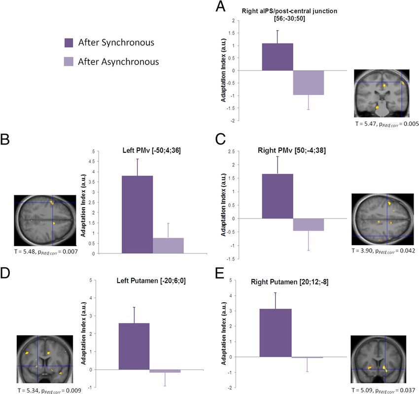
Brozzoli, Gentile et al. • Hand-Centered Space in Frontoparietal Cortices                                             J. Neurosci., October 17, 2012 • 32(42):14573–14582 • 14575

felt as if my real hand were turning rubbery”) and statement 4 (“It ap-
peared visually as if the rubber hand were drifting toward the right”)
served as control questions. The order of the stimulation periods was
counterbalanced across participants. The questionnaire was only given to
13 of 16 participants because of the limitation in booked scanning time.
Immediately after the questionnaire, we obtained objective behavioral
evidence of the illusion by using an intermanual pointing task that
probed changes in the perceived location of the hidden real hand (in a
total of eight participants). Participants were exposed to six 1-min inter-
vals of stimulation, divided into three synchronous and three asynchro-
nous blocks, the order of which was randomized. Immediately before
(“pre”) and after (“post”) each stimulation interval, the participants
closed their eyes and had their left index finger at a fixed starting position
on a panel aligned vertically over the location of the real right hand and
the rubber hand. After a go signal, they performed a swift sliding move-
ment with their left index finger stopping at the perceived location of the
right index finger. A ruler, invisible to the participant, was used to record
the end position of each movement. For each trial, the difference between
post- and pre-measurements was interpreted as follows: a value ⬍0 rep-
resented a drift toward the location of the rubber hand, whereas a value
⬎0 corresponded to an overshoot beyond the location of the real hand.
   Behavioral data analyses. All data acquired in the behavioral assess-
ments were tested for normality using the Kolmogorov–Smirnov test.
The data obtained from the questionnaires did not pass the test; hence,          Figure 1. Setup for experiment 1. A, The participant’s right hand was placed on a tilted table
nonparametric statistics were used. Comparisons were made for each of            in front of them, on either the left or the right of a central fixation point (black cross). A three-
the four statement judgments between the two conditions (synchronous             dimensional object was presented to either the left or the right of the fixation point. The result-
and asynchronous) using Wilcoxon’s signed-rank tests. In contrast, the           ing 2 ⫻ 2 factorial design allowed direct testing for the selective encoding of the object within
data obtained from the pointing localization task did pass the test for          the peri-hand space (yellow halo around the hand). B, In each trial, the object appeared in one
normality. Comparisons were made between the two conditions using                of the two locations for 6 s, with a jittered intertrial interval (7 ⫾ 4 s) with no stimulation.
two-tailed paired-sample t tests. Each post–pre difference was also con-
trasted against 0.
   fMRI setup and design. We used the same experimental setup for fMRI           rubber hand (corresponding to the OLHL stimulation in experiment 1).
acquisition as in the behavioral experiments, with a few minor modifica-         Finally, a 7 s baseline interval separated consecutive trials. The order of
tions as described below. An MR-compatible light-emitting diode was              synchronous and asynchronous trials was randomized. All participants
attached to the edge of the table at a distance of 12 cm from the middle         performed three experimental sessions each containing 24 trials, equally
finger of the real hand and at a distance of 10 cm from the middle finger        divided into synchronous and asynchronous trials.
of the rubber hand. The diode served as the fixation point, and the                 Data analyses. Regressors modeling the instances of visual stimulation
participants were instructed to maintain their gaze on this point. A             close to the rubber hand were defined according to the protocols devel-
trained experimenter used the same paintbrushes to deliver synchronous           oped for experiment 1. Specifically, each 6 s stimulus was divided into
or asynchronous stimulation. All visual stimuli close to the rubber hand         two 3-s events, leading to the definition of four different regressors.
were delivered by a second experimenter using the same object as in              Synch first and Asynch first modeled the first 3 s of each visual stimulus
experiment 1.                                                                    after the synchronous and asynchronous stimulation periods, respec-
   A mixed block- and event-related design was used in the fMRI exper-           tively, and Synch second and Asynch second modeled the last 3 s of object
iment. A trial started with a period of visuotactile stimulation during          presentation in each condition.
which the experimenter applied brushstrokes to corresponding locations              To identify all voxels displaying a stronger adaptation effect for the
on the fingers of the real hand and the rubber hand, either synchronously        visual stimulation after the induction of the illusion than the control, we
or asynchronously at 1 Hz (with iso-directional strokes as in the behav-         defined the following contrast: [(Synch first vs Synch second) vs (Asynch first
ioral experiment described above). To help the experimenter applying             vs Asynch second)]. This contrast is fully balanced in terms of all sensory
the same number of brushstrokes in the different conditions, he listened         properties of the stimuli, including the position of the visual stimulation
to an auditory metronome at 1 Hz over earphones. In the case of syn-             and the rubber hand.
chronous stimulation, the participant was asked to report the onset of the          Regression analyses. We also investigated the relationship between the
illusion (Ehrsson et al., 2004) by pressing a button with the left hand,         fMRI data and the behavioral measurements collected before scanning.
which was placed in a resting position underneath the tilted support. In         Therefore, we ran two independent whole-brain regression analyses. For
the case of asynchronous trials, out-of-synch brushstrokes were delivered        the proprioceptive drift, we computed individual scores by taking the
to the real and the rubber hands for a period of time (AsynchPRE ) iden-         difference of the average drifts between the synchronous and asynchro-
tical in duration to one of the preceding synchronous trials (SynchPRE ).        nous conditions. The individual values were entered as a covariate in a
This ensured that the duration and amount of visuotactile stimulation            regression analysis to identify significant positive correlations between
was perfectly balanced across synchronous and asynchronous trials. To            the proprioceptive drift and the differential adaptation to visual stimuli
match the key response, after an interval identical to one of the preceding      after synchronous or asynchronous conditions. For the questionnaire,
onset times for synchronous trials, a 500 ms flash was emitted from the          we computed the difference in subjective ratings between synchronous
diode during the asynchronous trials. Participants were instructed to            and asynchronous blocks for statement 1 (“referral of touch onto the
press a button with their left hand as soon as they noticed this. The            rubber hand”) and statement 2 (“sense that the rubber hand is one’s own
maximum duration of the induction period (for both SynchPRE and                  hand”) separately. The individual scores were entered as covariates in
AsynchPRE ) was set to 24 s; trials in which the participant did not press       separate regression analyses with the same adaptation effect described
the button within this period were aborted and modeled as conditions of          above. These regression analyses are independent because they assessed
no interest. After the participant’s response, synchronous (or asyn-             the correlation between BOLD adaptation and the two behavioral mea-
chronous) stimulation continued for a period ranging from 13 to 17 s             sures separately. They are unbiased because they tested the correlation
(SynchPOST or AsynchPOST ). One second after the end of this stimulation         between the behavioral measures and the BOLD adaptation at every
period, the ball was presented 2 cm from the tip of the index finger of the      voxel in the whole brain. Thus, this approach allows statistical inferences
14576 • J. Neurosci., October 17, 2012 • 32(42):14573–14582                                                                                  Brozzoli, Gentile et al. • Hand-Centered Space in Frontoparietal Cortices

Figure 2. Selective BOLD adaptation in response to an object presented near the hand for different hand positions. The IPS bilaterally (A, B), left PMd (D), and bilateral PMv (C, E) showed
significantly stronger adaptation to the object presented near rather than far from the hand ( p ⬍ 0.05 corrected; contrast: {[(OR first vs OR second)HR ⫹ (OL first vs OL second)HL] vs [(OR first vs OR second)HL
⫹ (OL first vs OL second)HR]; see Materials and Methods). The bar graphs report the average adaptation index, calculated as the difference in contrast estimates between the first and the second
presentation of the object, for near and far conditions (dark and light color, respectively; error bars represent SEM). The activation maps thresholded at p ⬍ 0.001 uncorrected for display purposes
and superimposed onto the average anatomical high-resolution T1-weighted MRI image of the participants’ brains. The detailed statistics after correction for multiple comparisons are reported for
each key area. aIPS, Anterior IPS; FWE, familywise error.

                                                                                                            Table 1. Experiment 1: hand-centered encoding of space
to be made, avoids any circularity in the statistics, and does not suffer                                                                 MNI coordinates                                             Peak
from the inherent selection bias of region-of-interest-based approaches.
                                                                                                            Anatomical location                         x                y               z            t value          p value
Results                                                                                                     L. anterior part of IPS                     ⫺30              ⫺42             58           5.13              0.003
Experiment 1: hand-centered encoding of space                                                               R. IPS                                       22              ⫺60             52           4.02              0.025
We first probed the cortical mechanisms underlying the encod-                                               L. precentral gyrus (PMd)                   ⫺36              ⫺10             52           4.12              0.027
ing of visually presented objects in coordinates centered on the                                            R. precentral gyrus (PMv)                    42              ⫺4              38           4.48              0.013
upper limb. To this end, BOLD adaptation was assessed when an                                               L. precentral sulcus (PMv)                  ⫺46               12             24           3.46             ⬍0.001*
object was presented visually either near or far from the partici-                                          R. lateral parietal operculum                44              ⫺34             24           4.49             ⬍0.001*
                                                                                                            R. putamen                                   20                2             14           3.31              0.001*
pant’s right hand for two different arm postures. We could thus
                                                                                                            *Uncorrected for multiple comparisons. Interaction contrast: {关(OR first vs OR second)HR ⫹ (OL first vs OL second)HL兴 vs
test the hypothesis that parietal and premotor areas remap peri-                                            关(OR first versus OR second)HL ⫹ (OL first versus OL second)HR兴}.
hand space along with the hand as it is moved to a different
location. Such a finding would constitute compelling evidence
for the dynamic encoding of space in hand-centered coordinates                                              (PMd; Fig. 2 D) exhibited adaptation specific to the encoding of
in the human brain.                                                                                         visual stimuli in hand-centered coordinates. At a lower threshold
    The participants’ right hand was placed fully visible on a table,                                       ( p ⫽ 0.001 uncorrected), a similar modulation was observed in
either to the right or to the left of a central fixation point. We                                          the right putamen, which is worth reporting descriptively be-
measured BOLD-adaptation responses to an object that was pre-                                               cause this structure is thought to contain peripersonal space neu-
sented either on the right or the left side (Fig. 1 A, B), i.e., near or                                    rons in human (Brozzoli et al., 2011b) and non-human
far from the hand depending on the arm’s position. The strength                                             (Graziano et al., 1993) primates. These areas showed greater ad-
of this 2 ⫻ 2 factorial design is that the interaction term corre-                                          aptation when the visual stimulus was presented near the hand
sponds to adaptation responses specifically related to visual stim-                                         compared with far away across the two arm positions (Table 1), a
uli near the hand. This contrast effectively rules out all possible                                         pattern of responses that is also evident from the effect size plots
effects related to the absolute position of the visual stimuli in                                           in Figure 2.
external space, proprioceptive and visual feedback from the arm                                                 Additional post hoc analyses confirmed near-hand adaptation re-
in different postures, and low-level visual processing associated                                           sponses in all key areas during both conditions in which the object was
with the moving object.                                                                                     presented near the hand (all p ⬍ 0.001 uncorrected, for [(OR first vs
    The interaction showed that the bilateral cortices lining the                                           OR second)HR] and [(OL first vs OL second) HL] contrasts). This is also
intraparietal sulcus (IPS; Fig. 2 A, B), the bilateral ventral premo-                                       evident in Figure 3 in which we descriptively plot the effect size of the
tor cortices (PMv; Fig. 2C,E), and the left dorsal premotor cortex                                          BOLD adaptation for the individual conditions. Furthermore, in the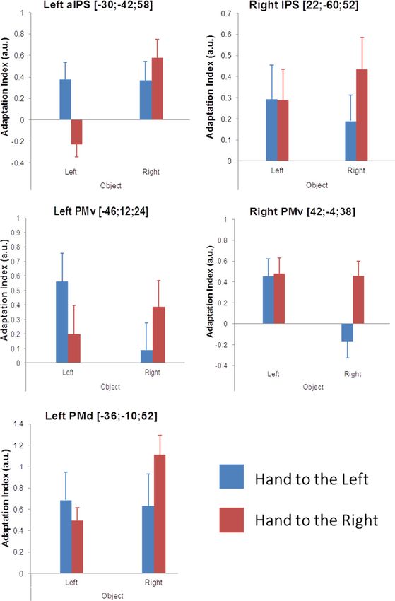
Brozzoli, Gentile et al. • Hand-Centered Space in Frontoparietal Cortices                                                         J. Neurosci., October 17, 2012 • 32(42):14573–14582 • 14577

                                                                                                        distinction between these areas showing absence of hand-
                                                                                                        centered processing and the frontoparietal areas exhibiting hand-
                                                                                                        centered encoding is further illustrated descriptively in Figure 4.
                                                                                                        In this figure, we display the effect sizes of the interaction contrast
                                                                                                        (hand-centered encoding; yellow) and main effect contrast (ab-
                                                                                                        sence of hand-centered encoding; blue–purple) as a single color-
                                                                                                        coded map for all voxels in the whole brain.
                                                                                                            In summary, the results of the first experiment clearly dem-
                                                                                                        onstrate adaptation responses to visual stimuli in the premotor
                                                                                                        and parietal cortices and the putamen that are restricted to the
                                                                                                        space near the hand and that are anchored to the hand as it is
                                                                                                        moved in space. In other words, these areas display responses
                                                                                                        distinctly characteristic of neuronal encoding of objects in hand-
                                                                                                        centered coordinates.

                                                                                                        Experiment 2: remapping of hand-centered space onto a
                                                                                                        rubber hand that feels like one’s own
                                                                                                        We moved on to test the hypothesis that the remapping of hand-
                                                                                                        centered space is centrally mediated and directly related to the
                                                                                                        subjective perception of one’s hand. To this end, we used the
                                                                                                        rubber hand illusion (Botvinick and Cohen, 1998) to experimen-
                                                                                                        tally manipulate the perceived ownership and localization of the
                                                                                                        hand. In this illusion, participants experience ownership over a
                                                                                                        prosthetic hand in direct view after a period of synchronous
                                                                                                        brushstrokes applied to the rubber and real hands, with the latter
                                                                                                        being hidden from view. Brushstrokes applied to the two hands
                                                                                                        asynchronously do not elicit the illusion and serve as a control for
                                                                                                        otherwise equivalent conditions. In the present experiment, we
                                                                                                        induced the illusion and then directly examined the dynamic
                                                                                                        remapping of hand-centered space onto the prosthetic hand im-
                                                                                                        mediately after the induction period (Fig. 5 A, B). Importantly,
                                                                                                        the position of the prosthetic hand in the left hemifield and the
                                                                                                        position of the hidden real right hand matched the two hand
                                                                                                        positions used in experiment 1 (Fig. 5A; see Materials and Meth-
Figure 3. Effect size of BOLD adaptation for individual conditions. The bar graphs report the           ods). Finally, because the illusion involves both a subjective feel-
adaptation index, calculated as the difference in contrast estimates between the first and the
                                                                                                        ing of owning the prosthetic hand and a drift in the perceived
second presentation of the object, for each of the four conditions used to define the interaction
contrast in the factorial design (blue bars refer to the conditions with the hand to the left and red
                                                                                                        location of the hand (Botvinick and Cohen, 1998), we investi-
bars to the conditions with the hand to the right; error bars represent SEM; the corresponding          gated how these two key percepts related to the neural hand-
activation map is shown in Fig. 2). aIPS, Anterior IPS.                                                 centered remapping responses.

                                                                                                        Behavioral and BOLD evidence confirming the illusion
bilateral PMv and right IPS, we observed a similar degree of near-                                      To confirm the successful induction of the rubber hand illusion
hand adaptation in the two arm postures (no significant differences                                     in the scanner environment, we conducted a questionnaire and
for neither [(OR first vs OR second)HR] vs [(OL first vs OL second)HL] nor                              an intermanual pointing experiment directly before the scanning
[(OL first vs OL second)HL] vs [(OR first vs OR second) HR] contrasts), but                             session in a subset of participants (see Materials and Methods).
in the left PMd and the left anterior IPS, the near-hand adaptation                                         The results of the questionnaire showed that the participants
effect was greater when the arm was placed to the right (p ⬍ 0.001                                      experienced a significantly stronger sense of ownership over the
uncorrected for [(OR first vs OR second)HR] vs [(OL first vs OL second)                                 prosthetic hand after the synchronous condition than the asyn-
HL] contrast; Fig. 3). We speculate that the latter could reflect a                                     chronous one (Fig. 6 B, Q2; Wilcoxon’s signed-rank test, n ⫽ 13,
greater representation of space surrounding the right hand in these                                     Z ⫽ 2.80, p ⫽ 0.005). The participants also affirmed the illusory
left-lateralized areas when the right hand is placed in the right hemi-                                 referral of touch to the rubber hand and, again, more strongly
field as opposed to the left one. It is important to point out that one                                 so after the synchronous condition compared with the asyn-
should interpret the plotted adaptation responses for the individual                                    chronous one (Fig. 6 B, Q1; Wilcoxon’s signed-rank test, n ⫽
conditions with great caution because they are not controlled for a                                     13, Z ⫽ 3.18, p ⫽ 0.002). Finally, the synchronous condition was
number of factors (for example, arm posture, visual stimulation in                                      accompanied by a significantly greater drift in the perceived lo-
different hemifields, encoding in head- or body-midline centered                                        cation of the right hand toward the left side (i.e., toward the
coordinates).                                                                                           prosthetic hand) than the asynchronous condition (Fig. 6 A; two-
    The experimental design also allowed us to test for spatial                                         tailed t test, n ⫽ 8, t ⫽ 4.03, p ⫽ 0.005).
encoding of the visual stimuli regardless of the position of the                                            Next, we took advantage of the fact that we could also provide
hand (main effects of object position). As expected, these con-                                         fMRI evidence for the successful induction of the illusion. Direct
trasts produced significant adaptation mainly in early visual ar-                                       comparison between the synchronous visuotactile stimulation
eas, consistent with the repeated activation of retinotopically                                         period and the asynchronous control revealed significant activa-
organized visual representations of the object. The anatomical                                          tion in the bilateral ventral premotor and right intraparietal cor-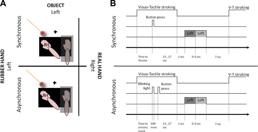
14578 • J. Neurosci., October 17, 2012 • 32(42):14573–14582                                                      Brozzoli, Gentile et al. • Hand-Centered Space in Frontoparietal Cortices

tices and additional activation in the
bilateral inferior parietal cortices and right
anterior insular cortex. Previous fMRI stud-
ies revealed that the rubber hand illusion is
associated with increased BOLD activation
in these areas (Ehrsson et al., 2004). There-
fore, the combination of the behavioral and
imaging data presented above confirms that
the participants were experiencing the rub-
ber hand illusion during the scan sessions.

Adaptation responses indicate
remapping of peri-hand space onto the
rubber hand
We predicted that adaptation responses to Figure 4. Descriptive mapping of contrast estimates for the spatial encoding of visual stimuli. The figure displays areas showing
the visual stimulus near the rubber hand BOLD adaptation to the visual stimulus using a gradient indexing the degree of hand-centered encoding (0 represents absence of
would be significantly stronger after periods hand-centered responses, whereas larger values represent stronger hand-centered encoding). Although early visual areas adapted
of synchronous rather than asynchronous to the object independently of its position relative to the hand, the posterior parietal and premotor cortices showed a high degree
                                                of hand-centered encoding. The anatomical labels correspond to the regions in which the hand-centered responses were statisti-
stimulation. Such findings would be com- cally significant (interaction contrast; shown in Fig. 2). The map was derived as follows. We computed the difference between the
patible with remapping of the peri-hand contrast estimates for the interaction and each of the main effects of visual stimulation on the left or right side (see Materials and
space representation onto the location of Methods). We then selected the minimum value of the two differences and assigned that to the corresponding voxel. The values
the rubber hand only when the latter is were then rescaled into a color map starting from zero and overlaid onto the inflated cortical surfaces of the standard brain. We
perceived as one’s own, in analogy with restricted this analysis to voxels showing a basic response to the presentation of the visual stimuli (by using the inclusive mask from
the dynamic remapping we observed in the contrast “all visual stimuli vs baseline,” thresholded at p ⬍ 0.001 uncorrected). SMG, Supramarginal gyrus.
experiment 1 when the participant’s own
hand was moved to the same location.                                             etal cortex, with the peak centered on the superior segment of the
    As predicted, the presentation of the object near the prosthetic             supramarginal gyrus (Fig. 6C). The cluster extended toward the
hand led to stronger BOLD adaptation in the premotor, posterior                  anterior part of the IPS, and the location of the peak in MNI space
parietal, and putaminal regions after the synchronous compared                   corresponded well with the adaptation response observed in the
with the asynchronous stimulation periods (for all significant                   main analysis of experiment 2 (compare with Fig. 7A). In con-
peaks, see Fig. 7, Table 2). It is noteworthy that this contrast was             trast, we found that the stronger the participants rated the feeling
matched in terms of the visual input from the rubber hand and all                of ownership over the rubber hand, the stronger the rubber hand-
low-level features of the visual stimulus. The peak of activation in             centered adaptation in the left PMv (Fig. 6 D). Again, the location
the right posterior parietal cortex was centered on the most su-                 of this peak corresponded well with the adaptation responses
perior part of the supramarginal gyrus, with the cluster extending               observed in this area in the main analysis of experiment 2 (com-
into the cortices lining the junction of the intraparietal and the               pare with Fig. 7B).
postcentral sulci (Fig. 7A). In the premotor cortices, significant
adaptation responses were found in bilateral PMv (Fig. 7 B, C).                  Discussion
We also observed bilateral adaptation in the putamen (Fig.                       This study has two main findings. First, we showed that neuronal
7 D, E).                                                                         populations in the human intraparietal, premotor, and inferior
    In summary, the pattern of frontoparietal adaptation re-                     parietal cortices and in the putamen construct a dynamic repre-
sponses observed is fully compatible with the encoding of the                    sentation of peri-hand space in coordinates centered on the up-
object in a spatial reference frame remapped onto the rubber                     per limb. Second, we revealed the link between the hand-centered
hand, only when participants experience it as their own hand.                    encoding of space and the perception of the hand with respect to
                                                                                 its location and identity. These findings are relevant because they
Contribution of hand-centered space remapping to sense of                        associate the encoding of peripersonal space with the perceived
position and ownership of the hand                                               ownership and localization of limbs, which has important bear-
Next, we looked for areas displaying a systematic relationship                   ings on models of bodily self-perception (Graziano and Botvin-
between the degree of neural hand-centered spatial remapping                     ick, 2002; Botvinick, 2004; Makin et al., 2008; Tsakiris, 2010;
and the degree of perceptual changes experienced during the il-                  Ehrsson, 2012).
lusion. To this end, we ran two independent whole-brain linear-
regression analyses in experiment 2 in which we looked for the                   Dynamic remapping of the hand-centered representation
following: (1) correlations between the subjectively rated                       of space
strength of ownership (questionnaire data) and the effect size of                A set of premotor–parietal–putaminal regions showed selective
the BOLD-adaptation response indicative of hand-centered re-                     BOLD adaptation to visual stimuli presented near the partici-
mapping of space onto the rubber hand; and (2) correlations                      pant’s hand across different postures. Such adaptation responses
between the proprioceptive drift toward the rubber hand and the                  are more closely related to the receptive field properties of neu-
same BOLD-adaptation effect size (for details, see Materials and                 rons than a traditional analysis (Grill-Spector et al., 2006; Weigelt
Methods).                                                                        et al., 2008; Doeller et al., 2010; Malach, 2012). Crucially, we can
    The results showed that the more individual participants mis-                rule out general effects related to the arm crossing the body mid-
localized the location of their right hand toward the location of                line (Lloyd et al., 2003) and encoding of visual stimuli in refer-
the rubber hand, the stronger the adaptation responses indicative                ence frames not centered on the upper limb, e.g., head centered
of hand-centered remapping of space in the right posterior pari-                 (Fischer et al., 2012), eye centered (Bernier and Grafton, 2010), or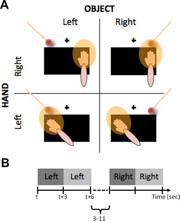
Brozzoli, Gentile et al. • Hand-Centered Space in Frontoparietal Cortices J. Neurosci., October 17, 2012 • 32(42):14573–14582 • 14579
Figure 5. Setup for experiment 2. A, The participant’s right hand was placed on the same tilted support as used in experiment 1. A rubber hand was placed to the left of the fixation point, in a
position equivalent to the left position of the real right hand in experiment 1. A light-emitting diode served as the fixation point (black cross). Two paintbrushes were used to deliver synchronous or
asynchronous visuotactile stimulation (top and bottom part, respectively). The spherical object (identical to that in experiment 1) was presented close to the rubber hand. B, A mixed block- and
event-related design was used (see Materials and Methods) V-T, Visuotactile.
allocentric. Furthermore, the object was
always presented within reaching space.
Consequently, our findings are specifi-
cally related to peri-hand space rather
than to a general representation of reach-
ing space, providing compelling evidence
for encoding in hand- or arm-centered
coordinates.
In experiment 1, the peri-hand space re-
mapping across the two arm positions was
mediated by the integration of afferent vi-
sual and proprioceptive signals. The ventral
premotor and intraparietal cortices receive
such afferent sensory information via pro-
jections from somatosensory and visual ar-
eas (Rizzolatti et al., 1998; Graziano and
Botvinick, 2002), forming with the putamen
a visual–somesthetic network that processes
the space on and near the body surface (Gra-
ziano et al., 1997). In contrast, during the
rubber hand illusion, the peri-hand space
remapping onto the rubber hand occurred
as a result of a central process. Here, an ini-
tial conflict between the seen and felt posi-
tions of the hand is resolved by the
recalibration of the peri-hand space repre-
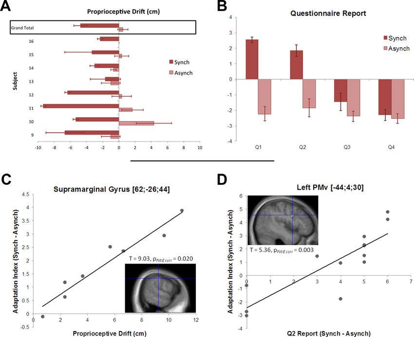
Figure 6. Correlations between behavioral percepts of the illusion and neural hand-centered remapping. A, Individual propri- sentation so that tactile, visual, and propri-
oceptive drift and group average. The bars represent the difference between post- and pre-measurements (negative values more oceptive signals fuse perceptually. Crucially,
to the left, positive values more to the right), in the synchronous and the asynchronous condition (average of 3 trials; error bars specific peri-hand space BOLD adaptation
represent SEM). The drift was significantly larger in the synchronous than in the asynchronous condition (drift in the synchronous was detected in the premotor and posterior
condition: ⫺4.75 ⫾ 2.6 cm, 2-tailed t test against 0, t ⫽ 4.53, p ⫽ 0.001; drift in the asynchronous condition: 0.5 ⫾ 1.78, 2-tailed parietal cortices when both the real hand
t test against 0, t ⫽ 0.12, p ⫽ 0.45). B, Participants were asked to rate four statements on a scale from ⫺3 (“completely disagree”) was physically moved and the arm was per-
to ⫹3 (“agree completely”) after 1 min of synchronous or asynchronous stimulation. They reported stronger illusory referral of ceived to have moved by means of the illu-
touch to the rubber hand after the synchronous condition compared with the asynchronous one (Q1, Wilcoxon’s signed-rank test, sion. This suggests that peri-hand space
n ⫽ 13, Z ⫽ 3.18, p ⫽ 0.002). They also experienced a significantly stronger sense of ownership over the prosthetic hand after the remapping arises from the dynamic integra-
synchronous than the asynchronous condition (Q2, Wilcoxon’s signed-rank test, n ⫽ 13, Z ⫽ 2.80, p ⫽ 0.005). C, A whole-brain
tion of visual and proprioceptive signals at
second-level regression model revealed a significant linear relationship ( p ⬍ 0.05 corrected) between the proprioceptive drift
toward the rubber hand and the effect size of the BOLD-adaptation response indexing hand-centered remapping to the rubber the level of multimodal frontoparietal areas.
hand across individuals (contrast described in Fig. 7 and Materials and Methods). D, Significant linear regression ( p ⬍ 0.05 Our results also shed light on the neu-
corrected) between the subjectively rated strength of ownership (questionnaire data) and the BOLD-adaptation response indexing ral mechanism underlying behavioral and
hand-centered remapping to the rubber hand across individuals. FWE, Familywise error. neurophysiological findings reporting se-14580 • J. Neurosci., October 17, 2012 • 32(42):14573–14582 Brozzoli, Gentile et al. • Hand-Centered Space in Frontoparietal Cortices
lective processing of peri-hand visual
stimuli (di Pellegrino et al., 1997; Farnè et
al., 2000; Pavani and Castiello, 2004;
Spence et al., 2004; Makin et al., 2009). An
attentional account has been proposed as
a possible interpretation for part of these
behavioral findings (Spence et al., 2000,
2004; Kennett et al., 2001), in line with the
notion that peripersonal space and cross-
modal spatial attention might share com-
mon mechanisms (Maravita et al., 2003;
Driver and Noesselt, 2008). The hand-
centered spatial remapping allows the en-
coding of visual stimuli within the same
reference frame as somatosensory infor-
mation, leading to more robust multi-
sensory interactions that can facilitate
behavior.
Peri-hand space and sense of position
Our findings clarify that information about
arm position is present in the bilateral ven-
tral premotor, the right anterior intrapari-
etal, and the bilateral inferior parietal
cortices (Young et al., 2004; Naito et al.,
2005), providing more direct evidence for
the link between the arm position sense and
the representation of peri-hand space than
previous studies. In previous research, vi-
sual stimuli have been presented near the Figure 7. Differential BOLD adaptation to an object presented near the prosthetic hand after the synchronous and asynchro-
hand placed in a single position (Makin et nous induction periods. The right IPS (A), PMv bilaterally (B, C), and the putamen bilaterally (D, E) exhibited significantly stronger
al., 2007; Brozzoli et al., 2011b). Other stud- BOLD adaptation to the repeated presentation of the ball near the prosthetic hand after the synchronous compared with the
ies have manipulated the posture of the arm asynchronous condition. The bar graphs plot the effect size of the adaptation, for the synchronous and asynchronous conditions,
but without assessing the selective encoding separately (dark and light color, respectively; error bars represent SEM). aIPS, Anterior IPS; FWE, familywise error.
of peri-hand space (Lloyd et al., 2003). Here,
we revealed remapping of peri-hand space by testing for selective Table 2. Experiment 2: remapping of hand-centered space onto the owned rubber
encoding in combinations with a postural manipulation. hand
The strongest support for a link to the perceived hand position MNI coordinates Peak
was found in the posterior parietal cortex, in which peri-hand Anatomical location x y z t value p value
space remapping onto the rubber hand correlated significantly
R. anterior IPS/postcentral junction 56 ⫺30 50 5.47 0.005
with the proprioceptive drift toward the rubber hand. This is in L. precentral gyrus (PMv) ⫺50 4 36 5.48 0.007
keeping with the known neurophysiological functions of the pos- R. precentral gyrus (PMv) 50 ⫺4 38 3.90 0.042
terior parietal cortex and its role in supporting the body-schema L. putamen ⫺20 6 0 5.34 0.009
representation (Head and Holmes, 1911; Kammers et al., 2009) R. putamen 20 12 ⫺8 5.09 0.037
and the planning of manual actions (Culham and Valyear, 2006; R. lateral parietal operculum 48 ⫺10 24 5.40 ⬍0.001*
Gallivan et al., 2011). Neurons in area 5 of the macaque brain L. IPS ⫺28 ⫺74 50 3.90 ⬍0.001*
L. supramarginal gyrus ⫺60 ⫺28 34 3.73 0.001*
encode the hand position by integrating visual and propriocep-
*Uncorrected for multiple comparisons. 关(Synch first vs Synch second) vs (Asynch first vs Asynch second)兴.
tive signals (Graziano et al., 2000; Graziano and Botvinick, 2002).
Similarly, the human intraparietal cortex integrates visual and
proprioceptive information about the upper limb (Lloyd et al., grated with visual, auditory, and vestibular signals in a common
2003; Ehrsson et al., 2004). Here, we provide evidence for the role reference frame. The result is a representation of the arm position
of this area in constructing a “proprioceptive skeleton” for the encoded in the same coordinates used for nearby objects, facili-
representation of peripersonal space, onto which selective visual tating object-directed actions (Jeannerod et al., 1995; Tunik et al.,
responses can be grounded (Cardinali et al., 2009). 2005; Makin et al., 2012). This is consistent with an involvement
These findings concur with a hierarchical view on propriocep- of parietal and premotor regions in action planning and execu-
tion whereby afferent signals from muscles, skin, and joints first tion (Fogassi and Luppino, 2005; Culham and Valyear, 2006;
reach their cortical targets in primary somatosensory (Iwamura Bernier and Grafton, 2010).
et al., 1983; Pons et al., 1992; Naito et al., 2005) and motor
(Lemon and Porter, 1976; Naito et al., 2002) cortices . The infor- Peri-hand space and limb ownership
mation is then transferred via direct connections to areas in the Our data suggest that, during the rubber hand illusion, the central
intraparietal (Cavada and Goldman-Rakic, 1989; Lewis and Van representation of peri-hand space is remapped onto the owned
Essen, 2000) and premotor (Luppino et al., 1999; Rizzolatti and model hand. Interestingly, we found the strongest association
Luppino, 2001) cortices in which proprioceptive signals are inte- between the feeling of limb ownership and the coding of hand-Brozzoli, Gentile et al. • Hand-Centered Space in Frontoparietal Cortices J. Neurosci., October 17, 2012 • 32(42):14573–14582 • 14581
centered space in the PMv. In this area, the degree of hand- Botvinick M (2004) Neuroscience. Probing the neural basis of body owner-
centered spatial encoding correlated with the subjective sense of ship. Science 305:782–783.
Botvinick M, Cohen J (1998) Rubber hands “feel” touch that eyes see. Na-
hand ownership. This is consistent with previous studies that
ture 391:756.
related ventral premotor activity to the subjective level of owner- Brozzoli C, Makin T, Cardinali L, Holmes N, Farnè A (2011a) Peripersonal
ship (Ehrsson et al., 2004, 2005; Petkova et al., 2011). This sheds space: A multisensory interface for body-objects enteractions. In: Fron-
light on the nature of the multisensory mechanisms mediating tiers in the neural basis of multisensory processes (Murray MM, Wallace
body ownership. In fact, despite the current consensus that we MT, eds). London: Taylor and Francis.
come to experience limbs (Botvinick and Cohen, 1998; Ehrsson Brozzoli C, Gentile G, Petkova VI, Ehrsson HH (2011b) fMRI-adaptation
et al., 2004; Ehrsson, 2007) and whole bodies as ours (Lenggen- reveals a cortical mechanism for the coding of space near the hand. J Neu-
rosci 31:9023–9031.
hager et al., 2007; Ionta et al., 2011; Petkova et al., 2011; Schmalzl
Cardinali L, Brozzoli C, Farnè A (2009) Peripersonal space and body sche-
and Ehrsson, 2011) as a result of interactions between vision, ma: two labels for the same concept? Brain Topogr 21:252–260.
touch, and proprioception, the precise mechanisms have re- Cavada C, Goldman-Rakic PS (1989) Posterior parietal cortex in rhesus
mained unclear. Based on changes in visual sensitivity of peri- monkey: I. Parcellation of areas based on distinctive limbic and sensory
hand neurons when objects were presented close to a visible fake corticocortical connections. J Comp Neurol 287:393– 421.
arm, Graziano speculated that remapping of peri-hand space Costantini M, Haggard P (2007) The rubber hand illusion: sensitivity and
could support the embodiment of prosthetic limbs (Graziano, reference frame for body ownership. Conscious Cogn 16:229 –240.
Culham JC, Valyear KF (2006) Human parietal cortex in action. Curr Opin
1999; Graziano et al., 2000), although he could not test this.
Neurobiol 16:205–212.
Ehrsson et al. (2004) theorized that the premotor activity associ- di Pellegrino G, Làdavas E, Farné A (1997) Seeing where your hands are.
ated with the feeling of limb ownership in humans might reflect Nature 388:730.
multisensory integration in hand-centered coordinates. The Doeller CF, Barry C, Burgess N (2010) Evidence for grid cells in a human
present results speak to these hypotheses because they inform us memory network. Nature 463:657– 661.
about the body-part-centered reference frame used in the neural Driver J, Noesselt T (2008 Jan 10) Multisensory interplay reveals cross-
computations supporting self-attribution of limbs. This is an im- modal influences on “sensory-specific” brain regions, neural responses,
and judgments. Neuron 57:11–23.
portant conclusion because it constrains models of body owner-
Duhamel JR, Colby CL, Goldberg ME (1998) Ventral intraparietal area of
ship (Makin et al., 2008; Tsakiris, 2010; Ehrsson, 2012), explains the macaque: congruent visual and somatic response properties. J Neu-
the spatial limits described in behavioral studies (Tsakiris and rophysiol 79:126 –136.
Haggard, 2005; Costantini and Haggard, 2007; Lloyd, 2007; Tsa- Ehrsson HH (2007) The experimental induction of out-of-body experi-
kiris et al., 2007; Folegatti et al., 2012), and provides a framework ences. Science 317:1048.
within which to study the peripersonal space as a crucial bound- Ehrsson HH (2012) The concept of body ownership and its relation to mul-
ary zone between self and non-self. tisensory integration. In: The new handbook of multisensory processes
(Stein BE, ed). Cambridge, MA: Massachusetts Institute of Technology, in
press.
Concluding remarks Ehrsson HH, Spence C, Passingham RE (2004) That’s my hand! Activity in
Previous studies investigated the representation of the periper- premotor cortex reflects feeling of ownership of a limb. Science 305:875–
sonal space by examining behavioral responses in patients with 877.
brain lesions (Farnè et al., 2000, 2005), probing reaction times Ehrsson HH, Kito T, Sadato N, Passingham RE, Naito E (2005) Neural sub-
during presentation of cross-modal stimuli near the body strate of body size: illusory feeling of shrinking of the waist. PLoS Biol
(Spence et al., 2004), and by registering changes in the excitability 3:e412.
Farnè A, Pavani F, Meneghello F, Làdavas E (2000) Left tactile extinction
of the primary motor cortex to the presentation of objects near
following visual stimulation of a rubber hand. Brain 123:2350 –2360.
the hands (Makin et al., 2009). Here, we have taken a more direct Farnè A, Demattè ML, Làdavas E (2005) Neuropsychological evidence of
approach and measured the BOLD-adaptation signatures of modular organization of the near peripersonal space. Neurology 65:
hand-centered encoding of space in the human brain. Unlike 1754 –1758.
previous fMRI studies (Lloyd et al., 2003; Sereno and Huang, Fischer E, Bülthoff HH, Logothetis NK, Bartels A (2012) Human areas V3A
2006; Makin et al., 2007; Macaluso and Maravita, 2010; Brozzoli and V6 compensate for self-induced planar visual motion. Neuron 73:
et al., 2011b), we directly tested for hand-centered encoding by 1228 –1240.
Fogassi L, Luppino G (2005) Motor functions of the parietal lobe. Curr
manipulating the position of the hand in view. Furthermore, we
Opin Neurobiol 15:626 – 631.
provided evidence that such encoding is directly related to Fogassi L, Gallese V, Fadiga L, Luppino G, Matelli M, Rizzolatti G (1996)
changes in body perception. Thus, the present data bridge a gap Coding of peripersonal space in inferior premotor cortex (area F4). J Neu-
between neurophysiological studies on non-human primates and rophysiol 76:141–157.
behavioral and neuropsychological observations in humans and Folegatti A, Farnè A, Salemme R, de Vignemont F (2012) The rubber hand
extend our knowledge of the brain mechanisms involved in the illusion: two’s a company, but three’s a crowd. Conscious Cogn 21:799 –
representation of the peripersonal space. 812.
Gallivan JP, McLean DA, Smith FW, Culham JC (2011) Decoding effector-
dependent and effector-independent movement intentions from human
Notes parieto-frontal brain activity. J Neurosci 31:17149 –17168.
Supplemental material for this article is available at http://130.237.111. Gentile G, Petkova VI, Ehrsson HH (2011) Integration of visual and tactile
254/ehrsson/pdfs/Brozzoli&Gentile_et_al._SI.pdf. This material has not signals from the hand in the human brain: an fMRI study. J Neurophysiol
been peer reviewed. 105:910 –922.
Graziano MS (1999) Where is my arm? The relative role of vision and pro-
References prioception in the neuronal representation of limb position. Proc Natl
Andersen RA (1997) Multimodal integration for the representation of space Acad Sci U S A 96:10418 –10421.
in the posterior parietal cortex. Philos Trans R Soc Lond B Biol Sci 352: Graziano MS, Gross CG (1993) A bimodal map of space: somatosensory
1421–1428. receptive fields in the macaque putamen with corresponding visual recep-
Bernier PM, Grafton ST (2010) Human posterior parietal cortex flexibly tive fields. Exp Brain Res 97:96 –109.
determines reference frames for reaching based on sensory context. Neu- Graziano MS, Yap GS, Gross CG (1994) Coding of visual space by premotor
ron 68:776 –788. neurons. Science 266:1054 –1057.You can also read