Computed Tomography Study of the Mummy of King Seqenenre Taa II: New Insights Into His Violent Death - Frontiers
←
→
Page content transcription
If your browser does not render page correctly, please read the page content below

ORIGINAL RESEARCH
                                                                                                                                              published: 17 February 2021
                                                                                                                                          doi: 10.3389/fmed.2021.637527

                                              Computed Tomography Study of the
                                              Mummy of King Seqenenre Taa II:
                                              New Insights Into His Violent Death
                                              Sahar N. Saleem 1* and Zahi Hawass 2
                                              1
                                               The Department of Radiology, Kasr Al Ainy Faculty of Medicine, Cairo University, Cairo, Egypt, 2 Former Minister of
                                              Antiquities of Egypt, Cairo, Egypt

                                              Seqenenre-Taa-II, The Brave, (c.1558–1553 BC) ruled Southern Egypt during the
                                              occupation of Egypt by the Hyksos. The mummy was physically examined and X-rayed
                                              in the 1960s, which showed severe head wounds that have prompted various theories
                                              about the circumstances of his death. We postulated that Computed Tomography (CT)
                                              study of Seqenenre-Taa-II’s mummy would give insights into the circumstances of his
                                              death. We examined Seqenenre’s mummy using CT and compared the findings with
                         Edited by:
                 Stephanie Panzer,
                                              the archaeological literature as well as with five Asian weapons found in Tell-el-Dabaa.
Berufsgenossenschaftliche Unfallklinik        CT findings indicate that Seqenenre died in his forties. The mummies deformed hands
                 Murnau, Germany              suggest that the King was likely imprisoned with his hands tied. CT images provided
                         Reviewed by:         detailed analysis of Seqenenre’s previously reported injuries to the forehead, right
                     Nunziata Barbera,
             University of Catania, Italy     supra-orbital, nose-right orbit, left chick, and skull base. This study revealed additional
                       Luca Mastracci,        craniofacial fractures in the right lateral side of the skull that had been concealed by
              University of Genoa, Italy
                                              the embalmers beneath layers of material. Analysis of the morphology of the injuries
                  *Correspondence:
                     Sahar N. Saleem
                                              enabled a better understanding of the mechanism of trauma, possible number of the
            saharsaleem1@gmail.com            attackers, and their relative position to the King. The size and shape of the fractures
                                              correlated well with the studied Hyksos weapons. The lethal attack was aimed at the
                    Specialty section:
                                              King’s face, likely in an attempt to disgrace him. Mummification of Seqenenre’s body
          This article was submitted to
                              Pathology,      was limited to evisceration without brain removal. The desiccated brain is shifted to the
                a section of the journal      left side of the skull. This may indicate that the King’s dead body stayed on its left side for
                   Frontiers in Medicine
                                              some time—long enough for decomposition start before the mummification began. This
       Received: 03 December 2020
        Accepted: 19 January 2021
                                              suggests that the King likely died at a location distant from the funeral place, possibly on
       Published: 17 February 2021            a battlefield. The embalmers attempted to conceal the King’s injuries; the methods used
                             Citation:        suggest that the mummification took place in a royal mummification workshop rather than
    Saleem SN and Hawass Z (2021)
                                              in a poorly equipped location. CT findings of Seqenenre’s mummy helped us to better
 Computed Tomography Study of the
   Mummy of King Seqenenre Taa II:            understand the circumstances of his violent death. His death motivated his successors
  New Insights Into His Violent Death.        to continue the fight to unify Egypt and start The New Kingdom.
              Front. Med. 8:637527.
    doi: 10.3389/fmed.2021.637527             Keywords: mummy, Egypt, mummification, Hyksos, computed tomography (CT), royal mummies, Seqenenre Taa II

Frontiers in Medicine | www.frontiersin.org                                          1                                           February 2021 | Volume 8 | Article 637527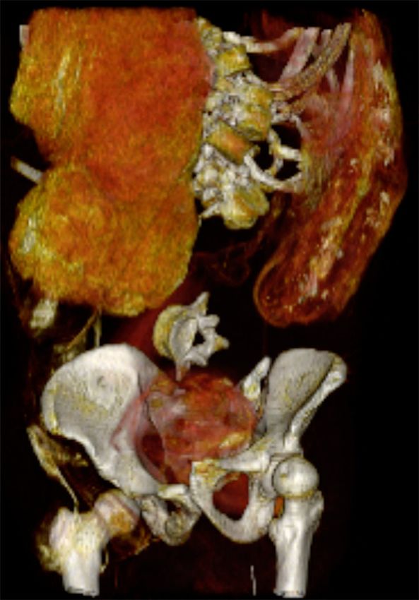
Saleem and Hawass                                                                                                                   CT Study of Seqenenre’s Mummy

INTRODUCTION
Seqenenre-Taa-II was the Egyptian king who ruled Southern
Egypt at the end of the 17th Dynasty (approximately1558–1553
BC), during the Hyksos occupation of Egypt. In the ancient
Egyptian language, “Heqau-Khasut” meant “the rulers of the
foreign lands”; the word was transmitted by Greek sources as
“Hyksos.” The Hyskos were likely a group of Asian shepherds
who occupied the Northern part Egypt and took Avaris (modern
Tell el Dabaa) as the capital for a period of time called the “second
intermediate period” (c. 1650–1550 BC). Although the Egyptian
                                                                                   FIGURE 1 | Picture of mummy Seqenenre-Taa-II. (A) Picture of head and
rulers maintained power over the South (capital Thebes), all of
                                                                                   upper torso of the mummy Seqenenre-Taa-II shows severe multiple
Egypt had to pay tributes to the Hyksos King (1, 2).                               craniofacial injuries. (B) Picture of right lateral view of the head and upper torso
   An ancient Papyrus, The Sallier I; Papyrus I, stated that                       of mummified Seqenenre shows the deformed upper limbs with flexed hands
there was hostility between Seqenenre and the king of Hyksos,                      at the wrists and spastic fingers.
Apophis. According to the Papyrus, the Hyksos king Apophis
sent a hostile message to Seqenenre, stating that noisy
hippopotamuses in a pool in Thebes were disturbing his sleep in
Avaris (644 Km away), and demanding that the Theban sacred                        described the mummy’s head injuries in detail, and noted the
pool be destroyed. The end of the Papyrus is lost, but the                        absence of wounds on the arms or on the rest of the body (9). In
preserved text ends with the statement that Seqenenre called                      the 1960s, the X-ray study of Seqenenre’s mummy confirmed five
his counselors, which probably indicates an introduction to a                     separate traumatic injuries confined to the head, and the absence
battle (2).                                                                       of any fractures to the rest of the skeleton (10).
   Deir el-Ballas, a settlement just north of Thebes, was likely                     Several scenarios were proposed to explain how Seqenenre’s
the base for military campaigns against the Hyksos. Text from an                  head injuries, though questions remained regarding the
ostraca which dates to the 17th dynasty, found at Deir el-Ballas,                 circumstances of Seqenenre’s death. Had the King died in battle?
indicates that a large number of men, large quantities of goods,                  Was he a victim of a palace conspiracy? (6, 11). Was Seqenenre’s
and ships with their crews were brought to the site. A lintel of                  body hastily mummified in a poorly equipped location away
Seqenenre, also found at Deir el-Ballas, confirmed that the site                  from the royal mummification workshop? (6).
was founded during his reign. A stela known as The Carnavaron                        Computed Tomography (CT) is a non-invasive modality that
Tablet, found in Thebes Karnak Temple, recorded the battles that                  has been used to examine the mummies of several ancient
Kamose, Seqenenre’s son, fought against the Hyksos in the North                   Egyptian royals and has allowed for greater insight into the
(3–5). Kamose fell dead during the war against the Hyksos and                     mysterious circumstances under which they died (6).
it was Ahmose, the second son of Seqenenre, who completed                            We postulated that a CT study of mummy Seqenenre, in
the expulsion of the Hyksos, chased them to Sharuhen (modern                      correlation with the historical data, could give more insights
Ghaza in Palastine), and unified Egypt (6). However, there has                    on his death circumstances and could shed new light on this
been no record of the fate of Seqenenre.                                          important chapter in Egypt’s history.
   In 1881 at the Deir el-Bahari cache in Thebes, a mummy was
discovered in its original linen wrappings and was transferred to                 MATERIALS AND METHODS
Cairo Museum. On June 9th, 1886, the mummy was unwrapped
by Gaston Maspero (the general director of antiquities in                         The mummy of Seqenenre is located at Cairo Egyptian Museum
Egypt) accompanied by Daniel Fouquet (a physician). Maspero                       with the catalog code [JE 26209(b) CG 61051 SR1/10192]
identified the mummy as Seqenenre-Taa by the inscriptions on                      (Figure 1).
the original wrappings. Taa (or Tao) was the birth name and                          On the 4th of May 2019 we transferred the mummy of
meant “Thoth is great”; while “Seqenenra” was the throne name                     Seqenenre to the multi-detector Computed Tomography (CT)
and meant “The one whom Ra has made brave” (7). Maspero and                       scanning machine (Somatom Emotion 6; Siemens Medical
Fouquet reported a foul odor of the putrefied mummy, limited                      Solutions, Malvern, Pennsylvania) installed on a truck in the
body mummification, as well as severe head injuries suggestive                    garden of the Cairo Egyptian Museum.
of a violent death (7, 8). On September 1st, 1906, Grafton Elliot                    We used the following CT parameters: kVp = 130 effective
Smith, the professor of anatomy at Kasr Al Ainy School of                         mAs ranged from 23 to 63; pitch ranged from 0.83 to 1.8;
Medicine in Cairo, examined the mummy of Seqenenre. Smith                         FOV from 350 to 500; slice thickness from 0.6 to 1.25 mm;
                                                                                  and reconstruction from 0.4 to 0.8 mm. Axial images were
                                                                                  created. We used a separate workstation (Leonardo Workstation,
Abbreviations: AD, Anno Domini; BC, Before Christ; cm, centimeter; CT,            Siemens Medical Solution) to reconstruct the CT images in
Computed Tomography; DNA, deoxyribonucleic acid; FOV, Field-Of-View; HU,
Hounsfield Unit; KV, Kilovolt; mAS, milliampere-seconds; MIP, Maximum
                                                                                  multiple two-dimensional planes, as well as a three-dimensional
Intensity Projection; mm, Millimeter; MPR, Multiplanar Reconstruction; SSD,       reconstruction. We analyzed the CT images of the mummy to
Surface shaded display; VRT, volume rendering technique.                          assess the preservation status, age at death, and pathologies. We

Frontiers in Medicine | www.frontiersin.org                                   2                                            February 2021 | Volume 8 | Article 637527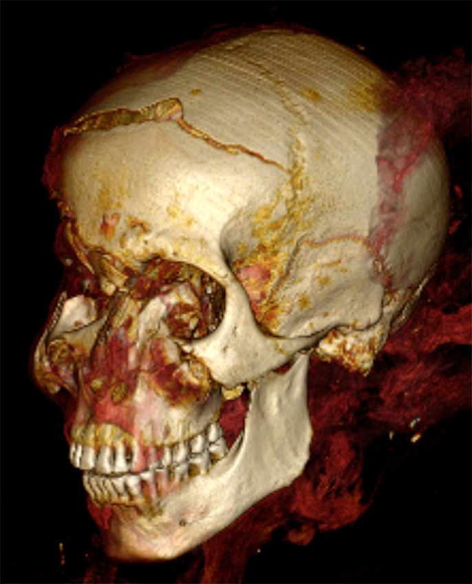
Saleem and Hawass                                                                                                                        CT Study of Seqenenre’s Mummy

TABLE 1 | Weapons found in Tell E-Dabaa (Ancient Avaris) belonged to Hyksos (Middle Bronze Culture II-MB II) at the time of Seqenenre’s reign.

Registered code                 Type                     Description                                                                 Measurements

J-91173                         Dagger                   Bronze dagger corroded and fractured. Dagger blade has                      Full Length of object: 230 mm;
                                                         five lines and diamond-shaped in cross section. Three nails                 Breadth: 38 mm; Length of blade
                                                         on the handle-thorn are broken                                              18.3 mm
J-91174                         Battle ax                Narrow bladed shaft-hole bronze battle-ax in corroded                       Length of the object: 142 mm,
                                                         condition. The handle is in line with the blade. The hollow                 Breadth of the cutting edge: 27.5 mm
                                                         handle is rounded at the upper end and oval at the lower
                                                         side. The middle of the ax is having hexagonal cross section.
                                                         The cutting edge is straight.
J-91175                         Spear head               A triangular blade with well-defined mid rib that extends the               Full Length of the object: 104 mm,
                                                         length of the blade to a thick shank. The blade is diamond in               Length of the blade 60 mm,
                                                         shape in cross section.                                                     maximum Breadth 15 mm
J-91683                         Dagger                   Long dagger. Lenticular in cross section                                    Length 215 mm, breadth 35 mm
J-91691                         Dagger                   Long dagger. Lenticular in cross section                                    Length 282 mm, breadth 30 mm

 FIGURE 2 | Photographs of bronze Asian weapons MB II (Middle Bronze II era) found in Tell el Dabaa (ancient Avaris). N.B. The accompanying codes are those given
 by Cairo Egyptian Museum where the weapons are currently housed. Each square of the measuring ruler equals to 10 mm in length. (A) Dagger (J-91173): A corroded
 fractured dagger with five-lined blade. Three nails are seen on the broken handle-thorn. (B,C) Battle-ax (J-91174) front and side views of a battle-ax. The cutting edge
 of the blade is straight. The hollow handle is in line with the cutting blade; this gives the weapon more stability and force. (D) Spearhead (J-91175) A triangular blade
 with a well-defined mid rib that extends along the full length of the blade to a thick shank. The blade has a rhomboid cross section. A large blade, shank, and tang
 form (tripartite). Tang to get into the shaft, and a shank to be used as stop and partial support for binding. (E) Bronze dagger (Code: J-91683) from MB II (Middle
 Bronze II era) found in Tell el Dabaa-Egypt (ancient Avaris). (F) Bronze dagger (Code: J-91691) from MB II (Middle Bronze II era) found inTell el Dabaa (ancient Avaris).

also examined five Asian bronze weapons housed in the Cairo                             daggers, a battle-ax, and a spearhead (Figure 2). Previous studies
Egyptian Museum, originally found in Tell el Dabaa region by the                        indicated that these weapons belong to the Asian Middle Bronze
Austrian Archaeological Mission. The weapons originated during                          Age Culture II, which coincided with Seqenenre’s reign (12).
the Hyksos period (Middle Bronze II), which was concurrent
with Seqenenre’s reign (12). Each weapon was photographed
and measured. We retrieved the available data on each weapon                            CT Study of the Mummy
from the Cairo Egyptian Museum’s files as well as from previous                         Preservation Status
research studies (12). We correlated CT findings of the mummy                           The mummy of Seqenenre is in poor condition; the head
of Seqenenre with the weapons as well as with the available                             and many bones are loose and misplaced. The mummy is
archaeological data.                                                                    disarticulated in most body regions. The head is separate from
                                                                                        the body. Most of the vertebrae and ribs are loose. The sternum,
                                                                                        clavicles and scapulae are disarticulated and located inside
RESULTS                                                                                 the torso cavity. The upper limbs are disarticulated from the
                                                                                        shoulders with minimal soft tissues or muscles remaining on the
Table 1 gives the results of our examination of five Asian bronze                       bones. Both femora are disarticulated from the hip sockets. The
weapons housed in the Cairo Egyptian Museum that were                                   bones of the left forefoot are disarticulated. The symphysis pubis
originally found in Tell el Dabaa region. We inspected three                            and the right sacro-iliac joints are disrupted.

Frontiers in Medicine | www.frontiersin.org                                         3                                           February 2021 | Volume 8 | Article 637527
Saleem and Hawass                                                                                                               CT Study of Seqenenre’s Mummy

 FIGURE 3 | Coronal CT image of the head and neck of mummy
 “Seqenenre-Taa-II.” The desiccated brain is seen shifted and occupying the
 left side of the cranial cavity.

Age at Death                                                                       FIGURE 4 | Coronal 3-dimensional CT image of lower torso of
We estimated the age at death to be around 40 years based                          “Seqenenre-Taa-II” shows evisceration and embalming material within the
on epiphyseal closure of all long bones, fusion of the meta-                       abdomino-pelvic cavity. Note the disarticulated vertebrae, and disrupted
and meso-sterna, and symphysis pubis surface features (stage 6                     symphysial articulation.
corresponding to 35–39 years) (13). There is a complete set of
teeth inside the mouth, including all of the third molars. Mild
to moderate attrition of the teeth is observed: grade G in the
                                                                                  hyper-extended at the metacarpo-phalangeal joints and flexed
mandibular teeth (corresponding to 35–40 years), and grade H
                                                                                  at the inter-phalangeal joints. No evidence of bony fractures in
in the maxillary teeth (corresponding to 40–50 years) (14, 15).
                                                                                  upper limbs was seen (Figure 5). Both lower limbs are extended;
Stature                                                                           the left foot is dislocated but there is no evidence of bony
We could not measure the length of the skeleton in its articulated                fractures. The head and face are severely injured. The upper and
anatomical position because of its partly disarticulated status.                  lower lips are retracted, showing the teeth.
However, we measured the maximum length of the tibia
(384 mm) to calculate stature using the regression equation                       CT of Head Injuries, Causative Weapons,
derived by Raxter et al. for ancient male Egyptians: (Stature =                   and Position of the Assailants
2.554 × 38.4 +69.12 = 167.2 cm ± 3.002). We thus estimated the                    Figures 6–8 and Supplementary Figures 1, 2 show the head
stature of Seqenenre to be about 167 cm ± 3 cm (16).                              injuries.

Mummification                                                                     A Cut Fracture in the Upper Forehead Region
The desiccated shrunken brain occupies the left side of the                       Description
cranial cavity. There is no intracranial resin or other evidence of               The upper region of the frontal bone shows a transverse cut
other embalming materials. The cribriform plate is intact with                    fracture that starts 5 mm to the left of the midline and extends
no CT evidence of attempt of brain removal (Figure 3). There                      transversely to the right for 70 mm. The gapping fracture is of
is no evidence of heart or viscera inside the body cavity. The                    irregular width that varies between 4 and 14 mm, being wider on
abdominal and pelvic cavities are stuffed with packs of variable                  the left side. A secondary fissure fracture extends horizontally for
CT densities consistent with linen (−100 HU) and resin (70–120                    43 mm from the left end of the original fracture line.
HU) (Figure 4). A left flank opening measuring 157 mm in length
was likely used to remove the viscera and insert the packs. No                    Suggested Weapon
amulets or jewelry could be detected within the mummy or on                       A heavy sharp object like a sword or an ax. The blow was most
its surface.                                                                      likely fatal.

Pathological Changes                                                              Position of the Assailant
Both arms are flexed at the elbows, the hands are flexed                          The site of the fracture at the top of Seqenenre’s head indicates
at the wrists (more on the left side), while the fingers are                      that the assailant was positioned above the King. This relative

Frontiers in Medicine | www.frontiersin.org                                   4                                         February 2021 | Volume 8 | Article 637527
Saleem and Hawass                                                                                                                        CT Study of Seqenenre’s Mummy

 FIGURE 5 | Three-dimensional frontal CT image of upper limbs of mummy of                   FIGURE 7 | Three-Dimensional CT image of Seqenenre’s head in left oblique
 Seqenenre-Taa-II. The arms are dislocated at the shoulder and bent at the                  projection shows an oblique cut wound of the left zygoma, fracture of left
 elbows. Note the deformity of the bent hands bent indicating they were tied at             coronoid process of the mandible.
 the wrists.

 FIGURE 6 | Three-Dimensional CT image of head of Seqenenre in frontal
 projection showing multiple craniofacial injuries: a gaping fracture of the frontal
 bone; fracture above the right eyebrowe; blunt trauma caused comminuted
 fractures of the nose, right orbit, and right zygoma; and a small perforating              FIGURE 8 | Axial thick slab three-dimensional reconstructed computed
 wound overlying the right cheek caused by fragments of the fractured zygoma.               tomography image of the external view of the skull base of Seqenenre’s
                                                                                            mummy showing fracture of the left lip of foramen magnum inflicted by a
                                                                                            penetrating injury of the left mastoid bone.
positionality may have been achieved by the assailant being in
a raised position, e.g., riding a horse, or by the King being in a
lowered position, either sitting down or kneeling. The assailant
was most probably in front of the King on his left side. This                              Right Supra-Orbital Puncture Wound
assumption is based on the width of the cut fracture and its site;                         Description
the cut fracture being wider at the left side indicates that the                           A spindle-shaped gaping fracture is seen just above the right
stronger impact came from the left side.                                                   supra-orbital margin. It extends almost horizontally for 32 mm.

Frontiers in Medicine | www.frontiersin.org                                            5                                         February 2021 | Volume 8 | Article 637527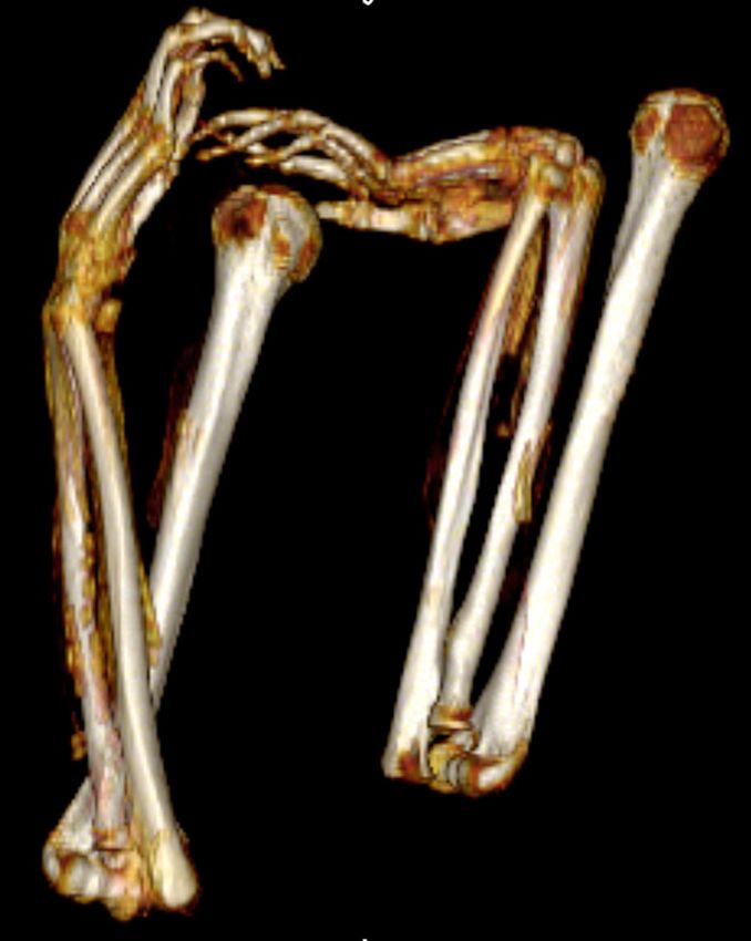
Saleem and Hawass                                                                                                CT Study of Seqenenre’s Mummy

The fracture has narrow ends and a wider central part that                of the left zygoma as well as the tip of the coronoid process of
measures 12 mm. This gapping fracture is associated with a short          the left mandible. The corresponding cut wound in the skin is
fissure fracture that extends from its medial end obliquely toward        spindle in shape with two pointed ends and a gapping center for
the fractured glabella.                                                   5 and 17 mm in depth.

Suggested Weapon                                                          Suggested Weapon
A double-edged blade weapon, its dimensions correspond to the             A heavy sharp weapon such as a sword or an ax. The bronze
wound’s measurements (about 32 mm wide and 12 mm thick).                  battle-ax found in Tel-el Dabaa (Figure 2B) fit into the size and
The injury may fit with the bronze battle-ax (MB II) found in             the spindle shape of the described wound.
Tel el-Dabaa (Figures 2B,C).
                                                                          Position of the Assailant
Position of the Assailant                                                 The blow came from the front of the King and to his left side.
The blow came perpendicularly from an assailant standing in
                                                                          Injury to the Left Side of the Skull Base
front of Seqenenre toward his right side. The blow was most
                                                                          Description
likely fatal.
                                                                          Irregular transverse fracture of the lower part of the mastoid
Injuries of the Nose, the Right Orbit, and the Right                      (with the tip of the mastoid missing), fractured left occipital
Cheek Bone                                                                condyle, and the left side of the margin of the foramen magnum.
                                                                          The fracture extends for 35 mm.
Description
Fractured glabella. An almost horizontal fissure fracture of the          Suggested Weapon
glabella measures 23 mm in length and 3 mm in width. The                  A pointed long sharp weapon similar to the Hyksos spearhead
fracture involves the anterior and posterior tables of the frontal        found at Tel el Dabaa (Figure 2D). The sharp spearhead
sinus. The right edge of the glabella fracture joins the medial end       penetrated the left mastoid transversely toward the foramen
of the previously described right supraorbital fracture.                  magnum. This wound could be fatal as it may have caused injury
                                                                          of the brainstem-spinal cord.
Fractured nose. A transverse fracture of the nasal bridge 5 mm
caudal to the naso-frontal suture. The fracture lines gap for 6 mm.       Position of the Assailant
Both nasal bones are displaced posteriorly for 8 mm, deviating            The blow came horizontally from the left side of Seqenenre below
to the right, and show multiple comminuted fractures. Also the            his left ear.
cartilaginous elements and soft tissues of the nose are distorted
and shifted to the right side.                                            Injuries of the Right Lateral Side of the
Fractured zygoma. A gapping vertical fracture 22 mm in length             Skull
splits and displaces the body of the right zygoma posteriorly.            CT images show three fractures in the right temporal,
It is possible that the displaced fractured zygoma prevented              sphenoid, and parietal bones. These fractures are mostly
translation of the coronoid process and resulted in a locked jaw.         covered by embalming material and cannot be detected by
A zygomatico-maxillary complex fracture is also seen, composed            physical inspection.
of fractures of the zygomatic arch, inferior orbital rim, anterior        Right Temporal Fracture
and posterior maxillary sinus walls, as well as the lateral orbital       Description
rim. The sharp fragments of the comminuted fracture of the                A transverse cut fracture of the squamous part of the right
right fronto-zygomatic process penetrated the overlying skin and          temporal bone runs parallel and about 5 mm inferior to the
resulted in an oval hole (measures 12 mm craniocaudal and 7               parieto-temporal suture. It joins anteriorly with the fractured
mm transversely).                                                         sphenoid and extends posteriorly for about 55 mm. The fracture
                                                                          is 5–6 mm wide with a short central constriction that measures
Suggested Weapon
                                                                          about 2 mm. A short vertical fracture 23 mm in length extends
The described injuries (in the nose and the right side of the face)
                                                                          caudally from the anterior edge of the fracture.
were likely caused by blunt forcible blow (or multiple blows)
using a heavy blunt object (e.g., a thick stick/the handle of ax).        Suggested Weapon
                                                                          A sharp weapon with a cut section matching the fracture’s shape
Position of the Assailant                                                 and size such as a dagger in Figures 2E,F.
The blow came from the front of the King toward his right side,
diagonally and from above.                                                Position of the Assailant
                                                                          The blow came horizontally from the right side of the King.
Left Cheek Region
Description                                                               Fractured Greater Wing of Right Sphenoid
An oblique cut fracture of the left side of the face extends for          Description
35 mm from the left orbit lower margin in the direction of the            The CT images reveal comminuted fracture of the greater wing
zygomatic-maxillary suture. The cut fracture involves the body            of the right sphenoid bone. The fracture shows as a roughly

Frontiers in Medicine | www.frontiersin.org                           6                                  February 2021 | Volume 8 | Article 637527
Saleem and Hawass CT Study of Seqenenre’s Mummy
triangular region lateral and posterior to the right orbit. The The most striking feature in the mummy of Seqenenre is
narrow apex points downwards, with the top of the triangle being the presence of severe injuries in the head (1, 9, 10). Multiple
the base, measuring 17 mm. The cranio-caudal height is about craniofacial injuries in Seqenenre’s mummy were previously
20 mm. described in the literature based on physical inspection and X-
rays (9, 10). In 1898, Maspero described three injuries by physical
Suggested Weapon inspection involving the upper forehead, the right cheek and the
A heavy sharp object like a dagger like in Figure 2E or Figure 2F. left cheek (7). GE Smith in 1912 described two other injuries: one
near the nose region and one on the left side of the skull base (9).
Position of the Assailant
CT imaging using two and three-dimensional reconstruction
The blow came horizontally from the right side of the King.
facilitates in-depth visualization of the bony and soft tissue
A Right Parietal Fracture elements of a mummy (17). In this study, CT imaging allowed
Description for detailed visualization of Seqenenre’s previously reported head
A vertical fissure fracture runs from above downward in the injuries, beyond what was possible using only x-rays or under
anterior part of the right parietal bone; it measures 30 mm in physical inspection. The injuries involved five different regions:
length and 3 mm in width. At the lower end, the fracture turns a transverse fracture of the forehead, a gaping fracture above the
posteriorly for a short segment 13 mm in length and 2 mm in right eyebrow, a blunt trauma to the nose and right eye region, a
width (possibly a secondary fracture). sharp cut wound of the left cheek, and a penetrating wound below
the left ear through the skull base.
Suggested Weapon CT also revealed additional injuries to the right side of the
The blow came vertically from the right side of the King. Possibly skull that were not described in the previous studies (7, 9, 10).
the king was lying down on his left side. These injuries were partially hidden beneath layers of embalming
material. We suggest that the embalmers deliberately concealed
Position of the Assailant these injuries, likely as a desperate attempt to beautify the injured
Most probably the weapon used to inflict the injury was a heavy corpse of the King.
blunt object like a thick stick. A blunt trauma or a sharp force trauma may result in skeletal
There is no CT evidence of healing in any of the described fractures. Analysis of the morphology of the fracture is often
craniofacial fractures. Few small broken bone chips are seen, informative to the type of trauma, and the direction of the
having fallen within the cranial cavity. impact. The shape and size of the cut fracture reflects the blade
of the weapon that induced it: the wider the cut fracture, the
DISCUSSION larger the blade. Variation in the width of the cut fracture
indicates the direction of the impact: the wider part of the
The CT study of the mummified Seqenenre shows that he was cut fracture points to the side of the stronger impact (18).
in his forties when he died. This age estimate is in accordance The morphology and location of Seqenenre’s injuries suggest
with the historical resources (10). The physical examination that several aggressors attacked him using different weapons.
of Seqenenre’s mummy done by GE Smith in 1906 (9), as However, the scholars have not been in agreement regarding the
well as the 1960s X-rays, both also estimated that the King type of weapons that caused Seqenenre’s injuries. According to
died at age 40 (10). However, as neither physical examination Maspero, the left cheek injury was caused by a heavy stick (a
nor plain x-ray allow for examination of the mummy’s pubic club), the right cheek injury was inflicted by a dagger, and the
symphyseal face, age estimation was based on less reliable criteria forehead by an ax (7). In contrast, GE Smith believed that an
such as degenerative bony changes and closure of the cranial ax or a sword caused the forehead, right supra-orbital, and left
sutures (9, 10). In this study, we were able to make a more cheek injuries, a heavy stick (or the handle of an ax) inflicted
accurate estimation of the mummy’s age at death using the the nose injury, and a spear was used to penetrate the skull
three-dimensional CT morphology of the pubic symphyseal face. base (9).
The undulating surface of the pubic symphyseal face becomes Scholars did agree that the morphology of Seqenenre injuries
smoother with age, allowing for improved accuracy of age matched those that would be inflicted with weaponry from
estimation (15). the Hyksos period (2, 12, 19). Bietak and Strouhal studied
Because of the disarticulated skeleton of the mummy, it is Asian bronze weapons found in Tell El Dabaa (ancient Avaris)
not possible to directly measure vertex to heel stature. Early and matched them to Seqenenre’s head injuries. The weapons
in the twentieth century, GE Smith reassembled Seqenenre’s belonged to the Middle Bronze Culture II (MB II), and were
mummy and estimated the stature to be about 170 cm (9). In similar to those used by Hyksos authorities at the time of
this CT study, we measured the tibial length and extrapolated Seqenenre’s reign (12). We studied five of these Asian bronze
the stature of Seqenenre using a regression equation derived weapons, currently housed at Cairo Egyptian Museum. We agree
for data for ancient male Egyptians to be 167 ± 3cm (16). with Bietak and Strouhal’s suggestion that the left cheek and
King Seqenenre Taa’s stature is comparable to the height of right supra-orbital fractures match with the size and shape
most of the New Kingdom Kings: Thutmose III’s stature was of MB II battle-ax (Figures 2B,C). The cutting edge of the
165 cm, Tutankhamun 167 cm, Seti I 167 cm, Ramesses II 172 cm, Hyksos battle-ax is smaller than its Egyptian counterpart. The
Merenptah 172 cm, and Ramesses III 165 cm (17). force of the impact of the Hyksos ax is more focused and
Frontiers in Medicine | www.frontiersin.org 7 February 2021 | Volume 8 | Article 637527Saleem and Hawass CT Study of Seqenenre’s Mummy
impacts a narrower area, explaining the deep bone punctures we that this attacked likely took place while he slept in his palace
observed (2, 12, 20). (8, 10). They hypothesize that because all of the craniofacial
We believe the forehead fracture was caused by a weapon with injuries were inflicted horizontally from aggressors at the King’s
a broader blade than the battle-ax which caused the left cheek and left side, he must have been sleeping on his right side at the
the right supra-orbital injuries. According to Bietak and Strouhal, time of the attack (9, 12). In 1974, Bietak and Strohal used the
a broad-edged ax caused the forehead facture, which could have results of their physical examination and 1960’s skull x-ray to
been an Egyptian weapon (12). argue that the King was riding a chariot into battle at the moment
This study suggests that a thick stick (the handle of a battle ax) of his death. They assumed that Seqenenre received the first blow
caused the blunt fracture around the King’s nose. We believe that to his left cheek from an attacker below him. When Seqenenre
the left mastoid and skull base injury may have been caused by lowered his head, possibly to look at his attacker, he received the
the studied MB II spearhead. We confirm in this CT study that second blow that landed just above his right eye. The King’s head
Seqenenre’s craniofacial injuries were inflicted around the time of sagged down as a result, which allowed the third blow to hit his
the King’s death (perimortem) as there is no evidence of healing. forehead and the fourth blow to smash the base of the nose (12).
Such severe craniofacial trauma could have caused fatal shock, A final explanation was posited by Shaw in 2009, who argues
blood loss, and/or intracranial trauma. that Seqenenre was imprisoned and executed by the victorious
The variety in attack angle, as well the wide range of weapons Hyksos (6).
we believe to have caused the King’s injuries, indicate that We believe that a situation in which the King was attacked
Seqenenre was killed in battle by numerous enemy attackers. while sleeping is unlikely, as the locations of the injuries
The match between weapons and the morphology of the injuries demonstrate that the blows came not only from the left side
strongly suggests that Seqenenre was killed during a war between of the King but also from other directions. A blow from the
the Egyptians and the Hyksos (2). The location of Seqenenre’s right side resulted in the right supra-orbital fracture, one from
craniofacial injuries indicates that he was likely facing his above caused the forehead fracture, and strikes from the front left
attackers. Facial injuries in combat are usually paired with fractured the left cheek and the left mastoid (12). Furthermore,
fractures of the forearms (parry fractures), as it is a natural reflex if the King died in the palace, then the body would have
to throw up the arms to protect the face (21). However, this CT been preserved properly. However, when Maspero unwrapped
study confirms that Seqenenre’s forearms were not fractured and Seqenenre’s mummy, he reported greasy bandages and bad odor,
that there are no injuries elsewhere in the body. This conclusion leading to his suggestion that the body must have been already
has been also suggested by previous X-ray studies done in the decomposing by the time of mummification (1). Scholars have
1960s (10). therefore reasoned that the corpse of Taa decomposed during
Seqenenre’s mummified hands are flexed at the wrists and the journey to Thebes from the location of his death and was
the fingers are spastic. This arm position is not consistent with mummified as soon as it arrived (6). This theory is supported
the usual crossed-pectoral position of the royal ancient Egyptian by the location of the King’s brain, shown in this CT study. The
mummies. This hand position suggests that the King’s wrists desiccated brain is usually seen occupying the posterior region of
were tied together, likely behind the body, when he died. If the cranial cavity in mummies lying down in the supine position
the King’s died while his hands were bound, the muscles that (24). However, in this case, as decomposition of the dead body
were in intense contraction just before death would have become progressed, the brain shifted toward the most dependent region.
rigid immediately after death, unable to relax; this condition is CT images in this study show Seqenenre’s desiccated brain shifted
known as cadaveric spasm. It typically affects the hands and limbs toward the left side of the cranial cavity. This is consistent with
of individuals who were subjected to violent deaths and whose the results of a recent CT study of a mummy with a head tilted
nervous systems were disturbed at the moment death, e.g., a to the right side, which showed that the shrunken brain was
drowning victim’s hand clenching on weeds from the waterbed. also shifted to the right of the skull cavity (25). We therefore
A deceased body demonstrating a cadaveric spasm will maintain suggest that the King’s dead body must have remained in a left
that position until the body decomposes (22). Cadaveric spasm is lateral position for some time; perhaps on the battlefield where
not part of rigor mortis, the progressive stiffness of the deceased he died, or while the mummy was being transported to Thebes. It
muscles that is seen over the course of a few hours after death due is unlikely that Seqenenre’s body was placed in the lateral position
to natural biomechanical changes (22–24). Cadaveric spasm has during the process of mummification, as embalmers at that time
been considered as a possible explanation in other archeological usually placed the body in a neutral supine position (2).
contexts: in an ancient Roman fortification (300–400 AD), a male Though we agree with Bietak and Strohal (12) that the King
with a head trauma was found holding a pig molar between his likely died as a result of a battle with the Hyksos, we believe
clenched fingers (23). A scenario in which Seqenenre’s hands that Shaw’s theory (2) best explains the results of this CT study.
were tied during his attack and subsequent death would also We argue that Seqenenre fought the Hyksos, was captured, and
explain the absence of defensive injuries on the King’s arms, as that his hands were cuffed (6). We agree with other scholars
he would have been unable to shield or protect himself from his that there is no evidence for the definite order of the blows that
assailants (6). King Seqenenre received, or which one killed him (2, 6, 9, 12).
Scholars have proposed several possible scenarios for the Seqenenre received several lethal wounds: the forehead, the right
King’s death. In 1912, GE Smith suggested that the King was supra-orbital, and possibly the skull base. Any one of those
killed while lying down on his right side (9). Others have added injuries is a potential cause of death, but the three of them
Frontiers in Medicine | www.frontiersin.org 8 February 2021 | Volume 8 | Article 637527Saleem and Hawass CT Study of Seqenenre’s Mummy
together is almost certainly fatal. We assume that the King was justification to declare war. When compared to CT scanning of
at a lower position than his assailant(s), possibly kneeling at other ancient Egyptian warriors with great military reputations,
least for some time during the attack. This position explains the such as Thutmose III and Ramesses II, only Seqenenre had severe
high forehead injury that could have been the first blow the king injuries inflicted by weapons (17). Such injuries suggest that
received, inflicted by a sword or an ax. The strong hit must have Seqenenre was on the front line, risking his life to liberate Egypt.
caused the King to fall down, possibly on his back. The King Seqenenre’s death motivated his successors to continue the fight
may have received several attacks from the assailant with the to unify Egypt and start the New Kingdom.
Hyksos battle ax, possibly using its blade to inflict the fracture
above the right eyebrow (right supra-orbital). Then a thick stick
(possibly the handle of the ax) was used to smash the nose and
CONCLUSION
the right eye of the King. The assailant hit the King’s left side This study of the mummified body of King Seqenenre-Taa-II
of the face with the ax. Another assailant at the left side used a helped to develop a better understanding of the circumstances
spear horizontally to pierce deeply the lower part of the left ear of his death. This study suggests that King Seqenenre was likely
(mastoid) and reached the foramen magnum. We assume that killed while leading the Egyptian military against the Hyksos
the King was dead at this point, and that his body was rolled to lie army. He was captured in battle and was killed in the struggle.
at his left side where he received several blows to the right side of The reconstructed CT images enabled a detailed analysis of
the skull possibly by a dagger. The dead King likely stayed lying the King’s craniofacial injuries, helped match the injuries with the
down on his left side for some time enough for the body to start weapons used to inflict them, and suggest a possible scenario of
decomposition as the brain shifted to this dependent side. the attack.
The exact location of the King’s death is unknown, as there is
no definite historic evidence for the location of a battle between
Seqenenre and the Hyksos (3). However, Deir el-Ballas, located DATA AVAILABILITY STATEMENT
just north of Thebes, was a base built by Seqenenre to launch
military campaigns against the Hyksos (4, 5). We assume that The original contributions presented in the study are included
a battlefield would have been located somewhere between the in the article/Supplementary Material, further inquiries can be
Egyptian stronghold at Deir el-Ballas and the Hyksos capital directed to the corresponding author/s.
Avaris, and that Seqenenre’s dead body was likely transferred
from the battlefield to Thebes via Deir el-Ballas. The location ETHICS STATEMENT
at which Seqenenre’s body was then mummified is a question
of great scholarly focus. Some scholars have suggested that The studies involving human participants were reviewed and
Seqenenre’s body was mummified hastily on the battlefield (1, 9). approved by the Egyptian Ministry of Antiquities and Tourism
Another plausible explanation would place his mummification Committee. Written informed consent for participation was not
at Deir el-Ballas, as excavations at the site have uncovered a required for this study in accordance with the national legislation
palace and tombs which date to Seqenenre’s time (26). However, and the institutional requirements.
there is no evidence that intentional mummification procedures
took place at Deir el-Ballas: there were no canopic jars found
there and all of the human remains were rather skeletons (26).
AUTHOR CONTRIBUTIONS
This CT study demonstrates that the embalmers went to great SS was responsible for the conception and design, acquisition of
lengths to properly mummify the body of Seqenenre. They data, analysis and interpretation of data, as well as drafting of
attempted to conceal some of Seqenenre’s head injuries using the manuscript, generation of the figures, and was accountable
a paste of embalming materials, a purely cosmetic procedure for accuracy and integrity of the work. ZH made substantial
which highlights the care with which the King was mummified. contributions to the design, interpretation of the results, drafting
Though the embalmers did not attempt to remove the brain of of the manuscript, critical revisions for important intellectual
Seqenenre, this seems to have been the norm for other royals, content, and agreed to be accountable for the integrity of any part
such as those who dated to the early 18th Dynasty like Thutmose of the work. Both authors read and approved the manuscript.
II (1493–1479 BC) and Thutmose III (1479–1425 BC) (26). The
embalmers would have been unable to place the mummy in the
usual neutral supine position with the arms crossed at the chest FUNDING
due to the partial decomposition and the cadaveric spasm of
his hands. We therefore do not agree with previous scholarship The research was self-funded by the authors.
that Seqenenre’s body was hastily or improperly mummified. We
believe that Seqenenre’s corpse was primarily mummified in the ACKNOWLEDGMENTS
Theban royal mummification workshop, and not at in a poorly
equipped or temporary setting as others have argued. We would like to show our gratitude to: Andrew Nelson,
It seems that Seqenenre was well-prepared for the war against Professor of Archaeology/Bioarchaeology-University of
Hyksos as he founded the huge military fortress at Deir el- Western Ontario, London (ON) Canada; Ronald G Beckett,
Ballas. He probably took the provocation of Hyksos King as a Professor Emeritus, Biomedical Sciences, Quinnipiac
Frontiers in Medicine | www.frontiersin.org 9 February 2021 | Volume 8 | Article 637527Saleem and Hawass CT Study of Seqenenre’s Mummy
University, Co-director, Bioanthropology Research Institute, manuscript that included improving its readability and fitness to
Quinnipiac University (CT)-USA; and Abla Ateyya Professor its purpose.
of Forensic Faculty of Medicine Cairo University-Egypt,
for sharing their insight and expertise that greatly assisted SUPPLEMENTARY MATERIAL
the research, and for revising and providing comments
that markedly improved the manuscript. The authors The Supplementary Material for this article can be found
would like to recognize Miss Maya Levin, Independent online at: https://www.frontiersin.org/articles/10.3389/fmed.
Researcher, (CA)-USA for her great efforts in copy editing the 2021.637527/full#supplementary-material
REFERENCES 15. Pasquier E, De Saint Martin Pernot L, Burdin V, Burdin V, Mounayer C,
Le Rest C, et al. Determination of age at death: assessment of an algorithm
1. Maspero G. History of Egypt, Chaldaea, Syria, Babylonia, and Assyria. of age prediction using numerical three-dimensional CT data from pubic
London: William Clowes And Sons, Limited. Volume 4. Project Gutenberg bones. Am J Phys Anthropol. (1999) 108:261–8. doi: 10.1002/(SICI)1096-
EBook, Release Date: December 16, 2005. EBook #17324 (1903). Available 8644(199903)108:33.0.CO;2-B
online at: http://www.gutenberg.org/files/17324/17324-h/17324-h.htm# 16. Raxter MH, Ruff CB, Azab A, Erfan M, Soliman M, El-Sawaf A. Stature
link2HCH0001 (accessed May 12, 2020). estimation in ancient Egyptians: a new technique based on anatomical
2. Shaw I. The Oxford History of Ancient Egypt. New York, NY: Oxford University reconstruction of stature. Am J Phys Anthropol. (2008) 136:147–55.
Press (2000). doi: 10.1002/ajpa.20790
3. Lacovara P. Deir el-Ballas. Overview of the Palace city of 17. Hawass Z, Saleem SN. Scanning the Pharaohs: CT Imaging of the New Kingdom
Deir el-Ballas Presented at the Conference on Palaces and Royal Mummies. New York, NY: AUC Press (2016).
Residences in Ancient Egypt. London (2013). Available online 18. Love JC, Wiersema JM. Skeletal trauma: an anthropological review. Acad
at: https://www.academia.edu/36177396/Deir_el-Ballas?fbclid= Forensic Pathol. (2016) 6:463–77. doi: 10.23907/2016.047
IwAR3Crjd3tfuJoV0oGIxppsSVrFCpWbBuCspBZR8lHIQOTbw0BLLMWrC- 19. Davies V, Friedman R. Egypt. London: British Museum Press (1998).
X5w (accessed October 25, 2019). 20. Dodson A, Dyan H. The Complete Royal Families of Ancient Egypt. London:
4. Lacovara P. Deir el-Ballas full history. The Ancient Egypt Thames & Hudson (2004).
Heritage and Archaeology. (2018). Available online at: http://www. 21. Judd MA. The Parry problem. J Archaeol Sci. (2008) 35:1658–66.
ancientegyptarchaeologyfund.com/deir-el-ballas-full-history/ (accessed doi: 10.1016/j.jas.2007.11.005
October 25, 2019). 22. Mann RW, Bass WM, Meadows L. Time since death and decomposition of
5. Dirminti E. Between Kerma and Avaris: The first Kingdom of Kush and the human body: variables and observations in case and experimental field
Egypt during the Second Intermediate Period. In: Anderson JR, Welsby studies. J Forensic Sci. (1990) 35:103–11 doi: 10.1520/JFS12806J
DA editors. Proceedings of the 12th International Conference for Nubian 23. Fierro MF. Cadaveric spasm. Forensic Sci Med Pathol. (2013) 9:253.
Studies. Leuven-Paris-Walpole, MA: British Museum Publications on Egypt doi: 10.1007/s12024-013-9414-x
and Sudan. Peeters (2014). p. 337–45. 24. Knusel CJ, Janaway RC, King SE. Death, decay, and ritual reconstruction:
6. Shaw GJ. The death of King Seqenenre Tao. J Am Res Center Egypt. archaeological evidence of cadaveric spasm. Oxf J Archaeol. (1996) 15:121–8.
(2009) 45:159–76. Available online at: http://www.jstor.org/stable/25735452 doi: 10.1111/j.1468-0092.1996.tb00079.x
(accessed January 29, 2021). 25. Hawass Z, Saleem SN. Computed tomography examination of the screaming
7. Maspero G. Les momies royale de Deir el-Bahari. Paris: Ernest Leroux (1889). mummy “Unknown-Woman-A”. Egypt J Radiol Nucl Med. (2020) 51:139.
8. Ikram S, Dodson A. Royal Mummies in the Egyptian Museum. Cairo: doi: 10.1186/s43055-020-00255-6
American University in Cairo Press (1997). 26. Jensen VI. The Cemeteries of Deir el-Ballas: Non-elite Burials of
9. Smith G. The Royal Mummies: Cairo: Catalogue general des Antiquites 17th-19th Dynasties and Their Relationship to the Royal Palace.
Epyptiennes du Musee du Caire. Nos 61051-61100. The Royal Mummies. (2019). Available online at: https://search.proquest.com/openview/
Cairo: L’Institut d’Archeologie Orientale (1912). 965999097128e9ea7a93b408d7d8ed8d/1?pq-origsite=gscholar&cbl=18750&
10. Harris JE, Wente EF. An X-Ray Atlas of the Royal Mummies. Chicago: diss=y (accessed May 18, 2020).
University of Chicago Press (1980).
11. ten Berge RL, van de Goot FR. Seqenenre Taa II, the violent death of a pharaoh. Conflict of Interest: The authors declare that the research was conducted in the
J Clin Pathol. (2002) 55:232. doi: 10.1136/jcp.55.3.232 absence of any commercial or financial relationships that could be construed as a
12. Bietak M, Strouhal E. Die Todesumstände des Pharaos Seqenenre (17. potential conflict of interest.
Dynastie). Ann Naturhistor Mus Wien. (1974) 78:29–52.
13. Todd T. Age changes in the pubic bones, I: the white Copyright © 2021 Saleem and Hawass. This is an open-access article distributed
male pubis. AJPA. (1920) 3:285–334. doi: 10.1002/ajpa.13300 under the terms of the Creative Commons Attribution License (CC BY). The use,
30301 distribution or reproduction in other forums is permitted, provided the original
14. Lovejoy CO. Dental wear in the Libben population: Its functional pattern and author(s) and the copyright owner(s) are credited and that the original publication
role in the determination of adult skeletal age at death. Am J Phys Anthropol. in this journal is cited, in accordance with accepted academic practice. No use,
(1985) 68:47–56. doi: 10.1002/ajpa.1330680105 distribution or reproduction is permitted which does not comply with these terms.
Frontiers in Medicine | www.frontiersin.org 10 February 2021 | Volume 8 | Article 637527You can also read