The Comparative Skeletal Structure of Blenniella bilitonensis (Skippers) and Bathygobius fuscus (Remainers)
←
→
Page content transcription
If your browser does not render page correctly, please read the page content below

Journal of Physics: Conference Series

PAPER • OPEN ACCESS

The Comparative Skeletal Structure of Blenniella bilitonensis (Skippers)
and Bathygobius fuscus (Remainers)
To cite this article: RA Putri and Sukiya 2019 J. Phys.: Conf. Ser. 1241 012005

View the article online for updates and enhancements.

                               This content was downloaded from IP address 46.4.80.155 on 25/02/2021 at 07:31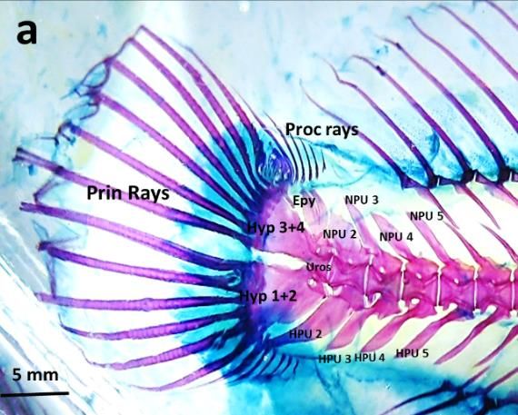
The International Seminar on Bioscience and Biological Education                                                IOP Publishing
IOP Conf. Series: Journal of Physics: Conf. Series 1241 (2019) 012005                     doi:10.1088/1742-6596/1241/1/012005

The Comparative Skeletal Structure of Blenniella bilitonensis
(Skippers) and Bathygobius fuscus (Remainers)

                     RA Putri, Sukiya
                     Department of Biology Education, Faculty of Mathematics and Natural Sciences,
                     Yogyakarta State University, Indonesia

                     rizka_apriani@uny.ac.id

                     Abstract. Intertidal Zone is an area with high biological diversity including vertebrates. In order
                     to survive in area with rapid change of environmental condition, animals need to be adapted
                     accordingly. One type of adaptations that mostly employed is structural adaptation specifically
                     ones that related to locomotor performances. Differences on how to navigate during high tides
                     or when the water level subsided as well as types of locomotion between skippers and remainers
                     suggested that there might also be structural differences in their skeleton. This research is aimed
                     to study the skeletal structure of two species that use different strategy in their locomotion. Two
                     species of fish from two main groups of intertidal fish (skippers and remainers) were caught and
                     preserved in 96% Ethanol. The skeletal preparation was made using Inouye’s Alizarin Red
                     Alcyan Blue Method. Data were collected by comparing skeletal structure between two species
                     particularly the axial (vertebrae) and appendicular (pelvic, pectoral, caudal fin) skeleton. Based
                     on the observation, skippers and remainers skeletal features has been adapted to different
                     function during locomotion. Modifications on pelvic, pectoral and caudal bones were found in
                     skippers and remainers. In B. bilitonensis, there are fusion of bones, reduction of some structures
                     and also smaller bone area to provide agility. In remainers, modification mostly found in pelvic
                     and pectoral fin. Pelvic fins possess larger and more rigid basal lepidotrichia as it was used as
                     an attachment apparatus while fish cling to the substrate during high tide. In B. fuscus, pectoral
                     fins are used as main propellers during swimming instead of caudal fins, hence larger bones area
                     to provide more muscle attachment are observed in this species

                       Keywords : Remainers, Skipper, skeletal structure, intertidal zone

1. Introduction
Tropical intertidal zone is rich in species diversity including fish. This area is characterised by the
fluctuation of sea level, in which causing extreme periodical changes of environmental factors during
24 hours . To be able to cope with rapid changes of abiotic factors, either behavioural, physiological,
morphological adaptation (or combination of three) is needed, therefore only certain species can thrive
and survive in this zone[1].
    There are three groups of amphibious fish inhabiting tropical intertidal zone, which are : Remainers,
Skippers, and Tidepool emerger [2]. Remainers are group of amphibious fish characterised by their small
range of mobility during lowtide. Remainers tend to stay in the crevices of the rock, under the boulders
or tidepools with only millimetres deep when sea level subsided. Tidepool emergers only emerge from
water when physical condition of water becomes inhospitable during low tide. Tidepool emergers

              Content from this work may be used under the terms of the Creative Commons Attribution 3.0 licence. Any further distribution
              of this work must maintain attribution to the author(s) and the title of the work, journal citation and DOI.
Published under licence by IOP Publishing Ltd                          1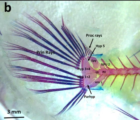
The International Seminar on Bioscience and Biological Education                              IOP Publishing
IOP Conf. Series: Journal of Physics: Conf. Series 1241 (2019) 012005   doi:10.1088/1742-6596/1241/1/012005

crawled out from one tidepool to another to find more suitable place to hide particularly during low tide.
On the other hand, skippers can actively move out of water to forage, defend territories or mate [3].
Different strategy employed by remainers, tidepool emergers and skippers suggested that there are
different morphological structures related to locomotor performances.
    Locomotion in land and water requires distinct morphological features particularly in
musculoskeletal system [4]. According to Hsieh [5], morphological innovation in locomotor system
enables aquatic organisms to find and invade new ecological niche, allowing them to thrive in new
habitat. Gibb et. al [4] distinguishes three types of fish locomotion when they are out of water, which are:
Trashing, tail-flip jump and prone jump. Trashing is uncontrolled and uncoordinated movement of fish
in land. Experimental observation by Gibb, et. al [6] showed that in flat surface, trashing does not result
in clear orientation. However, when fish were placed in a surface with 30° of slope, trashing can be
effectively used to descent the terrain. Tail flip jump usually can be found in fish that occasionally move
out of water particularly to escape from predator.
     Difference between tail flip jump and trashing lies on the direction of the movement. In a flat surface,
trashing does not result in a clear direction, however tail flip jump usually produces a movement with
clear directionality, which is toward the posterior part of the body. Another type of fish locomotion in
land called prone jump. Prone jump is an active movement from water to land and can easily be observed
in fish of family Gobiidae and Blenniidae. This type of movement requires good level of coordination
and effectively results in clear direction of movement [4].
    To perform coordinated movements, well adapted locomotor system is needed. One part of the
locomotor systems that might undergone evolutionary changes to enable fish moving in land is skeletal
system. According to [7], group of fish with ability to navigate themselves on land are differs from fully
aquatic fish by having dorsoventral body form, with ratio between height to total length is more than
4.5. This means that amphibious fish possess more vertebrae compared to fully aquatic fish with the
same size.
    Aside from vertebrae, fins, particularly pectoral and caudal fins play important roles during fish
locomotion on land. Harris (1960) [8] mentioned that there is enlargement of cleithrum bone in
Periophthalmus koelreuteri ‘s pectoral fin. This modification allows more place for muscle to attach
hence more agility and power to navigate on land.
    Other type of modification in fish is Pelvic Sucker Organ (PSO). Pelvic sucker organ, a modification
of pectoral fins, used by fish to attach themselves on substrate. PSO can be found in fish that lives in the
area with high turbulence such as waterfall and intertidal zone [9]; [10] ; [11].
    Remainers are known to stay inside tidepools during low tide while skippers tend to jump on to the
land. Therefore, how are the structure of skeleton differing in Remainers and skippers? What kind of
skeletal structure modification can be found in these two group of fish? This research is aimed to study
the skeletal structure of two groups of fish namely remainers and skippers that use different strategy in
their locomotion.

2. Research Method

2.1 Specimen Sampling
Fish specimen were collected from Siung Beach, one of the beaches with rocky intertidal zone in the
southern part of Yogyakarta. Two species of amphibious fish namely Bathygobius fuscus and Blenniella
bilitonensis, were collected as representation of remainers and skippers, respectively. They were
collected by hands or small nets. Fish then sacrificed and fixed in Ethanol 96% for 1 week.

                                                       2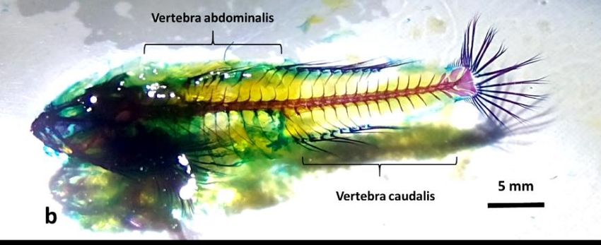
The International Seminar on Bioscience and Biological Education                              IOP Publishing
IOP Conf. Series: Journal of Physics: Conf. Series 1241 (2019) 012005   doi:10.1088/1742-6596/1241/1/012005

2.2 Skeletal Staining
After a week, fish skin and visceral organs were removed. Skeletal preparation was made using Alizarin
Red Alcyan Blue skeletal staining method by Inouye [12] with modification. Coloured skeletal specimen
were analysed descriptively. Description were made particularly in the structure and number of vertebrae
and also in appendicular skeleton namely: pectoral, pelvic and caudal fins.

3. Results and Discussion

3.1. Axial Skeleton
Based on observation, most of fish species that lives intertidal zone possess elongated body form. Fish
with this type of body rely on the movement of the posterior part and tail to swim. Force were generated
by the contraction of the muscles in caudal part of the body, which also causing the movements of tail.
Other fins like pelvic and pectoral fins are used for manoeuvring such as changing direction, making
turn or stopping. Since posterior part of the body are used in generating force for swimming, more
numbers of vertebrae are needed for muscle attachments. These pictures represent the full body skeleton
of two species used in this research:

        Figure 1a. Skeleton of Blenniella bilitonensis, species belongs to skippers group

        Figure 1b. Skeleton of Bathygobius fuscus, species belongs to remainers group

         Axial skeleton comprises three parts, which are: cranium, vertebrae and ribs. In fish, vertebrae
play pivotal role in locomotion, particularly caudal vertebrae. The main functions of the vertebrae are

                                                       3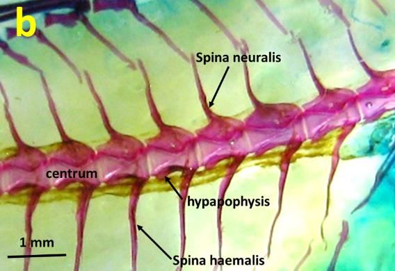
The International Seminar on Bioscience and Biological Education                              IOP Publishing
IOP Conf. Series: Journal of Physics: Conf. Series 1241 (2019) 012005   doi:10.1088/1742-6596/1241/1/012005

to provide attachment for muscles fibres also to support fish’s body. Based on the observation, the
number of caudal vertebrae of B. bilitonensis is 21 while B. fuscus’s body supported by 15 vertebrae.
The difference in the number of the vertebrae suggested different number of muscles fibres attached to
them. In fish, body muscles are arranged in rows or segments called Myomers. Myomer as a contractile
unit of the muscle generate power during locomotion. With more numbers of vertebrae, more segments
or rows of muscle can be attached to them therefore more power can be produced.
           Figure 2 depicted the detailed features of vertebra structure on both species. Each vertebra
consists of structures such as centrum, neural spine, haemal spine and hypapophyses. However, the
difference between species observed can be point out in the angle and direction of the spine. In B.
bilitonensis, direction of the spines is toward the posterior part of the body with 60° angle between
centrum and spine. Meanwhile in B. fuscus, angle formed by each centrum and spine is 90° towards
dorsal and ventral side of the body. Yet, the impact of this arrangement toward the contraction of the
muscles fibres isn’t fully understood and more research needs to be done.

             Figure 2. Lateral view of B. bilitonensis (a) and B. fuscus (b) vertebrae

3.2. Appendicular Skeleton
Appendicular skeleton in fish comprises of bones that construct fins on the specific regions of the body.
Fins are named based on their locations and can easily be distinguished from one to another. In this
research we focused on three fins that differ in function namely Pectoral, Pelvic and Caudal Fins.

                                                       4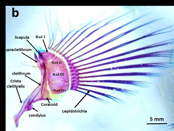
The International Seminar on Bioscience and Biological Education                               IOP Publishing
IOP Conf. Series: Journal of Physics: Conf. Series 1241 (2019) 012005    doi:10.1088/1742-6596/1241/1/012005

  Table 1. Comparison of appendicular bones structures in B. bilitonensis and B. fuscus
     Appendicular                 B. bilitonensis                              B. fuscus
       Skeleton
 Pectoral Fins
 Os Supracleithrum    Enlargement on the dorsomedial part No enlargement on dorsomedial parts of
                      of the bone, blunt end                   the bone, sharp end
 Os Cleithrum              Large christa cleithralis                    Narrow christa cleithralis
 Os Coracoideum            Oval shaped, attached to Os Radialia         Triangle shaped , attached to Os
                           III dan IV                                   Radialia IV

 Os Radialia               4, shape is varied between each bone         4, uniform in shaped, no cleft between
                           ; cleft between each radial bone             each bone
 Lepidotrichia             14, no branch at the end , segmented ,       18, branched at the end (2-6 branch ),
                           all parts consist of osteon                  unsegmented, medial parts consist of
                                                                        osteon while distal end consist of
                                                                        cartilage
 Pelvic Fins
 Basypterygium             Consists of three bones with triangle        Consist of two bones with rectangular
                           shape                                        shape
 Lepidotrichia             Reduction on the number                      5 pairs of lepidotrichia, branched (2-6
                           lepidotrichia→ 3 pairs, consists of          branches ), unsegmented, medial parts
                           osteon , unbranched, segmented               consist of osteon while distal part are
                           basal lepidotrichia is modified into a       made of cartilage
                           ball and socket joint → more flexible        Basal lepidotrichia attached to
                           movement                                     lepidotrichia by hinge joint →
                                                                        movement of lepidotrichia is restricted
 Caudal Fins
 Os Pleural Centrum        5 bones, posterior part modified into        2 bones, posterior part modified into
                           urostyle                                     urostyle

 Os Hypural                4, fusion between Hypural I and II           5, fusion between Hypural I and II
                           (Hypural 1+2), and also between              (Hypural 1+2), and also between
                           Hypural III and IV (Hypural 3+4)             Hypural III and IV (Hypural 3+4).
                                                                        Hypural V located between Hypural 3+4
                                                                        and Os Epural
 Os Epural                 2, separated                                 2 → fused into one disc-shaped bone
 Lepidotrichia             - Number of principal rays 14,               - Number of principal rays 14,
                           segmented , each posterior end               unsegmented, each posterior end
                           branched into two branches consist of        branched into 6 branch (hair like –
                           cartilage. Segmented (anterior parts         branch) , basal lepidotrichia are made of
                           of the fin) are made of osteon               osteon
                           - 6 Proccurent rays , made of osteon,        - 4 Proccurent rays , made of osteon,
                           unbranched, unsegmented                      unbranched, unsegmented

3.2.1 Pectoral Fins
Pectoral Fins are one of two paired fins found in lateral side of the body. In both species these fins are
required during swimming mainly in manoeuvring, changing direction or slowing down and stopping.

                                                       5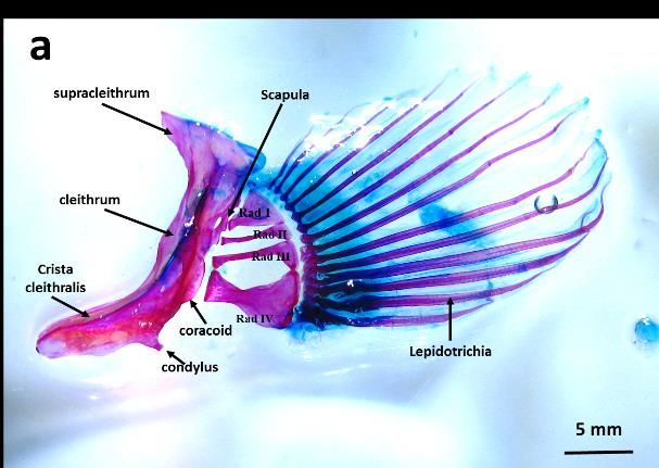
The International Seminar on Bioscience and Biological Education                              IOP Publishing
IOP Conf. Series: Journal of Physics: Conf. Series 1241 (2019) 012005   doi:10.1088/1742-6596/1241/1/012005

However, other functions of the fins can be observed in B. bilitonensis. In B. bilitonensis pectoral fins
can be utilized as hook and stabilizer when fish clings to rocks. According to [13], pectoral fins of fish
from Family Blennioidea consists of two regions called Fanfield and Hookfield. During swimming,
fanfield and hookfield on both sides can be expanded to create larger surface area. This method is applied
when fish needs to slow down and finally stop. However, in B. bilitonensis, hookfield can also be used
as “hook” when fish attach themselves on the surface of the rocks. Hookfield (fig 3) can be distinguished
from fanfield by having sharp end and dorsally oriented lepidotrichia. Branched end of lepidotrichia is
not observed in B. bilitonensis, but present in B. fuscus, which main function of pectoral fins is as
mentioned above. During manoeuvre, surface area of the fins is regulated by expanding or contracting
the lepidotrichia. Branched end of lepidotrichia allows fish to maintain the expanding state of the fins
for as long as it needed.
       Aside from lepidotrichia, differences in pectoral fins structures can also be found in other bones
such as Cleithrum, Radialia and condyles of pelvic and pectoral girdle. List of the detailed comparison
between B. bilitonensis and B. fuscus bone structures provided in table 1.

          Figure 3. Lateral View of Pectoral fins in B. bilitonensis (a) and B. fuscus (b)
3.2.2 Pelvic Fins
Pelvic fins in B. bilitonensis are used mainly in land, as a structure to supports fish body as well as
“walk” in solid surfaces such as sand and rocks. However, different function of these paired fins is
observed in B. fuscus. In B. fuscus, pelvic fins have been modified into Pelvic Sucker Organ (PSO) (fig
4), which used to attach fish body onto the substrate when there’s high turbulence. PSO allows fish to
maintain its position even during hightide. In rare occasions, PSO can also be used by fish to cling on a
vertical substrate. Since Pelvic fins in both species were operated in different circumstances, different
structures were observed. In B. bilitonensis, there are fusions of lepidotrichia and also flexible joint
connecting lepidotrichia and basal lepidotrichia. Combination between the rigid structure of
lepidotrichia (because of the fusions) and flexible joint allow fish to stand on its pelvic fins as well as
walk on land.
       In B. fuscus, lepidotrichia were separated into 5 rays on each side and each lepidotrichium was
branched and segmented. The joint between lepidotrichia and basal leidotrichia were rigid, allowing less
movement of lepidotrichia. Nonetheless, this modification allowing B. fuscus to create a vacuum system
that can be utilized as a sucker organ, which help this fish to survive in a highly turbulent environment.
Vacuum system was created by the assistance of membrane between each lepidotrichia (fig. 4).

                                                       6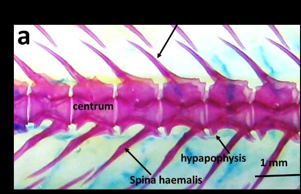
The International Seminar on Bioscience and Biological Education                              IOP Publishing
IOP Conf. Series: Journal of Physics: Conf. Series 1241 (2019) 012005   doi:10.1088/1742-6596/1241/1/012005

            Figure 4. Dorsal view of Pelvic girdle in B. bilitonensis (a) and B. fuscus (b)
3.2.3 Caudal Fin
The function of caudal fin in general are to maintain direction also to generate force so fish can move
forward during swimming. In B. fuscus, tail mostly utilized to generate power for swimming while in B.
bilitonensis, aside of swimming, tail can also be used when fish moves out of water as well as navigate
on land. According to [8] for fish that able to move in land, additional function of tail has been observed.
These additional functions including maintaining body’s balance and also acting as the propeller when
the fish jump [ 4]. Based on the observation, there are slight differences in caudal fin structure between
B. bilitonensis and B. fuscus, mostly related to the number of the bones. Comparison between caudal
fin’s bones can be seen in table 1 and figure 5. Since bones provide place for muscle attachments,
additional number of bones can be used as the place of attachment of more muscle fibres. Blenniella
bilitonensis possess more caudal vertebrae as well as bones that construct caudal fins. It can be suggested
that more type of muscle can be found in B. bilitonensis as part of its adaptation to navigate on land.
Tail serves its function not only for swimming but also jumping, therefore agility, flexibility and
robustness of the tail is required.

  Figure 5. Lateral view of Complex Caudal Vertebrae - Caudal Fin of B. bilitonensis (a) and
                                       B. fuscus (b)

                                                       7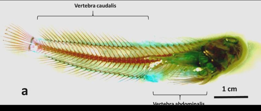
The International Seminar on Bioscience and Biological Education                              IOP Publishing
IOP Conf. Series: Journal of Physics: Conf. Series 1241 (2019) 012005   doi:10.1088/1742-6596/1241/1/012005

4. Conclusion

    Based on our observation, it can be concluded that there are differences between axial and
appendicular skeleton of Blenniella bilitonensis and Bathygobius fuscus. Blenniella bilitonensis was
adapted to navigate on land in which modification can be observed in the number of vertebrae, fusions
of lepidotrichial bones in pelvic fin and enlargement of some bones, mainly to provide more muscle
attachments. In B. fuscus notable adaptation can be found in pelvic fins which modification is Pelvic
Sucker Organ, allowing this species to attach to its substrate in a turbulent environment.

References
[1] Yoshiyama, R.M. (1981). Distribution and abudance patterns of rocky intertidal fishes in Central
        California. Environmental Biology of Fishes Vol.6. No3/4 : 315-332.
[2] Martin, K.L.M. 1995. Time and tide wait for no fish: intertidal fishes out of water. Environmental
        Biology of Fishes 44: 165-181.
[3] Allen, L.G. and M.H. Horn. (2006). The Ecology of marine fishes : California and adjacent
        waters. University of California Press. pp 208-211
[4] Gibb, A.C., M.A. Ashley-Ross and S. T. Hsieh. (2013). Thrash, flip, or jump: The behavioral and
        functional continum of terrestrial locomotion in teleost fishes. Integ. Comp. Biol. Vol 53 (2):
        295-306.
[5] Hsieh S.TT.(2010). A Locomotor innovation enables water-land transition in a marine fish. PLoS
        ONE 5(6):e11197
[6] Gibb, A.C., Ashley-Ross, M.A., Pace, C.M., and Long, J.H. Jr. (2011). Fish out of water :
        Terrestrial jumping by fully aquatic fishes. J.Exp. Zool. 315A : 649-653
[7] Pace, C.M., and A.C. Gibb. (2014). Sustained periodic terrestrial locomotion in air-breathing
        fishes. J Fish Biol. 2014 Mar, 84 (3): 639-60.
[8] Harris, V.A.(1960). On the locomotion of the mud-skipper (Periophthalmus koelreuteri Pallas)
        (Gobiidae). Proc. of Zool. Soc. Lond. 134: 107-135.
[9] Schoenfuss, H.L and R.W Blob. (2003). Kinematics of watefall climbing in hawaiian freshwater
        fishes (gobiidae): Vertical propulsion at the aquatic-terrestrial interface. J. Zool. Lond.261:
        191-205.
[10] Blob, R.W., R. Rai, M.L. Julius and H.L Schoenfuss. (2006). Functional diversity in extreme
        environments : Effects of locomotor style and substrate texture on the waterfall-climbing
        performance of hawaiian gobiid fishes. J. of Zool. 268: 316-324.
[11] Budney, L.A. and B.K Hall.( 2010). Comparative morphology and osteology of pelfic fin-derived
        midline suckers in lumpfishes and gobies. J.Appl. Ichtyol. 26: 167-175.
[12] Inouye, M.(1976). Differential staining of cartilage and bone in mouse skeleton by Alcian Blue
        and Alizarin Red S. Cong. Anom. 16 : 171 - 173
[13] Brandstӓtter, B. Misof, C. Pazmandi, G.P Wagner.(1990). Micro-anatomy of the pectoral fin in
        Blennies (Blenniini, Blennioidea, Teleostei). J. of. Fish Biol. Vol 37 (5) : 729-743

Acknowledgement
We would like to convey our gratitude to FMIPA UNY for the funding of this research and also to
Cahyoaji P. Anggara, Bima Gana P and Aditya Wijaya for specimen collection.

                                                       8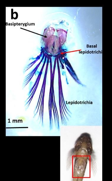
You can also read