The Latest Strategy for Keloid and Hypertrophic Scar Prevention and Treatment: The Nippon Medical School (NMS) Protocol - J-Stage
←
→
Page content transcription
If your browser does not render page correctly, please read the page content below
―Review―

       The Latest Strategy for Keloid and Hypertrophic Scar Prevention and
                     Treatment: The Nippon Medical School (NMS) Protocol

                                          Rei Ogawa, Teruyuki Dohi, Mamiko Tosa,
                                              Masayo Aoki and Satoshi Akaishi

                  Department of Plastic, Reconstructive and Aesthetic Surgery, Nippon Medical School Hospital, Tokyo, Japan

         In 2006, we established a scar/keloid-specialized unit in the Department of Plastic, Reconstructive, and
         Aesthetic Surgery at Nippon Medical School (NMS) in Tokyo, Japan. In the ensuing 15 years, we treated
         approximately 2,000 new scar/keloid patients annually. This extensive experience has greatly improved
         the efficacy of the treatments we offer. Therefore, we discuss here the latest NMS protocol for prevent-
         ing and treating keloids and hypertrophic scars. While this protocol was optimized for Japanese pa-
         tients, our experience with a growing body of non-Japanese patients suggests that it is also effective in
         other ethnicities. The extensive evidence-based experience underlying the NMS protocol suggests that it
         may be suitable as the foundation of a standard international prevention/treatment algorithm for
         pathological scars. (J Nippon Med Sch 2021; 88: 2―9)

         Key words: keloid, hypertrophic scar, wound

                          Introduction                                    tation, including trauma, burn, surgery, vaccination, skin
In 2006, we established a scar/keloid-specialized unit in                 piercing, acne, and herpes zoster. In general, the scars
the Department of Plastic, Reconstructive, and Aesthetic                  that spontaneously lose this dermal inflammation over
Surgery at Nippon Medical School (NMS) in Tokyo, Ja-                      time are defined as HSs, whereas the scars with enduring
pan. In the following 15 years, we treated approximately                  inflammation are defined as keloids. This difference in
2,000 new scar/keloid patients annually. This vast experi-                inflammation durability explains the disparate growth
ence has greatly improved the efficacy of the treatments                  habits of HSs and keloids. Thus, the temporary, waning
we offer. Therefore, we discuss here the latest NMS algo-                 inflammation in HSs means that they do not grow over
rithm for preventing and treating keloids and hy-                         the borders of the original wound. By contrast, the con-
pertrophic scars (HSs). While this algorithm has been op-                 tinuous, escalating inflammation of keloids causes them
timized for Japanese patients, our experiences with a                     to grow into the adjacent normal skin.
growing body of international patients suggest that it is                   In terms of histopathology, the inflammation and the
also effective in other ethnicities and could therefore                   resulting accumulation of extracellular matrix causes der-
serve as the starting point in the development of an in-                  mal nodules and thick eosinophilic (hyalinizing) collagen
ternational prevention/treatment algorithm.                               bundles called “keloidal collagen” to form over time2. If
                                                                          the dermal nodule is the main histological finding, it is
           Pathomechanisms of Keloids/HSs                                 diagnosed as an HS. This suggests that keloids/HSs are
To effectively prevent and treat keloids/HSs, it is neces-                manifestations of the same inflammatory fibroprolifera-
sary to understand the pathomechanisms that drive their                   tive condition and simply differ in the intensity and du-
formation and progression. The abnormal growth of                         ration of inflammation.
these scars is powered by chronic inflammation in the re-                   Multiple lines of evidence suggest that the intensity/
              1
ticular dermis that is triggered by dermal injury or irri-                duration of dermal inflammation in keloids/HSs is dic-

  Correspondence to Rei Ogawa, MD, PhD, FACS, Department of Plastic, Reconstructive and Aesthetic Surgery, Nippon Medical
  School, 1―1―5 Sendagi, Bunkyo-ku, Tokyo 113―8603, Japan
  E-mail: r.ogawa@nms.ac.jp
  https://doi.org/10.1272/jnms.JNMS.2021_88-106
  Journal Website (https://www.nms.ac.jp/sh/jnms/)

  2                                                                                                      J Nippon Med Sch 2021; 88 (1)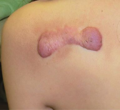
Keloid & Hypertrophic Scar NMA Protocol

tated by genetic, systemic, local, and lifestyle risk fac-                   lized by placing bandages or garments over the dressing.
tors1. Genetic causes include single nucleotide polymor-                        In normal wound healing, once epithelialization is
            3,4
phisms            and genetic diseases such as Rubinstein-Taybi              completed, the inflammation in the dermis/dermal-like
                  5,6
syndrome . There are probably many other, as yet un-                         granulation tissues under the epidermis starts to subside
identified, genetic factors. Systemic factors include preg-                  spontaneously and the dermal tissues begin maturing.
        7                       8
nancy , hypertension , high circulating concentrations of                    However, if strong/repetitive mechanical forces are ex-
inflammatory cytokines9, and possibly female sex10.                          erted on the immature scar, they can prolong the inflam-
  The key local risk factor is mechanical force on the                       mation, thereby fomenting pathological scarring. One of
wound/scar: considerable evidence shows that this                            the keys to preventing such scarring is to understand
strongly           influences       inflammation-driven       pathological   that the healing rates of the epidermis and dermis differ
             11―13                                             13
scarring           . Indeed, our finite-element analysis suggests            completely. In the case of sutured wounds, the epidermis
that keloid shape is largely determined by the direction                     can regenerate within 7-10 days, leading both the patient
of the tension on the skin around the wound/scar site.                       and the physician to believe that the wound has healed
We also showed that keloids are particularly prevalent                       completely. In fact, it can take 3 months before the der-
on body regions with strong skin-stretching force due to                     mis regains 90% of its normal strength14. This lengthy
body movements, such as the joints, anterior neck, chest                     vulnerability   to      inflammation-triggering   mechanical
wall, scapula/upper-arm, and suprapubic region. Re-                          forces means that it is essential to provide the immature
peated dermal injury and infection is also a local risk fac-                 scar with protracted external mechanical support (i.e. scar
tor.                                                                         stabilization) until maturity has been attained (i.e. the
  Lifestyle risk factors include strong/continuous body                      scar becomes soft, pliable, and flat).
movements (e.g. those of athletes/manual workers) that                          Therefore, all immature scars should be fixed continu-
stretch the wounds/scars. Other factors are excessive                        ously with tape. Silicone tape is the best choice. The tape
consumption of hot/spicy foods and alcohol and over-                         is retained until it detaches naturally. Alternatively,
long bathing in hot water, which may augment wound/                          silicone-gel sheets or cheaper paper tapes can be used on
scar inflammation. Obesity may also be a risk factor.                        a case-by-case basis.
                                                                                When an individual is at high risk of keloid formation
                        Prevention of Keloids/HSs                            and/or the scars are located on keloid-prone regions,
Because traumatic, surgical, and burn wounds and                             scar stabilization should be prolonged for at least 6-12
wounds caused by infectious agents, can turn into patho-                     months after maturation. It should also be combined
logical scars, it is important to assess the pathological                    with first steroid ointment and then tape/plaster admini-
scarring risk of patients with such wounds. If the patient                   stration. In Japan, two types of steroid tapes are avail-
or his/her family has a history of keloid and/or the                         able15: the strong deprodone-propionate plaster (EclarⓇ
wound is on a high-risk zone, the patient should be con-                     plaster; Hisamitsu Pharmaceutical Co., Inc., Tokyo, Ja-
sidered to be high risk and treated appropriately, as de-                    pan) should be used for adults while the weak fludroxy-
scribed below.                                                               cortide tape (DrenisonⓇ tape; Teikoku Seiyaku Co., Inc.,
  Trauma and Burn Wounds with a Raw Surface                                  Kagawa, Japan) is suitable for children. The steroid oint-
  In general, the longer wounds take to epithelialize, the                   ment should be applied for the first 2-3 weeks after epi-
greater the risk that keloids/HSs develop. Therefore,                        thelialization because the epidermis is still immature (full
trauma/burn wounds should undergo primary suturing                           epithelialization takes 4 weeks) and could be injured by
or skin grafting/flap transfer as soon as possible. How-                     daily steroid tape/plaster changes. Once steroid tape/
ever, if surgery cannot be planned, the raw surface must                     plaster replaces the ointment, it is continued until the
be encouraged to epithelialize rapidly by applying con-                      scar is soft and flat. Scar redness should be ignored: con-
servative therapies that limit infection and inflammation                    tinuing steroid tape/plaster just because of redness ele-
and accelerate granulation-tissue formation. These thera-                    vates the risk of telangiectasia because the steroid thins
pies, which should be applied on a case-by-case basis, in-                   the structures supporting the blood vessels. Once steroid
clude negative-pressure wound therapy, appropriate                           tape/plaster is discontinued, a non-steroidal ointment/
wound dressing materials, topical drugs (e.g. basic fibro-                   cream such as heparinoid ointment/lotion/spray (Hiru-
blast       growth       factor      spray),   antibiotics,    and   anti-   doidⓇ; Maruho Co., Inc., Osaka, Japan) or an NSAID
inflammatory agents. The wound should also be stabi-                         ointment/cream (Ibuprofen piconol: StadermⓇ; Torii

  J Nippon Med Sch 2021; 88 (1)                                                                                                       3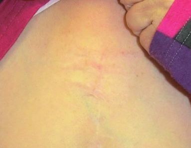
R. Ogawa, et al

Pharmaceutical Co., Ltd., Tokyo, Japan) should be ap-                           Treatment of Hypertrophic Scars and Keloids
plied to keep scar surface moist and inhibit inflamma-                    Diagnosis of Keloid/HS Risk
tion.                                                                     The Japan Scar Workshop (JSW) scar scale (JSS)16 helps
  In the case of low-risk individuals and/or wounded                    to differentially diagnose HS/keloid-like lesions. It also
regions (e.g. the face, lower leg), wound stabilization is              has indicators that suggest the scar may be or become in-
sufficient to prevent pathological scarring.                            tractable:
  Primarily Closed Wounds                                                 1. The patient has multiple keloid/HS-like lesions on
  Sutured wounds should be cleaned daily with tap                       the body
water/saline or disinfectant and covered with non-                        2. The keloid/HS-like lesion is large, even if it is soli-
adhesive materials until the sutures can be removed. This               tary
prevents infections and wound stretching, thereby block-                  3. Age at pathological-scar onset is
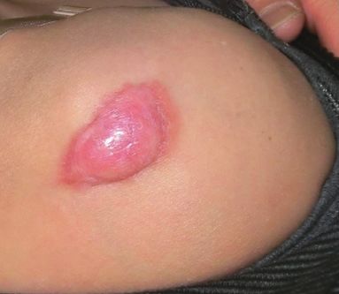
Keloid & Hypertrophic Scar NMA Protocol
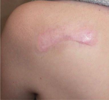
a b
Fig. 1 The left scapular keloid of a woman in her 30s was treated by deprodone-propionate
plaster.
(a) Pretreatment.
(b) Twenty-four months after starting treatment.
The tape was placed on the keloid 24 hours a day and changed daily. After 24 months,
the inflammation had completely resolved and the subjective and objective symptoms
improved.
a b
Fig. 2 The right shoulder keloid of a woman in her 50s was treated by triamcinolone acetonide
steroid injections.
(a) Pretreatment.
(b) Twenty-four months after starting treatment.
The keloid underwent steroid injection (5 mg triamcinolone diluted with 1.5 mL xylo-
caine 1% with epinephrine) every 2–3 months. After 24 months, the subjective and objec-
tive symptoms improved.
Steroid Injection ciently throughout the tissue. The consequent local pres-
Triamcinolone acetonide is a powerful tool because it sure may also cause pain. Rather, the target should be
rapidly reduces inflammation (Fig. 2). Each injection the scar border where it meets the normal skin: the nee-
should comprise 5-10 mg mixed with a local anesthetic dle should penetrate either shallowly to target the heav-
16
such as xylocaine 1% with epinephrine . Women may ex- ily inflamed leading edge of the scar or deeply towards
perience fewer menstrual irregularities if the dose does the center (the scar tissue along this deep plane is softer
not exceed 5 mg. We use thin needles such as 30 G and than the upper central core). Once one or more injections
27 G along with syringes with locks. The initial target of have softened the scar, the needle can be injected straight
the injection should not be the center of the mass: its into the core. Smaller keloids/HSs can improve markedly
hardness means the injection fluid will not disperse suffi- after just 1-2 injections. If these improvements can be
J Nippon Med Sch 2021; 88 (1) 5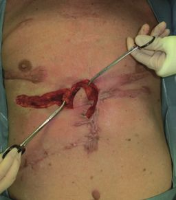
R. Ogawa, et al

  a                         b                         c                         d                         e
           Fig. 3 The left upper-arm keloid of a woman in her 30s was treated by z-plasty and postoperative radiotherapy.
 (a) Before surgery.
 (b) After keloid excision.
 (c) Z-plasty design.
 (d) Immediately after surgery.
 (e) Two years after surgery.
 The keloid developed from BCG vaccination. It was excised, tension was released by fascial sutures, and z-plasties were performed.
 Postoperative radiotherapy (18 Gy/3 fr/3days) was then conducted. No recurrence was observed 2 years after surgery.

continued/maintained with steroid tape/plaster, further              skin grafts, which often form pathological scars around
injections are unnecessary. If patients are afraid of                the graft. Compared to island flaps, skin-pedicled flaps
injection-induced pain, we recommend steroid tape/plas-              are superior because they extend more postsurgically and
ter until the scar has softened. Injections can then be              therefore release contractures more effectively19. Flap sur-
added because the risk of pain is much reduced.                      gery is especially suitable for severe keloids; however,
  Surgery                                                            multimodal therapy should be applied to both the exci-
  Surgical monotherapy associates with an atrocious rate             sion and donor sites to prevent recurrent and new le-
of keloid/HS recurrence. Therefore, surgical techniques              sions from forming.
that abrogate dermal tension, namely, subcutaneous/fas-                 Radiation
cial tensile-reduction sutures, z-plasties, and local flap              Post-excision keloid/HS recurrence can be effectively
transfer17,18, should be used in combination with postop-            controlled by adjuvant radiation. Radiation protocols are
erative radiation (described below) and steroid tape/                constantly improving. In the past, orthovoltage or super-
plaster.                                                             ficial or soft X-rays were used. However, the safety and
  Placing sutures on the superficial/deep fascia below               efficacy of radiotherapy protocols have improved mark-
the dermis causes the wound edges to juxtapose natu-                 edly recently; therefore, we now routinely use postopera-
rally. This allows dermal sutures to be placed with very             tive electron-beam irradiation after keloid/HS resection.
little tension, after which superficial sutures can be               This, when combined with the surgical and steroid tape/
added. It is essential to realize that without the deep su-          plaster modalities described above, markedly reduces the
tures, dermal sutures not only fail to reduce the dermal             recurrence rate: the keloid-recurrence rate after surgery is
tension, they may in fact exacerbate it.                             now less than 10% in our hospital18. Moreover, 15 years
  Zig-zag sutures, including z-plasties, are good for re-            of close monitoring and long-term follow-up has shown
leasing linear scar contractures and tension (Fig. 3). An-           that this protocol has no major side effects (Fig. 3, 4)18.
other advantage is that segmented scars mature faster                   Another, increasingly popular, radiation modality for
than long linear scars. Zig-zag incision/suturing is par-            keloids is high-dose rate-superficial brachytherapy. De-
ticularly indicated if scars cross a joint.                          pending on the shape of the surgical scar, an applicator
  Local flaps are also useful for releasing scar contrac-            can be used to ensure the evenness and localization of
tures (Fig. 4): they expand naturally after surgery and are          the radiation to the wound surface. However, we prefer
therefore not prone to postsurgical contractures, unlike             electron beams because they reach the reticular dermis

  6                                                                                                J Nippon Med Sch 2021; 88 (1)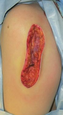
Keloid & Hypertrophic Scar NMA Protocol

  a                                b                                c                                  d
 Fig. 4   The chest and abdominal keloids of a man in his 70s were treated by flap surgery, z-plasties, and postoperative radiothera-
          py.
          (a) Before surgery.
          (b) Intraoperative view of the first operation, during which the chest keloid was excised and reconstructed with a flap.
          (c) Intraoperative view of the second operation, during which the abdominal keloid was removed and closed with z-plas-
          ties.
          (d) Two years after the second operation.
          The keloids were removed in two operations. The donor and recipient sites were irradiated. The post-treatment course was
          uneventful. After 2 years, recurrence was not observed and the remaining pathological scars had matured.

                   a                                                b
                       Fig. 5 The chest-wall keloid of a woman in her 70s was treated by radiation monotherapy.
                   (a) Before treatment.
                   (b) Eighteen months after treatment.
                   This mild chest-wall keloid was treated by high-dose rate-superficial brachytherapy (25 Gy/5
                   fr/5days). The inflammation resolved completely. Six months later, the subjective and objective
                   symptoms improved dramatically. Eighteen months later, the pathological scars matured.

more selectively; moreover, the beam attenuates after                   higher than the postoperative radiation doses, the radia-
passing through the skin and thus has little effect on the              tion should be delivered carefully to avoid secondary ra-
internal organs.                                                        diation carcinogenesis. It is also essential to secure in-
  To further improve the safety of our adjuvant radio-                  formed consent. However, the risks of primary radiother-
therapy protocol, we optimized them in 2013 such that                   apy should be measured against its immense benefits: it
fewer fractions are now used18. As a result, high-                      rapidly decreases subjective symptoms such as pain and
recurrence keloid-excision sites are now treated with 18                itch and, over the following year, progressively normal-
Gy/3 fr/3days while low-recurrence sites (e.g. earlobe)                 izes scar color and thickness.
and other body sites are now treated with 8 Gy/1 fr/1                     Laser Therapy
day and 15 Gy/2 fr/2days, respectively.                                   In our institution, a 1,064-nm long-pulsed Nd:YAG la-
  We also occasionally employ radiation monotherapy to                  ser is used for flattish keloids/HSs that have low levels
treat older patients or patients with huge keloids (Fig.                of inflammation20,21. It is mainly used to decrease the red-
5)1. However, since the total radiation dose needed is                  ness/telangiectacia   caused       by   steroid   therapy.   Flat

  J Nippon Med Sch 2021; 88 (1)                                                                                                      7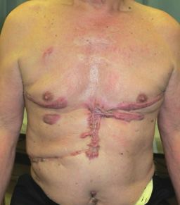
R. Ogawa, et al

keloids/HSs are particularly suitable for laser therapy           Conflict of Interest: The authors declare no conflicts of inter-
because the laser beam can reach the blood vessels in the         est.
dermis.
  Once scar inflammation has been completely quelled                                        References
                                                                   1.Ogawa R. Keloid and hypertrophic scars are the result of
by other methods, fractional abrative lasers can be used
                                                                     chronic inflammation in the reticular dermis. Int J Mol
for low-risk individuals. This induces a wound healing               Sci. 2017;18(3):606.
response that increases collagen III production, thereby           2.Matsumoto NM, Peng WX, Aoki M, et al. Histological
                                                                     analysis of hyalinised keloidal collagen formation in ear-
encouraging scar remodeling. Note: it is important to
                                                                     lobe keloids over time: collagen hyalinisation starts in the
cool the skin when conducting laser therapy to prevent               perivascular area. Int Wound J. 2017;14(6):1088―93.
superficial injury.                                                3.Nakashima M, Chung S, Takahashi A, et al. A genome-
                                                                     wide association study identifies four susceptibility loci
  However, these lasers have only limited efficacy with
                                                                     for keloid in the Japanese population. Nat Genet. 2010
thick, highly inflamed keloids/HSs.                                  Sep;42(9):768―71.
  Make-up Therapy                                                  4.Ogawa R, Watanabe A, Than Naing B, et al. Associations
                                                                     between keloid severity and single-nucleotide polymor-
  Medical make-up techniques such as “Rehabilitation                 phisms: importance of rs8032158 as a biomarker of keloid
Make-upⓇ” are recommended if the scars are on exposed                severity. J Invest Dermatol. 2014 Jul;134(7):2041―3.
areas (e.g. the face, neck, and upper limbs). These tech-          5.van de Kar AL, Houge G, Shaw AC, et al. Keloids in
                                                                     Rubinstein-Taybi syndrome: a clinical study. Br J Derma-
niques can be learned and self-applied by the patients.              tol. 2014;171(3):615―21.
Since our cosmetics are water-proof, they are not affected         6.Yagi Y, Kuwatsuka Y, Asai M, Honda M, Utani A. Coexis-
by humid/wet conditions. These techniques associate                  tence of keloids and pilomatricoma in a patient with
                                                                     Rubinstein-Taybi syndrome. Dermatol Online J. 2018;24
with improved mental health, acceptance of the scar ap-              (1):13030/qt4rq2k5fr.
pearance, and more positive attitudes to scar treatment.           7.Moustafa MF, Abdel-Fattah MA, Abdel-Fattah DC. Pre-
                                                                     sumptive evidence of the effect of pregnancy estrogens on
  Since it is difficult to improve the aesthetics of highly
                                                                     keloid growth. Case report. Plast Reconstr Surg. 1975 Oct;
elevated scars with make-up therapy, scar thickness                  56(4):450―3.
should be reduced with steroid plaster/injection before            8.Arima J, Huang C, Rosner B, Akaishi S, Ogawa R. Hyper-
                                                                     tension: a systemic key to understanding local keloid se-
commencing make-up therapy.
                                                                     verity. Wound Repair Regen. 2015;23(2):213―21.
  Slightly rough scars can be covered by thin tapes prior          9.Quong WL, Kozai Y, Ogawa R. A case of keloids compli-
to applying the foundation.                                          cated by Castleman’s disease: Interleukin-6 as a keloid
                                                                     risk factor. Plast Reconstr Surg Glob Open. 2017;5(5):e
                                                                     1336.
                 Follow-up of Keloids/HSs                         10.Noishiki C, Hayasaka Y, Ogawa R. Sex differences in
Keloid/HS patients who receive multimodal therapy                    keloidogenesis: An analysis of 1659 keloid patients in Ja-
                                                                     pan. Dermatol Ther (Heidelb). 2019;9(4):747―54.
should undergo prolonged follow-up and be taught how              11.Ogawa R, Akaishi S, Huang C, et al. Clinical applications
to manage their scar and any new wounds. Patient edu-                of basic research that shows reducing skin tension could
cation is a hugely important factor that dictates the suc-           prevent and treat abnormal scarring: the importance of
                                                                     fascial/subcutaneous tensile reduction sutures and flap
cess of our protocol.                                                surgery for keloid and hypertrophic scar reconstruction. J
                                                                     Nippon Med Sch. 2011;78(2):68―76.
                        Conclusions                               12.Ogawa R, Akaishi S, Kuribayashi S, Miyashita T. Keloids
                                                                     and hypertrophic scars can now be cured completely: Re-
Multimodal therapy, particularly surgery, radiation, and             cent progress in our understanding of the pathogenesis of
steroid tape/plaster, successfully manages keloids/HSs.              keloids and hypertrophic scars and the most promising
                                                                     current therapeutic strategy. J Nippon Med Sch. 2016;83
Our extensive experience, including with international
                                                                     (2):46―53.
patients, suggests that the NMS protocol may be suitable          13.Akaishi S, Akimoto M, Ogawa R, Hyakusoku H. The re-
as a foundation for a standard international prevention/             lationship between keloid growth pattern and stretching
                                                                     tension: visual analysis using the finite element method.
treatment algorithm for pathological scars. It should be
                                                                     Ann Plast Surg. 2008;60(4):445―51.
noted that the NMS protocol relies on close, prolonged            14.Levenson SM, Geever EF, Crowley LV, Oates JF 3rd, Ber-
follow-up, which is only feasible if keloid/HS treatments            ard CW, Rosen H. The healing of rat skin wounds. Ann
                                                                     Surg. 1965;161(2):293―308.
are covered by a national universal insurance system (as          15.Goutos I, Ogawa R. Steroid tape: A promising adjunct to
it is in Japan)22. We expect that treatments will improve            scar    management.      Scars    Burn     Heal.    2017 ; 3 :
significantly as our understanding of scar biology grows,            2059513117690937.
                                                                  16.Ogawa R, Akita S, Akaishi S, et al. Diagnosis and treat-
more good-quality institutional trials are conducted, and            ment of keloids and hypertrophic scars-Japan scar work-
new agents are found.

  8                                                                                             J Nippon Med Sch 2021; 88 (1)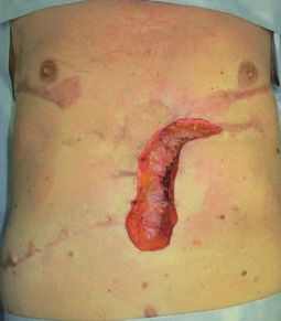
Keloid & Hypertrophic Scar NMA Protocol

   shop consensus document 2018. Burns Trauma. 2019;7:39.           5;Forthcoming 2020. doi: 10.1097/DSS.0000000000002235
17.Ogawa R. Surgery for scar revision and reduction: from        22.Takura T. An evaluation of clinical economics and cases
   primary closure to flap surgery. Burns Trauma. 2019;7:7.         of cost-effectiveness. Intern Med. 2018;57(9):1191―200.
18.Ogawa R, Tosa M, Dohi T, Akaishi S, Kuribayashi S. Sur-
   gical excision and postoperative radiotherapy for keloids.
   Scars Burn Heal. 2019;5:2059513119891113.                                                           (Received,       May 30, 2020)
19.Yoshino Y, Kubomura K, Ueda H, Tsuge T, Ogawa R. Ex-                                               (Accepted,        July 2, 2020)
   tension of flaps associated with burn scar reconstruction:
                                                                            (J-STAGE Advance Publication, August 1, 2020)
   A key difference between island and skin-pedicled flaps.
   Burns. 2018;44(3):683―91.
20.Koike S, Akaishi S, Nagashima Y, Dohi T, Hyakusoku H,           Journal of Nippon Medical School has adopted the Creative Com-
   Ogawa R. Nd:YAG laser treatment for keloids and hy-             mons Attribution-NonCommercial-NoDerivatives 4.0 International
   pertrophic scars: An analysis of 102 cases. Plast Reconstr      License (https://creativecommons.org/licenses/by-nc-nd/4.0/) for
                                                                   this article. The Medical Association of Nippon Medical School re-
   Surg Glob Open. 2015;2(12):e272.
                                                                   mains the copyright holder of all articles. Anyone may download,
21.Tsai CH, Kao HK, Akaishi S, An-Jou Lin J, Ogawa R.              reuse, copy, reprint, or distribute articles for non-profit purposes
   Combination of 1,064-nm neodymium-doped yttrium alu-            under this license, on condition that the authors of the articles are
   minum garnet laser and steroid tape decreases the total         properly credited.
   treatment time of hypertrophic scars: An analysis of 40
   cases of cesarean-section scars. Dermatol Surg. 2019 Nov

  J Nippon Med Sch 2021; 88 (1)                                                                                                      9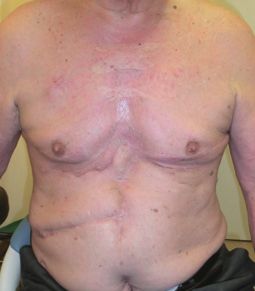

You can also read