The Ultrastructure of the Nuclear Envelope as Seen in Replicas from Normal and Neoplastic Nuclei
←
→
Page content transcription
If your browser does not render page correctly, please read the page content below
The Ultrastructure of the Nuclear Envelope as Seen in
            Replicas from Normal and Neoplastic Nuclei
                                                      A. A. BARTON
                      (Department   of Anatomy,   Rayai College of Surgeons of England, London, England)

                                                          SUMMARY
              A method of preparing carbon replicas of isolated liver and tumor nuclei is described
          which enables large areas of the nuclear surface to be examined. Two types of struc
          ture may be recognized: the first, large and irregular elevations thought to be due to
          nucleoli within the nucleus and, second, large numbers of small circular elevations
           0.25 ßin diameter covering the whole surface. The appearance is compared with the
          results obtained by sectioning isolated nuclei and the tissues from which they were
          derived.

   The appearance of the surface of isolated nuclei               acid-sucrose medium and centrifuged for 6 min
under the light microscope is known to yield in                   utes at 500Xg. The supernatant fluid was dis
complete information as to the degree of contami                  carded and the precipitate resuspended and spun
nation of the nucleus by cytoplasm, because of the                twice at 500 Xe.
containing 0.008 M citric acid. The homogenate                                          RESULTS
was strained twice through nylon gauze and cen                        Liver nuclei.—Alow-power view of several repli
trifuged at 0°C.for 10 minutes at 880X0- The
                                                                  cas obtained in the above manner from liver nuclei
supernatant fluid was discarded and the pre                        is seen in Figure 1. Each produced a circular im
cipitate resuspended in 4 volumes of the citric                    pression on the carbon, with an average diameter
   Received for publication August 1, 1960.                       of 8p, raised above the film of Formvar which
                                                               198

Downloaded from cancerres.aacrjournals.org on December 30, 2020. © 1961 American Association for Cancer
                                               Research.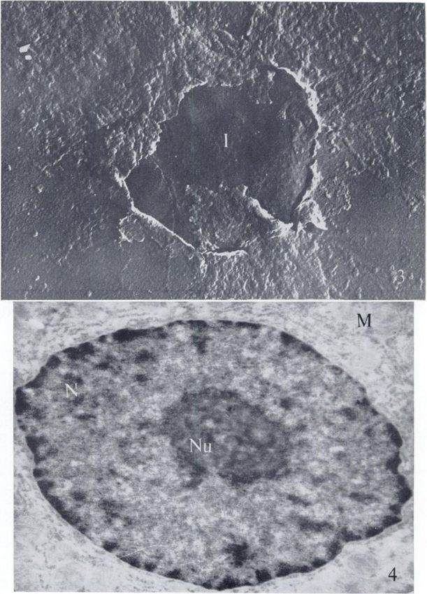
BARTON—Ultrastructureof Normal and Neoplastic Nuclei 199
originally supported it. The shadowing angle used Sßhigh at the edges. Two types of elevation may
was tan~1J, meaning that the shadow cast was be recognized in these casts of the nuclear surface :
8 times the height, so that it was possible to calcu the larger is indistinct but probably corresponds
late the thickness of dried, collapsed nuclei. to that seen in liver nuclei; the fine element covers
The color of the carbon deposit indicated that the nuclear surface.
the thickness of the covering film was about 250 A, The debris surrounding the nucleus consists of
so that it could be determined that the thickness of rounded masses of material j/u in diameter, pre
the nuclear rim was approximately 0.05 /¿;this senting the appearance of several smaller rounded
measurement remained constant whether the nu units surrounding a central depression. The sus
clei were fixed in formaldehyde vapor or dried pension of crushed cells was gelatinous, but this
without fixation. decreased with washing and centrifugation.
The more detailed appearance of the nuclear An ultrathin section of a single nucleus taken
surface is shown in Figure 2. It is seen that there from the same sample of nuclei is seen in Figure 7,
are two types of elevation. There are (^4), the when identical conditions of isolation as were em
large, somewhat irregular elements, often four in ployed in the case of liver were used. A consider
number, occupying the central part of the cast, able quantity of cell debris remains attached to
together with similar masses at the periphery; in the nucleus. This confirms the findings of Davison
addition, there are numbers of smaller, circular and Mercer (7) and Hawtrey and Silk (9) for tu
elevations (B), \p in diameter, which cover the mor nuclei.
whole surface of the nucleus, similar in appear
ance to those seen by Õngulo and Watson (1) on DISCUSSION
the surface of whole liver nuclei shadowed with An examination with the electron microscope of
chromium. thin sections of isolated nuclei has been used pre
When replicas are taken from the partially puri viously (6, 7) to check the cytoplasmic contami
fied fraction of the second centrifugation hi sucrose nation of nuclei subjected to chemical analysis.
mixture, there is no increase in the number of ele It is known that osmium tetroxide is reactive
vations on the nuclear surface. only with certain groups (2). As a result, high and
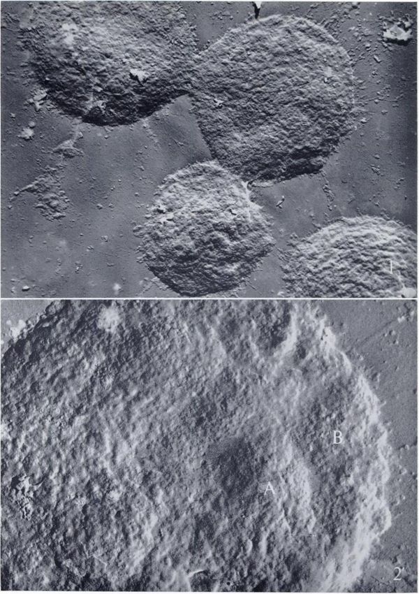
Surrounding the nucleus, and radiating from it, low polymers of DNA and RNA, nucleotides, car
are the replicas of strands of material which ex bohydrates, etc., cannot be demonstrated in the
tend several p into the surrounding support. Fig electron microscope if osmium fixation is em
ure 3 shows a nucleus which has burst, with ployed. The replica method, however, is a less
strands of cytoplasmic material attached to it. specific way of revealing structures, which, since
The inner surface of the nuclear membrane can it is based on their dry mass, may be expected to
be seen. It is free from the elevations associated give a more valid assessment of the contamination
with the outer nuclear surface. of samples of nuclei by cytoplasm and is important
The appearance of ultrathin sections of a sample in the analysis of nuclear fractions for those sub
taken from the preparation of liver nuclei fixed stances which cannot reveal themselves in thin
in 1 per cent osmium tetroxide buffered to pH 7.3 sections. It would be interesting to know what sub
is seen in Figure 5. In spite of the interval of time stance fills the relatively large spaces between iso
and manipulations between the death of the mouse lated nuclei as they appear in thin sections, since
and the fixation, the preservation of structure is the nuclei are widely separate, and, although it is
fairly good, which confirms the findings of Davi- not possible to pack them closer together, little
son and Mercer (7) and Chauveau et al. (6), who electron-dense material can be demonstrated be
compared isolated nuclei with those of intact liver tween them.
cells. The outline of the nuclear membrane is The elevations of the nuclear surface of both
shown, and the homogeneity of the nuclear con
normal and tumor cells could be due to the pres
tents can be seen and compared with that of an
intact liver cell (Fig. 4). ence of material either outside or within the
Tumor nuclei.—Figure6 is a replica of a tumor nucleus.
nucleus. Surrounding it are replicas of cytoplasmic The larger form of elevation (^4) is thought to
debris. The length of the shadow cast at tan"1, be derived from the presence of nucleoli within the
which is used here because of the relatively great nucleus. These are known to possess a density
height, indicates that this nucleus has dried to a greater than that of any other cell structure (10),
form which is higher than that of the liver nucleus, and, as the nucleus dries, they become trapped
though there is considerable variation in the within the nuclear membrane, leaving a raised
shadow cast by these nuclei, which may be up to impression.
Downloaded from cancerres.aacrjournals.org on December 30, 2020. © 1961 American Association for Cancer
Research.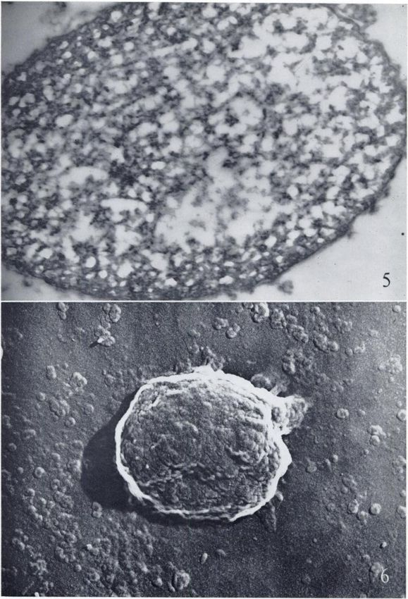
•200                                               Cancer Research                                Vol. 21, February 1961
   The similarity in ultrastructural appearance of                                        REFERENCES
replicas of cytoplasmic debris and the smaller ele                 1. ÕNGULO,  J. J., and WATSON,J. H. L. An Electron Micro
vations (B) which cover the surface of normal and                     scope Study of Isolated Nuclei of Liver Cells from Labora
                                                                      tory Animals. Science, 111:670-73, 1950.
tumor cell nuclei suggests that they are the same                  •i.
                                                                      BAHR, G. F. The Reactions of Osmium Tetroxide in Rela
structures and that a certain amount of cyto                          tion to Electron Microscopy. Proc. Third Internat. Conf.
plasmic material remains adherent to the nucleus.                     Electron Microscopy, pp. 144-47. London, 1954.
                                                                   3. BARTON,A.A. Replication Techniques in Electron Micros
An examination of thin sections reveals irregulari                    copy. J. Anat. (in press).
ties of the outer nuclear membrane, but not in the                 4. BRADLEY,D. E., and WILLIAMS,D. J. An Electron Micro
numbers that might be expected from the results                       scope Study of the Spores of Some Species of the Genus
given by the replica technic. It is believed, how                     Hacittus Using Carbon Replicas. J. Gen. Microbio!., 17:
                                                                      75-79, 1957.
ever, that this is because many of the adherent                    5. CAUSEY,G. Experimental Tumours of Peripheral Nerve in
particles are not osmiophilic and are invisible in                    Mice. Acta Union internat, contra cancrum, 16(1) : 142-48,
section. Sections of nuclei derived from neoplastic                   1959.
                                                                   6 CHAUVEAU,     J.; MOULÉ, Y.; and ROUILLER,C. Isolation of
cells are surrounded by a varying quantity of cyto                    Pure and Unaltered Liver Nuclei Morphology and Bio
plasm, greater in extent than is the case with nor                    chemical Composition. Exper. Cell Research, 11:317-21,
mal liver cells. In carbon replica these nuclei ap                     1956.
pear higher as a result of the material drying onto                7. DAVISON,P. F., and MERCER,E. H. Electron Microscopy
                                                                      of Cell Nuclei Isolated in Aqueous Media. Exper. Cell Re
their surface.                                                        search, 11:237-39, 1956.
                                                                   8. FRAZER,S. C., and DAVIDSON,J. N. Photometric Estima
                      ACKNOWLEDGMENTS                                 tions of Deoxyribonucleic Acid in Individual Cell Nuclei.
                                                                      Exper. Cell Research, 4:316-32, 1953.
    I wish to thank Professor G. Causey for valuable sugges
                                                                   9. HAWTREY,A. O., and SILK, M. H. Mitochondria and the
tions and for supplying the tumor material used throughout            Ehrlich Ascites-Tumour Cell. Biochem. J., 74: 21-26, 1960.
these investigations.                                             10. VINCENT,W. S. Some Studies on Differentiation and De
    I also wish to thank Mr. S. A. Edwards and Miss J. Arm            velopment of the Oocyte. In: The Beginnings of Embryon
strong for technical assistance and the British Empire Cancer         ic Development, pp. 1-22. American Association for the
Campaign for financial support.                                       Advancement of Science, Washington, D.C., 1957.

                                     FIG. 1.-—Acarbon replica, shadowed with gold/palladium,
                                obtained from the preparation of isolated nuclei. Each nucleus
                                has left an imprint of its surface. X8.000. (By permission of
                                Journal of Anatomy.)
                                     FIG. 2.—Areplica of part of the nuclear surface of a liver
                                cell showing its elevations. The larger (A) is believed to be
                                due to nucleoli trapped within the nucleus, the smaller (D) due
                                to cytoplasmic material drying on to the outer surface of the
                                nucleus. X20.000.

Downloaded from cancerres.aacrjournals.org on December 30, 2020. © 1961 American Association for Cancer
                                               Research.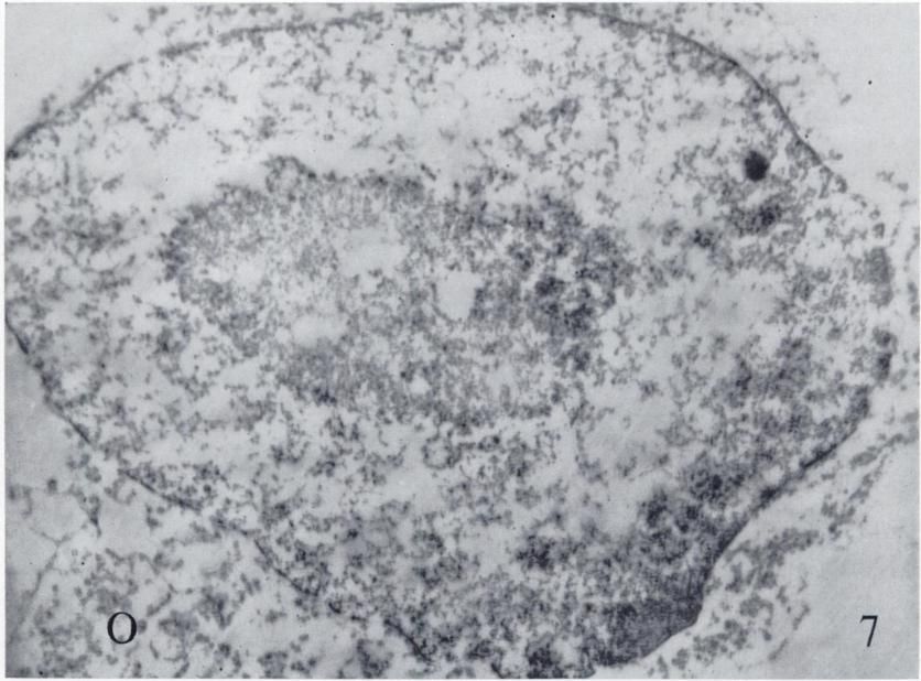
Downloaded from cancerres.aacrjournals.org on December 30, 2020. © 1961 American Association for Cancer
                                               Research.
FIG. 3.—A carbon replica of a liver nucleus which has
burst to reveal the smooth inner surface of the nucleus (/).
X 17,000.
FIG. 4.—An electron micrograph of an ultrnthin section
of liver fixed in osmium tetroxide showing the nucleus (fi),
nucleolus (Nu), and mitochondria (M). X20.000.
Downloaded from cancerres.aacrjournals.org on December 30, 2020. © 1961 American Association for Cancer
Research.Downloaded from cancerres.aacrjournals.org on December 30, 2020. © 1961 American Association for Cancer
Research.FIG. 5.—Electron micrograph of an ultrathin section of
a liver nucleus isolated using the methods of Frazer and
Davidson fixed in osmium tetroxide. The surface of the nucleus
is seen to l>e relatively free of osmiophilic material. XÕO.IHIO.
FIG. 6.—A carbon replica of a tumor nucleus shadowed
with gold palladium at an angle of tan"1. The length of the
shadow cast indicates that this nucleus dried to a form of
relatively greater height than that of the normal liver nucleus,
owing to the presence of cytoplasmic material adherent to
the nucleus. XSO.OOO.
Downloaded from cancerres.aacrjournals.org on December 30, 2020. © 1961 American Association for Cancer
Research.Downloaded from cancerres.aacrjournals.org on December 30, 2020. © 1961 American Association for Cancer
Research.Fio. 7.—Osmiophilicmaterial (O) attached to an isolated
tumor nucleus seen in an ultrathin section. X16.000.
Downloaded from cancerres.aacrjournals.org on December 30, 2020. © 1961 American Association for Cancer
Research.£ü
,
F--1
Downloaded from cancerres.aacrjournals.org on December 30, 2020. © 1961 American Association for Cancer
Research.The Ultrastructure of the Nuclear Envelope as Seen in Replicas
from Normal and Neoplastic Nuclei
A. A. Barton
Cancer Res 1961;21:198-200.
Updated version Access the most recent version of this article at:
http://cancerres.aacrjournals.org/content/21/2/198
E-mail alerts Sign up to receive free email-alerts related to this article or journal.
Reprints and To order reprints of this article or to subscribe to the journal, contact the AACR Publications
Subscriptions Department at pubs@aacr.org.
Permissions To request permission to re-use all or part of this article, use this link
http://cancerres.aacrjournals.org/content/21/2/198.
Click on "Request Permissions" which will take you to the Copyright Clearance Center's (CCC)
Rightslink site.
Downloaded from cancerres.aacrjournals.org on December 30, 2020. © 1961 American Association for Cancer
Research.You can also read