Three-dimensional ultrasonographic features of diamniotic conjoined twins with body stalk anomaly - BMC Pregnancy and Childbirth
←
→
Page content transcription
If your browser does not render page correctly, please read the page content below
Xiang et al. BMC Pregnancy and Childbirth           (2020) 20:221
https://doi.org/10.1186/s12884-020-02920-0

 CASE REPORT                                                                                                                                        Open Access

Three-dimensional ultrasonographic
features of diamniotic conjoined twins with
body stalk anomaly
Guishuang Xiang1†, Yanting Wen1*†, Li Zhang2, Xiaoqian Tong1 and Lu Li1

  Abstract
  Background: Since conjoined twins were thought to be monoamniotic in the past, diamniotic conjoined twins
  would be improbable theoretically. Body stalk anomaly is a severe defect of the body wall, which is rare among
  twins. Body stalk anomaly in monochorionic diamniotic conjoined twins has never been reported prenatally so far
  as we know.
  Case presentation: Here we present an extremely rare case of concordant body stalk anomaly in monochorionic
  diamniotic conjoined twins. Ultrasonography at 9 + 5 weeks revealed one chorionic and two amniotic cavities, close
  apposition of abdomen, limited movement, and common umbilical vessels. Follow-up ultrasonography at 11 + 6
  weeks and 13 + 2 weeks showed close apposition of the lower abdominal region with cystic structures and a small
  bowel-like mass between the two fetuses. Three-dimensional ultrasonography assisted in observing the entire
  appearance of both twins in earlier first trimester, including amnioticity, conjoined region and umbilical vessels. We
  attribute this diamniotic conjoined twin in our case to the fusion theory. A single yolk sac was observed, challenging
  the idea that yolk sac number predicts amnionicity. Identification of single yolk sac and its allantois may form a
  common body stalk during this fusion, leading to concordant body stalk anomaly in monochorionic diamniotic twins.
  Conclusions: Our case may provide important insights into both ultrasonographic features and embryogenesis of this
  extremely rare anomaly.
  Keywords: Conjoined twins, Monochorionic-diamniotic, Ultrasound, Yolk sac, Body stalk anomaly

Background                                                                             features in our case are more consistent with the fusion
Monochorionic diamniotic (MD) conjoined twins is rare,                                 hypothesis, which stipulates that the infraumbilical ab-
and only 9 previous cases were reported [1–9]. This                                    dominal wall forms in the area of allantois and the caudal
anomaly manifests on ultrasonography as one chorionic                                  part of yolk sac. Fusion in this area disturbs the develop-
and two amniotic cavities, union of peritoneal cavities                                ment of the infraumbilical abdominal wall and induces
through an abdominal wall defect, conjoined intestine,                                 conjoining of adjacent intestines, leading to MD conjoined
and anorectal malformation. In the literature, fusion and                              twins. Body stalk anomaly (BSA) is a severe defect of the
fission models have been proposed to explain the embryo-                               body wall, which occurs in approximately 1 per 7500 preg-
genesis of MD conjoined twins. The ultrasonographic                                    nancies [10] in the first trimester. BSA is rare among
                                                                                       twins, in which case it can be discordant or concordant.
* Correspondence: 1375825374@qq.com                                                    Here we describe a concordant BSA in MD conjoined
†
 Guishuang Xiang and Yanting Wen contributed equally to this work.                     twins treated at our hospital. We compare our case with
1
 Department of Ultrasound, The Fifth People’s Hospital of Chengdu, 33
Mashi Street, Chengdu 611130, Sichuan Province, China
                                                                                       similar ones in the literature to establish characteristic
Full list of author information is available at the end of the article                 ultrasonography features that may facilitate early prenatal
                                        © The Author(s). 2020 Open Access This article is licensed under a Creative Commons Attribution 4.0 International License,
                                        which permits use, sharing, adaptation, distribution and reproduction in any medium or format, as long as you give
                                        appropriate credit to the original author(s) and the source, provide a link to the Creative Commons licence, and indicate if
                                        changes were made. The images or other third party material in this article are included in the article's Creative Commons
                                        licence, unless indicated otherwise in a credit line to the material. If material is not included in the article's Creative Commons
                                        licence and your intended use is not permitted by statutory regulation or exceeds the permitted use, you will need to obtain
                                        permission directly from the copyright holder. To view a copy of this licence, visit http://creativecommons.org/licenses/by/4.0/.
                                        The Creative Commons Public Domain Dedication waiver (http://creativecommons.org/publicdomain/zero/1.0/) applies to the
                                        data made available in this article, unless otherwise stated in a credit line to the data.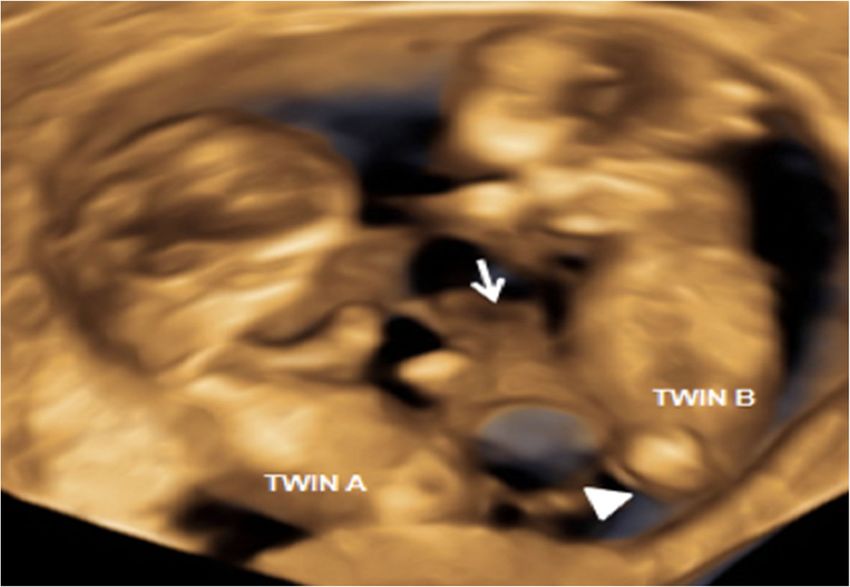
Xiang et al. BMC Pregnancy and Childbirth         (2020) 20:221                                                                                 Page 2 of 5

diagnosis, and we attempt to gain insights into the em-                         the small intestine of twin A (Fig. 3). Twin A was found
bryogenesis of these anomalies.                                                 to have bowel involving the small intestine and colon
                                                                                herniated into exocoelom. An urachal remnant was also
Case presentation                                                               found outside the lower part of the abdomen. This post-
A 27-year-old woman (gravida 1, para 0) who had be-                             abortion analysis confirmed the diagnosis of MD con-
come pregnant naturally was referred to our department                          joined twins with BSA.
at 9 + 5 weeks of gestation. She had no significant history
of health issues and no history of multiple gestations.                         Discussion and conclusions
Two-dimensional (2D) and three-dimensional (3D)                                 In the literature, MD conjoined twins are characterized by
ultrasonography at 9 + 5 weeks revealed one gestational                         one chorionic and two amniotic cavities, the union of
sac and an apparently dividing amniotic membrane sur-                           peritoneal cavities through an abdominal wall defect, con-
rounding each twin (Fig. 1a, b), which led to diagnosis of                      joined intestine, and anorectal malformation [1–9]. 5 out
MD conjoined twin pregnancy. Only one yolk sac was                              of 9 cases were described as shared or bifurcated umbilical
observed (Fig. 1a). It was difficult to determine whether                       cord, 3 out of 9 cases were separated, and 1 out of 9 cases
the abdominal structures were conjoined because of the                          was not mentioned. Furthermore, 5 out of 9 previous
twins’ relatively fixed position and limited movement.                          cases were described as a single yolk sac, and the rest were
2D/3D Ultrasonography at 11 + 6 weeks showed close                              not mentioned (Table 1). This case was diagnosed as MD
apposition of the lower abdominal region with cystic                            conjoined twins because of the conjoined intestine and
structures and a small bowel-like mass between the two                          fused skin bridge in lower part of abdomen.
fetuses (Fig. 2), and limited fetal movements for both                             BSA is a severe abdominal wall defect and is associated
twins. Doppler ultrasound showed no free-floating um-                           with abnormal embryonic folding in the 5th week of ges-
bilical cords for either twin, but several umbilical vessels                    tation [11]. It seems to be more common in twin preg-
coiled around the cystic structures and inserted into the                       nancies. Previous reports showed a large abdominal wall
placenta along the dividing membrane. Follow-up ultra-                          defect and herniation of the liver, bowels, or heart into
sonography at 13 + 2 weeks of gestation revealed the                            the exocoelom [12–14]. However, none of the cases
twins were in the same relative position from all angles                        showed conjoined intestine or fused skin bridge, in con-
and moved together, the cystic structures and bowel-like                        trast to the present case. Gastroschisis and intestine
mass seemed to lie in the exocoelom between the two                             partly herniated into exocoelom in our case are consist-
amniotic cavities, and hydrodermia in twin B. The blad-                         ent with characteristics of BSA.
der configuration of either fetus could not be observed.                           3D ultrasonography assisted in revealing the entire ap-
In the end, MD conjoined twins with BSA was diag-                               pearance of both twins, including amnioticity, conjoined
nosed. The parents requested induced abortion at 13 + 4                         region and the location of umbilical vessels in the first
weeks of gestation and consent to pathological analysis.                        trimester. However, this may not always prove feasible
  Post-abortion examination showed a single placenta                            early in the first trimester, which may show limited fetal
with dividing amniotic membranes attached to the cen-                           movement and unclear position of the herniated abdom-
ter of the placenta. Four umbilical vessels traveled along                      inal mass, as in our case. The diagnosis of BAS in MD
the dividing amniotic membrane and inserted together                            conjoined twins was not clear until we found the bowel-
into the placenta. Both twins had gastroschisis with con-                       like mass lying in the exocoelom at 13 + 2 weeks.
joined intestine and a fused skin bridge in lower part of                          Fission and fusion models have been proposed to ex-
abdomen. Twin B was found to have hydrodermia, stre-                            plain the embryogenesis of MD conjoined twins. The fis-
phenopodia and a small intestine exstrophy fused with                           sion hypothesis [1] speculates that there is only one

 Fig. 1 Diamniotic membrane (arrows) between the twins at 9 + 5 weeks in two-dimensional (a) and three-dimensional (b) ultrasonography, respectively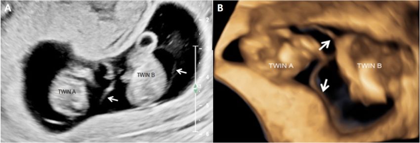
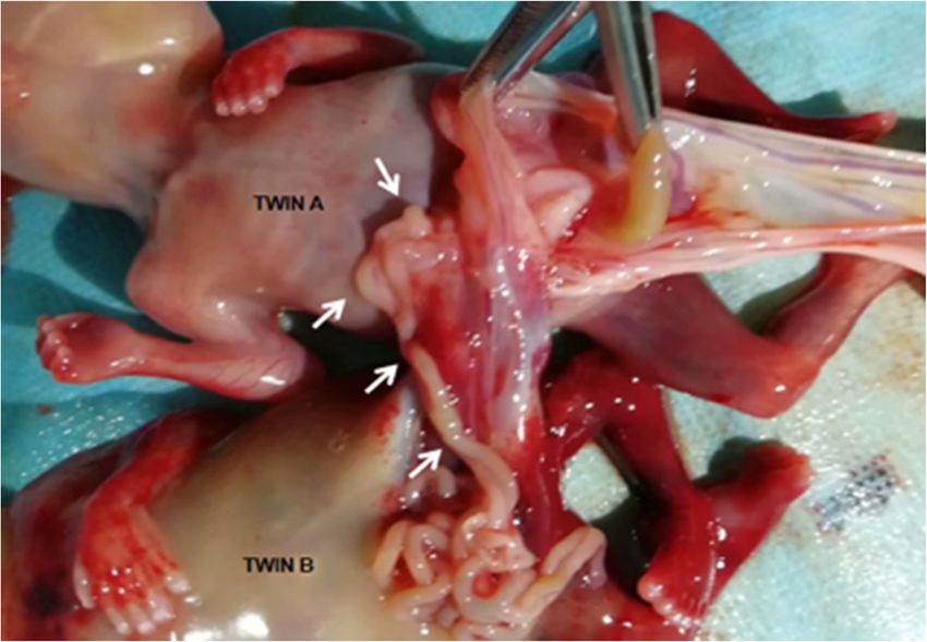
Xiang et al. BMC Pregnancy and Childbirth      (2020) 20:221                                                                          Page 3 of 5

 Fig. 2 Cystic structures (arrow head) and bowel-like mass (arrow) between the two fetuses at 11 + 6 weeks in three-dimensional ultrasonography

amniotic cavity with two embryos conjoined at specific                      developed. In the literature, yolk sac number can pre-
sites. During embryo folding, a crease develops between                     dict amnionicity [16], however, a single yolk sac in
the two embryos, and the amniotic cavity is divided into                    MD conjoined twins may be associated with a higher
two. This hypothesis cannot explain gastroschisis and                       risk of congenital defects [17]. Shen et al. [18] specu-
urachal remnant at the lower part of the abdomen                            late that yolk sac formation occurs before the differ-
visible by ultrasonography. The fusion hypothesis [15]                      entiation of amnion and after that of chorion. They
stipulates that the infraumbilical abdominal wall                           suggested that the splitting occurs after chorion and
forms in the area of allantois and the caudal part of                       before amnion differentiation. As a consequence, only
yolk sac. Fusion in this area disturbs the development                      one yolk sac develops, with one chorionic and two
of the infraumbilical abdominal wall and induces con-                       amniotic cavities. We speculate that the single yolk
joining of adjacent intestines, leading to MD con-                          sac in our case explains the single allantois, since the
joined twins. Our case had a single yolk sac, which                         allantois is a small diverticulum of the caudal part of
may provide insights into how those anomalies                               the yolk sac. We hypothesize that the single yolk sac

 Fig. 3 Post-abortion examination showed the intestinal tract of twin A was fused to that of twin B (arrows)
Xiang et al. BMC Pregnancy and Childbirth      (2020) 20:221                                                                           Page 4 of 5

Table 1 Clinical characteristics of diamniotic conjoined twins
Study                 Yolk sac Twin characteristics                                          Umbilical cord                             Gestational
                      no.                                                                                                               age, wk
Kapur R P 1994 [1]    single   Conjoined bowels in communicating                             Two separate umbilical cords, each         16
                               omphalocele sac, shared persistent cloaca                     containing 2 arteries and 1 vein
Costa S L 2006 [6]    NR       Conjoined bowels in communicating                             NR                                         NR
                               omphalocele sac, a shared bladder, anal atresia
Karnak I 2008 [4]     NR       Conjoined bowels in communicating omphalocele sac,            Two separate umbilical cords, each         NR
                               cloacal anomaly                                               containing 3 vessels
Tihtonen K 2009 [5]   NR       Conjoined bowels in communicating omphalocele sac             Shared umbilical cord with 4 arteries and 18
                                                                                             2 veins
Destephano C C        single   Joined in abdominal region only (omphalopagus)                A bifurcated umbilical cord                11
2010 [7]
Weston P J 2010       single   Conjoined bowels in communicating omphalocele                 Shared umbilical cord with 2 separate      After birth
                               sac, anal atresia, hypoplasia                                 sets of blood vessels
Wielgos M 2014 [2]    NR       Conjoined bowels in communicating omphalocele sac             Two separate umbilical cords               27
Maruyama H 2015       single   Conjoined bowels, anal atresia                                Shared short umbilical cord                12
[8]
Nupur Shah 2019 [9]   single   Joined at the periumbilical region                            A bifurcated umbilical cord                8
NR Not reported

 Fig. 4 Drawing of the embryogenetic model to explain our case. The common body stalk and single allantois contrast with the separated body
 stalk and two allantoises of other embryogenetic models. A, allantois; AC, amniotic cavity; BS, body stalk; CM, cloacal membrane; ED, embryonic
 disk; YS, yolk sac
Xiang et al. BMC Pregnancy and Childbirth (2020) 20:221 Page 5 of 5
and its allantois induce this fusion [15] and form 7. Destephano CC, Meena M, Brown DL, et al. Sonographic diagnosis of
common body stalk (Fig. 4). This may indicate com- conjoined diamniotic monochorionic twins. Am J Obstet Gynecol. 2010;
203(6):e4–6.
mon umbilical vessels and herniated intestines in the 8. Maruyama H, Inagaki T, Nakata Y, et al. Minimally conjoined Omphalopagus
exocoelom, suggesting a BSA in MD conjoined twins. twins with a body stalk anomaly. Ajp Reports. 2015;05(02):e124–8.
Considering the uniformly fatal nature of MD con- 9. Shah N. Monochorionic diamniotic conjoined twins: prenatal sonological
diagnosis at 8 weeks. Ultrasound Obstet Gynecol. 2019;54(5):699.
joined twins with BSA, early prenatal diagnosis is critical 10. Daskalakis G, Sebire NJ, Jurkovic D, et al. Body stalk anomaly at 10–14 weeks
for averting complications during pregnancy. Additional of gestation. Ultrasound Obstet Gynecol. 1997;10(6):416–8.
work is needed to explore the mechanisms behind this 11. Duhamel B. Embryology of exomphalos and allied malformation. Arch
DisChild. 1963;38:142–9.
anomaly and to understand its epidemiology and risk 12. Tavares MV, Domingues AP, Tavares M, Fonseca E, Moura P. Monoamniotic
factors. twins discordant for body stalk anomaly. J Matern-Fetal Med. 2015;28(1):113–5.
13. Iba T, Harada T, Iba Y, Nishikori K, Iwabe T, Harada T, et al. Concordant body
Abbreviations stalk anomalies in Dichorionic twins. J Ultrasound Med. 2016;35(12):2736–9.
2D: Two-dimensional; 3D: Three-dimensional; MD: Monochorionic diamniotic; 14. Rovida PL, Prefumo F, Frusca T, Fichera A. Concordant body stalk anomaly in a
BSA: body stalk anomaly monoamniotic twin pregnancy at 9 weeks. Prenat Diagn. 2015;34(9):915–6.
15. Spencer R. Minimally united ischiopagus twins: infraumbilical union with
Acknowledgements cloacal anomalies. J Pediatr Surg. 1996;31(11):1538–45.
Not applicable. 16. Bromley B, Benacerraf B. Using the number of yolk sacs to determine
amnionicity in early first trimester monochorionic twins. J Ultrasound Med.
1995;14(6):415–9.
Authors’ contributions
17. Poláková M, Zetová L, Vlk R. S V. Monochorionic biamniotic twins with a
GX and YW were major contributors in writing the manuscript. LZ was in
common yolk sac in the first trimester ultrasound scan - is there a higher
charge of post-abortion examination. XT and LL were in charge of revised
risk of a congenital defect? CeskaGynekol. 2012;77(6):521–3.
this manuscript substantively. All authors have read and approved the final
18. Shen O, Samueloff A, Beller U, Rabinowitz R. Number of yolk sacs does not
manuscript.
predict amnionicity in early first-trimester monochorionic multiple
gestations. Ultrasound Obstet Gynecol. 2006;27(1):53–5.
Funding
Not applicable.
Publisher’s Note
Availability of data and materials Springer Nature remains neutral with regard to jurisdictional claims in
Not applicable. published maps and institutional affiliations.
Ethics approval and consent to participate
Not applicable.
Consent for publication
Written informed consent was obtained from the patient for publication of
this case report and any accompanying images. A copy of the written
consent is available for review by the Editor-in-Chief of this journal.
Competing interests
The authors declare that they have no competing interests.
Author details
1
Department of Ultrasound, The Fifth People’s Hospital of Chengdu, 33
Mashi Street, Chengdu 611130, Sichuan Province, China. 2Department of
Pathology, The Fifth People’s Hospital of Chengdu, Chengdu 611130, China.
Received: 31 January 2020 Accepted: 2 April 2020
References
1. Kapur RP, Jack RM, Siebert JR. Diamniotic placentation associated with
omphalopagus conjoined twins: implications for a contemporary model of
conjoined twinning. Am J Med Genet. 1994;52(2):188–95.
2. Wielgos M, Bomba-Opon D, Kociszewska-Najman B, Brawura-Biskupski-
Samaha R, Kaminski A, Piotrowska A, et al. Minimally conjoined
monochorionic diamniotic twins – a case report. Eur J Obstet Gynecol
Reprod Biol. 2014;180:206–7.
3. Weston PJ, Ives EJ, Honore RL, et al. Monochorionic diamniotic minimally
conjoined twins: a case report. Am J Med Genet. 1990;37(4):558–61.
4. Karnak I, Sanlialp I, Ekinci S, et al. Minimally conjoined omphalopagi:
emphasis on embryogenesis and possibility of emergency separation. Turk J
Pediatr. 2008;50(5):503.
5. Tihtonen K, Lagerstedt A, Kirkinen P. Diamniotic Omphalopagus conjoined
twins in a diamniotic pregnancy. Fetal Diagn Ther. 2009;25(3):343–5.
6. Costa SL, Dunn L, Pantazi S, et al. P05.14: a novel case of monochorionic
diamniotic conjoined twins with genitourinary and gastrointestinal union.
Ultrasound Obstet Gynecol. 2006;28(4):562–3.You can also read