DIAGNOSTIC MEDICAL IMAGING - 1st Part - Introduction Ing. Tommaso Rossi
←
→
Page content transcription
If your browser does not render page correctly, please read the page content below
DIAGNOSTIC
DIAGNOSTICMEDICAL
             MEDICALIMAGING
                       IMAGING
    1st
     1stPart
        Part--Introduction
               Introduction

        Ing. Tommaso Rossi
     tommaso.rossi@uniroma2.it

   Tommaso Rossi - Modulo di SEGNALI , a.a. 2013/2014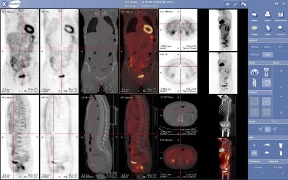
Overview                              2

How we can look on the inside of human body?

Invasive techniques: surgery, endoscope, etc.
• can cause damage or trauma to the body
• offer direct optical viewing
Non-invasive techniques: medical imaging
• some of these techniques are completely risk-free, for
  others there are risks associted with the radiation
  exposure
• allow us to see things not visible to the naked eye
             Tommaso Rossi - Modulo di SEGNALI, a.a. 2013/2014
Brief History                             3

The first published medical image was a radiograph
Of Wilhelm Conrad Roentgen wife’s hand (1895).
Using a Crookes’ tube Roentgen discovered a new
kind of rays, x-rays (wavelength between 10 nm and
10 pm), that could expose film even when optically
sheilded.
Few months later the first clinical use of x-rays
occurred. Later the medical use of x-rays became
common.

Nuclear medicine arose from the discovery of radioactivity by
Antoine Henri Becquerrel in 1896. The initial idea of using
radioactive tracers to study human physiology was introduced by
George de Hevesy in 1923.
The modern scintillation camera was developed in 1952.

                 Tommaso Rossi - Modulo di SEGNALI, a.a. 2013/2014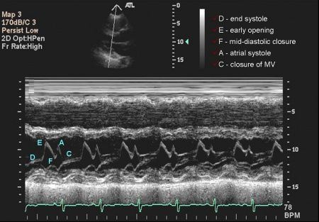
Brief History                                 4

The first interaction of acoustic waves with media was first described
by Lord John Rayleigh at the end of 1800.
Modern Ultrasound medical imaging was developed after the II World
War due to the development of Navy sonar technology.

Magnatic resonance imaging arises form the Nuclear magnetic
resonance phenomenon, discovered by Felix Bloch and Edward
Purcell that received the Nobel Prize in 1952.
In 1971 the use of this phenomenon in
medical imaging was suggested by
Raymond Damadian and this concept
was developed by Paul Lauterbur (who
won the Nobel Prize in Medicine in
2003) in 1973.

                 Tommaso Rossi - Modulo di SEGNALI, a.a. 2013/2014
Signals                                       5

    Physical signals studied in medical imaging arise from different processes

            a) Projection radiography            transmission of
                  and Computed                   photons (x-rays)
  use of                                         through the human
 ionizing    Tomography scanning
                                                 body
radiation                                        emission of photons           use of
            b) Nuclear medicine                  (gamma rays) from          electromagn.
                                                 radiotracers in the body      energy

                                                  precession of spin
            c) Magnetic resonance                 systems in a large
                                                  magnetic field

            d) Ultrasound imaging                reflection of ultrasonic   use of sound
                                                 waves within the body         waves

                       Tommaso Rossi - Modulo di SEGNALI, a.a. 2013/2014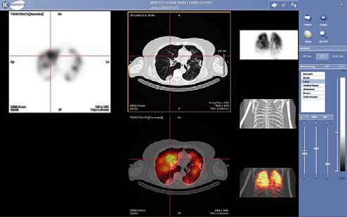
Projection Radiography                                   6

Projection of a 3D object or signal into a 2D image. The signal generator is
a x-ray a tube able to create a x-ray pulse in a uniform con beam.
The pulse, passing through the body, is attenuated by tissues. The signal
intensity profile becomes non uniform and shadows are created by dense
objects (i.e.: bones).

The x-ray signal intensity profile is revealed through
a scintillator that converts the signal to visible light,
that is finally captured (on a film, a camera or a solid-
state detector).

Structures located at different
deepts in the human body are
superimposed on a 2D image

                     Tommaso Rossi - Modulo di SEGNALI, a.a. 2013/2014
Computed Tomography                                          7

 CT uses x-rays not travelling in a 3D cone beam but collimated in a 2D
 “fan beam”.
Shadows are created by tissues in a 2D cross-section and the signal
intensity is detected by a large number of detectors. This measurement is
called projection.
Many projections are collected for different angular orientation of the tube
signal generator (and detectors that rotate around the human body).
Through these projections an image of
the human body cross-section is computed
(spatial resolution < 0.5 mm).

Different CT modalities:
•standard single-slice
•helical (whole body scan
 in less than a minute)
•multislice

                    Tommaso Rossi - Modulo di SEGNALI, a.a. 2013/2014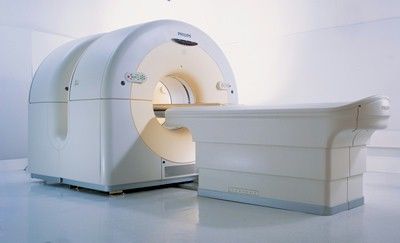
Nuclear Medicine 8
Images can be acquired only if appropriate radioactive substances
(radiotracers) are introduced into the body.
The image reflects the local concentration of a radiotracer within the body.
Being this concentration tied to the physiological behaviour, this method
is called functional imaging.
e.g. radioactive iodine is used to study tyroid functions.
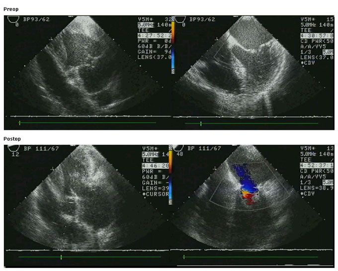
Three main modalities:
•conventional radionuclide imaging or planar scintigraphy
• single-photon emission computed tomography (SPECT) emission
computed
• positron emission tomography (PET) tomography
Planar scintigraphy and SPECT use radiotracres that are gamma emitters.
PET uses radiotracers that emit positrons.
SPECT and PET require tomographyc recnostruction while planar imaging
forms images by projection.
Tommaso Rossi - Modulo di SEGNALI, a.a. 2013/2014Nuclear Medicine 9
In contrast with projection radiography and computed tomography, the
biological behaviour of a substance’s biodistribution in the body is of
interest in nuclear medicine.
Each molecule of the substance is labeled with a radioactive atom. The
ionizing radiation emitted when this atom undergoes radioactive decay is
used to determine the location of the molecule within the body.
a) Projection radiograph,
image intensity reflects the
varying absorption of
transmitted x-rays through
the bones (structural
anatomical information)
b) Nuclear medicine “bone
scan”, image intensity
reflects the metabolic
activity of the bones
(metabolic information) a) b)
Tommaso Rossi - Modulo di SEGNALI, a.a. 2013/2014Nuclear Medicine 10
In nuclear medicine a 2D gamma ray detctor called Anger camera is
Used (invented in 1952 by Hal Anger of the Donner Laboratory at the
University of California).
Anger camera is able to detect single rays. This procedure combines the
effect of emission with effects of attenuation of rays due to body tissues.
Images are 2D projections of 3D distribution of radiotracers plus
Attenuation (spatial resolution 5-18 mm).
Nuclear medicine images are based on
the distribution of radiotracers, the
interest is not in total intesity (as
projection radiography and CT) but in
the detected decay rate of the source,
typically expressed as “counts” per
time. Anger camera
Tommaso Rossi - Modulo di SEGNALI, a.a. 2013/2014Nuclear Medicine 11
In convetional radionuclide imaging and SPECT a radioactive atom’s decay
produces a single gamma ray which may be detected by Anger camera (a
collimator is needed).
In PET a radionuclide decay produces a positron, which annihilates with an
electron producing two gamma rays flying off in opposite directions. PET
scanner looks for coincident detections from opposing detectios in its ring,
determining the line that passes through the site where the annihilation
occured.
SPECT scan that indicates the
baseline blood reaching the brain PET-CT
Tommaso Rossi - Modulo di SEGNALI, a.a. 2013/2014Ultrasound Imaging 12
Ultrasound imaging uses electrical-to-acustical transducers to generate high
frequency pulses (typically 1-10 MHz). These pulses travel through the body
and reflect back to the transducer.
gives information about
time of return of the reflected pulses
location of the reflector
intensity of the reflected pulses gives information about the
strength of the reflector
Since ultrasound imaging systems
have low image quality they are
used to analyse the anatomy (real-
time)
They are very cheap and small
Tommaso Rossi - Modulo di SEGNALI, a.a. 2013/2014Ultrasound Imaging 13
• A-mode imaging: (or amplitude-mode) one-dimensional pulse waveform,
used to generate detailed information about rapid or undetectable
motion, i.e.: hearth valve motion
• B-mode imaging: ordinary cross-sectional anatomical imaging (2D
image), created by a linear array of transducers scanning a plane
through the body
• M-mode imaging: (or motion-mode) a succession of A-mode signals,
each A-mode signal is a column in an image. Not an anatomical image
but important for measuring of time-varying displacements
• Doppler imaging: uses the property of frequency or phase shift caused
by moving objects to generate images that are colour coded by their
motion
M-mode image – mitral valve
Tommaso Rossi - Modulo di SEGNALI, a.a. 2013/2014Magnetic Resonance 14
Magnetic resonance scanners use the property of nuclear magnetic
Resonance (NMR) to create images
In a strong magnetic field the nucleus of hydrogen tends to align
itself with the field, creating a magnetization of the body.
It is possible to excite a selected region of the body, moving away
from the magnetic field direction groups of these “little magnets”.
Once protons return back to be aligned with the field they experience
a precession movement generating a radio-frequency wave that
is captured by an antenna.
MR produces high-resolution high-contrast cross-sectional anatomic
images and, like ultrasound imaging, is non-invasive.
Different kind of pulse sequences can be used to create different
images, a clever combination of pulse sequences can be used to
create dynamic series of images, which can be used to estimate
blood flow (Functional Magnetic Resonance Imaging)
Tommaso Rossi - Modulo di SEGNALI, a.a. 2013/2014Magnetic Resonance 15
All nuclei have positive charges (they are composed by protons and
neutrons). A nucleus with either an odd atomic number or an odd mass
number has an angular momentum – they have spin
If the nuclei of the hydrogen atoms (¹H) are subjected to a strong magnetic
field they tend to align with the field; being the number of hydrogen atoms
into the human body very high, this tendency results in a magnetization of
the body
Φ N
+ +
Nucleus angular Microscopic + +
momentum magnetization of +
nucleus + +
S
Tommaso Rossi - Modulo di SEGNALI, a.a. 2013/2014Magnetic Resonance 16
In normal conditions individual spins of ¹H nuclei have a random
JJG orientation,
JJG
has results no macroscopic magnetic field is produced M = µi = 0 ∑
µ is the magnetic moment vector
If a strong magnetic field, B0 , is applied, the components of µi vectors
parallel to the field produce a macroscopic magnetic field ≠ 0
Nuclei spin precess around an axis along the direction of the field. This
precession has a frequency, called Larmor frequency (rad/sec,proportional
to B0 ), of the order of MHz (radiofrequency)
If a microscopic sample of nuclei is excited using a electromagnetic
radiation having Larmor frequency, the radiation magnetic component
interacts with nuclei magnetic moment
A quantum of energy is absorbed changing the nuclei energy status
The proton magnetisation vector is rotated by an arbitrary angle
Tommaso Rossi - Modulo di SEGNALI, a.a. 2013/2014Magnetic Resonance 17
When these energy transitions occur, nuclei are resonant with applied
radiation
When the external electromagnetic radiation ends, nuclei emit
electromagnetic radiation at the same frequency in order to return to
their previous energy state.
The radio-frequency electromagnetic signature emitted by the nuclei
can be sensed with an antenna and used for image reconstruction
A magnetic resonance image has a medium spatial resolution but it is
possible to obtain high tissues discrimination. The operator can
choose in real-time to analyse different tissues characteristics
Paramagnetic contrast-agents /
tracers can be used to improve
MR imaging (enhanced contrast
and measurement of additional
functions)
Tommaso Rossi - Modulo di SEGNALI, a.a. 2013/2014PACS System 18
Picture Archiving and Communication System is a software and
hardware system for medical images archiving, transimssion and
visualization.
A PACS is composed by a file archive (able to manage data and images)
and visual display units, able to represent images on an high resolution
Monitor.
Images/data shall not be modified, hence usually the archiving process
is done using a legal archive.
The new generation of PACS is able to process the images, i.e. creating
3D reconstructions.
PACS is integrated with the RIS
(Radiology Information System) that
is the software for the management of
the radiology ward.
Tommaso Rossi - Modulo di SEGNALI, a.a. 2013/2014DICOM Standard 19
Digital Imaging and COmmunication in Medicine Standard is a standard
for the exchange of medical images in a digital format.
It has been created to solve the problem of information sharing
DICOM has been developed by National Electrical Manufacturers
Association (NEMA) in conjunction with the American College of
Radiology (ACR). The first version was released in 1985
DICOM is designed to ensure the interoperability of systems used to:
Produce, Store, Display, Process, Transmit, Handle or Print
medical images and derived structured documents as well as to manage
related workflow.
Tommaso Rossi - Modulo di SEGNALI, a.a. 2013/2014DICOM Standard 20
DICOM is used in:
· radiology · breast imaging · cardiology · radiotherapy · oncology
· ophthalmology · dentistry · pathology · surgery · veterinary
· neurology · pneumology
DICOM is an industrial standard (not an ISO standard)
In general the equipments are partially DICOM compliant
DICOM standard includes both a file format definition and a network
communication protocol; a large class of services can be provided
The communication protocol is an
application protocol that uses TCP/IP
to communicate between systems.
Tommaso Rossi - Modulo di SEGNALI, a.a. 2013/2014DICOM Standard
21
DICOM does not define new algorithms for image compression but a
standard for data encapsulation.
A DICOM image consists of a header and a content:
•the header is a long stream of textual information that specify the type
of content (patient identification attributes, data on the type of exam,
etc.) and other “administrative” info
•the content is the medical image data (it can be compressed or not)
Imaging modality Radiology Information System Workstation
DICOM Network
Other Networks Printer Digital Archive
Tommaso Rossi - Modulo di SEGNALI, a.a. 2013/2014You can also read