Ultrasound follow up, prediction of pregnancy loss and ectopic pregnancy - Shahla Ahmed MRCOG, MD St Mary's Hospital, Imperial NHS Trust London
←
→
Page content transcription
If your browser does not render page correctly, please read the page content below
23.02.2019

Ultrasound follow up,
prediction of pregnancy loss
and ectopic pregnancy
Shahla Ahmed MRCOG, MD
St Mary’s Hospital, Imperial NHS Trust
London
Summary

• 1. How to get the diagnosis RIGHT
   – Miscarriage
   – Ectopic
• 2. How to manage expectations
US measurements and
diagnosis of miscarriage
Ultrasound scanning in early pregnancy

• Localisation
• Accurate measurement of pregnancy structures
• Assessment of viability
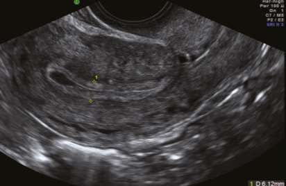
How to measure an early pregnancy
Gestation sac
   – MSD: Three orthogonal planes; two in sagittal plane, one in transverse.
     Largest sac diameters from inner borders of the sac.
   – Location
   – Regularity
   – Sub-chorionic haematoma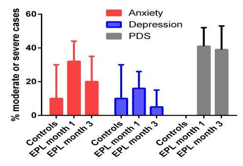
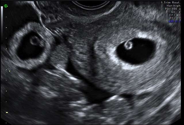
Gestation sac

Fluid in cavity - pseudosac
Yolk sac
    – Three orthogonal planes, from outer borders
CRL
– Greatest straight line length while caudal and cephalic cannot be
distinguished
– CRL once sufficiently deflexed (9 weeks)Fundamental issue:
A normal early pregnancy
may be indistinguishable from
an abnormal early pregnancy that has
arrested its developmentPregnancy of unknown viability
First visible on TVS   Growth
                (days from LMP)
Gestation Sac   31days (4+3 weeks)     1mm/day

YS              35 days (5 weeks)      Max at 10/40

Embryo          37 days (5+2 week)     0.7mm/day

Amnion          49 days (7 weeks)
Why you might not see what you expect to…
Patient External factors
• Incorrect dates • Experience of sonographer
– Erratic cycles (PCO) • Quality of machine
– Recent pregnancy/ breastfeeding • Inter/Intra-observer variation
– Contraception – 14-18% 6-9 weeks
– Ovulation to implantation interval`
• Sonographic view Pregnancy
– TA vs TV scan • Miscarriage
– BMI • Genetic abnormality
– Fibroids • Location
– Axial uterusAim for 100% specificity
• Key question: “Is there a chance of viable pregnancy?”
– A false positive diagnosis of miscarriage is much worse
than a false negative diagnosis
• False positive: Inadvertent ToP
• False negative: raised hopes, delay in intervention
First do no harmEvolution of criteria for the diagnosis of
miscarriage
2006:
ACR – empty GS MSD >16mm, CRL >5mm and no FH
RCOG – empty GS MSD >20mm, CRL >6mm and no FH
2011 (Abdallah et al):
4.4% false positive rate if cut-off MSD ≥16mm
0.5% false positive rate if cut off MSD ≥20mm
8.3% false positive rate for cut-off CRL 5mm
MSD >25mm, CRL >7mm RCOG 2011
2015 (Priesler et al):
Verified cut-offs proposed in 2011
Intervals between scansDefining safe criteria to diagnose miscarriage: prospective
observational multicentre study
Jessica Preisler,1 Julia Kopeika,2 Laure Ismail,1,3 Veluppillai Vathanan,4 Jessica
Farren,1Yazan Abdallah,1 Parijat Battacharjee,5 Caroline Van Holsbeke,6 Cecilia
Bottomley,4Deborah Gould,3 Susanne Johnson,7 Catriona Stalder,1 Ben Van
Calster,8 Judith Hamilton,2, Dirk Timmerman,6,8 Tom Bourne1,6,8
To validate change in guidelines
Prospective multicentre study; 2845 women presenting with bleeding, pain,
hyperemesis
Validated guidance on miscarriage diagnosis
Added evidence based guidance on repeat scans
BMJ2015;351:h4579doi:10.1136/bmj.h4579Criteria that are specific for miscarriage Initial scan • Empty GS MSD >25mm. • Embryo CRL 7mm with no FH Initial scan beyond 70 days (10w from LMP) • MSD >18mm with no embryo • Embryo CRL 3mm with no FH Repeat scan • CRL < 7mm: rescan in 7 days shows no FH • MSD 12mm, no embryo with or without a yolk sac: rescan in 7 days shows no CRL with FH Priesler et al BMJ 2015
If you or patient have any doubts….

                     RE-SCAN
Findings suspicious for pregnancy failure
Findings close to decision boundaries CRL 7mm
Small GS wrt embryo (Ectopic pregnancy
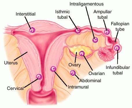
Ectopic pregnancy
•   Tubal 90%
•   Scar ectopic 6%
•   Interstitial 2-4%
•   Ovarian 1%
•   Cervix 0.15%
•   Broad ligament
•   Cornual
•   Abdominal 0.1-0.7%
Non tubal EP account for 20% of mortalities connected
to all ectopic pregnancies
Heterotopic pregnancy 1/7000 pregnancies (1/100 post
IVF)
Sonographic criteria for different types of tubal
ectopic pregnancy
       Sonographic criteria                % of ectopics seen on
                                                    US
Inhomogenous adnexal mass                  60%
(‘blob’ sign’)
Empty extrauterine gestation sac           20%
(‘bagel sign’)
Extrauterine gestation sac +/- yolk        20%
sac +/- fetal pole +/- fetal cardiac
activity
              Brown DL, Doubilet PM. J Ultrasound Med. 1994;13:259–266.
Interstitial pregnancy
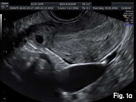
Interstitial pregnancy
• Pregnancy high in fundus, towards edge of uterus
• Endometrial stripe connecting to pregnancy site
• Thin myometrial mantle of < 5mm around the GS

• Interstitial pregnancy and cornual pregnancy are 2 separate entities:
• Interstitial pregnancy – GS in the muscular part of the tube that
  penetrates the uterine wall
• Cornual pregnancy – GS in a rudimentary horn of a unicornuate
  uterus, cornu of a bicornuate or septate uterus
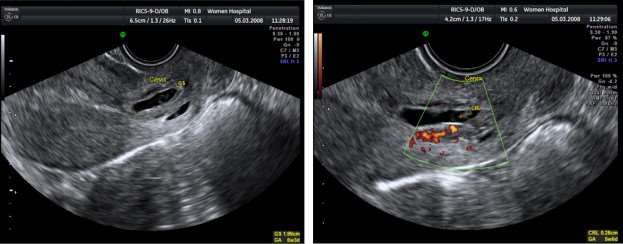
Caesarean scar
    ectopic

•   Incidence increasing. 1:2000 of all pregnancies, 6% of EP
•   Prompt diagnosis is crucial – uterine rupture, haemorrhage, bladder invasion
•   Sonographic criteria
•   Empty uterus, empty cervix
•   GS in the anterior wall of the lower segment of the uterus
•   Thin or no myometrium between bladder and gestation sac
•   Doppler flow
•   Discontinuity in uterine wall on sagittal view
Cervical ectopic
Sonographic criteria
• GS in the cervical tissue not the cervical canal
• Decidual ring, vascularity
• To differentiate from a GS in the cervical canal (inevitable miscarriage):
Sliding sign – when pressure is applied to cervix with TV probe, a GS in the
cervical canal will slide but a cervical EP does not moveHeterotopic pregnancy • Incidence: • 1:7 000 natural conceptions • 1- 3:100 assisted conception • Laparoscopy and salpingectomy
Ectopic pregnancy: Diagnostic pitfalls

• Intrauterine fluid collection – pseudosac
• “Incomplete miscarriage” with blood in uterus may not be
  a miscarriage - exclude an EP (unless IUP seen
  previously)
• “Complete miscarriage” is a PUL and possible EP (unless
  IUP seen previously)
• IUP does not exclude EP (heterotopic)
• Low GS may be scar EP
• Sac in the cervix may be a cervical EP rather than an
  inevitable miscarriage
A triple layer endometrium with a postive PT is likley to
be an ectopic pregnancy - beware
Managing expectations and
psychological considerations
The issue of early testing…

• Very early presentations due to ovulation apps, home
  ovulation tests, sensitive home pregnancy tests -
  Inconclusive scans and uncertainty
   – PUV and PUL

When should we do the first scan?
• Day 35 (5 weeks) – PUV rate 60%
• Day 42 (6 weeks) – PUV rate 29%
• If no clinical symptoms, USS to look for viability on Day 49
  (7 weeks)
Managing patient expectations
•   .Accurate prediction of pregnancy viability by means of a simple scoring
    system.
•   Bottomley C1, Van Belle V, Kirk E, Van Huffel S, Timmerman D, Bourne T.
•   Hum Reprod. 2013 Jan;28(1):68-76. doi: 10.1093/humrep/des352. Epub 2012
    Oct 30

•   The psychological effects and patient acceptability of a test to predict
    viability in early pregnancy: a prospective randomised study. / Davison,
    A. Z.; Appiah, A.; Sana, Y.; Johns, J.; Ross, Jackie.
•   In: European Journal of Obstetrics Gynecology and Reproductive Biology,
    Vol. 178, 07.2014, p. 95-99
Managing patient expectations:
Scoring systems (Bottomley et al)
• Maternal age
• Bleeding score
• Mean gestational sac size
• Fetal heart beat
• Mean yolk sac diameter
• Mean gestational sac size +/- fetal heart beat
– Estimated chance of a viable pregnancy
• Human Reproduction, Bottomley et al, Volume 28, Issue 1, 1 January 2013,
Pages 68–76,• Women offered information of potential pregnancy outcome based on a predictive model found it useful to manage their expectation and anxiety • Reassurance is difficult
The psychological impact….
In the future

•   Criteria for IVF
•   Criteria for 3-D USS
•   AI
•   Non-USS markers to predict miscarriage
Conclusion

• Always err on side of caution in diagnosis of
  miscarriage: if in doubt, re-scan
• Manage expectations – informally or formally
  (predictive models)
• High index of suspicion in the dx of EP
• Consider the psychological sequelae of
  pregnancy loss
• My thanks to
• Jessica Farren
• Shabnam Bobdiwalla
You can also read