Uncommon presentation of a ganglionic cyst: a case study of intra muscular ganglion cyst of rectus femoris
←
→
Page content transcription
If your browser does not render page correctly, please read the page content below

International Journal of Research in Orthopaedics
Singh R et al. Int J Res Orthop. 2021 Jan;7(1):168-170
http://www.ijoro.org

                                                  DOI: https://dx.doi.org/10.18203/issn.2455-4510.IntJResOrthop20205584
Case Report

      Uncommon presentation of a ganglionic cyst: a case study of intra
               muscular ganglion cyst of rectus femoris
                       Rithika Singh1*, P. Mohan Kumar1, B. Gavaskar1, E. praveena2

  1
   Department of Orthopedics, 2Department of Pathology, Government General Hospital, Nizamabad, Telangana, India

  Received: 10 October 2020
  Accepted: 12 November 2020

  *Correspondence:
  Dr. Rithika Singh,
  E-mail: drrithikasingh@gmail.com

  Copyright: © the author(s), publisher and licensee Medip Academy. This is an open-access article distributed under
  the terms of the Creative Commons Attribution Non-Commercial License, which permits unrestricted non-commercial
  use, distribution, and reproduction in any medium, provided the original work is properly cited.

   ABSTRACT

   Ganglionic cyst most commonly occurs in hand and wrist. When it presents in uncommon location like in lower limb,
   it causes a diagnostic dilemma. One such case is ours, an uncommon presentation of intra muscular cystic ganglion of
   rectus femoris. Due to its unlikely presentation in rectus femoris, diagnosis and management was delayed. A 12 years
   old boy presented with complaints of pain and swelling over left lower limb in suprapatellar region. On radiographic
   and ultrasound examination, swelling was found to be cystic lesion in rectus femoris. Histopathological examination of
   biopsied specimen was found to be intra muscular ganglion cyst of rectus femoris. The patient’s general condition
   improved with betterment in laboratory parameters, resolution of the lesion, without any sequelae, no residual deformity
   and excellent clinical outcome. To consider cystic ganglion as differential diagnosis, along with intra muscular myxoma,
   lipoma and synovial cyst in patients with lower limb intra muscular swelling.

   Keywords: Cystic ganglion, Intramuscular cyst, Dilemma

INTRODUCTION                                                            of multiple ganglion cysts involving unusual sites like the
                                                                        temporo mandibular joint affecting young patients, the so
Ganglion cyst are benign soft tissue tumors, usually found              called cystic ganglionosis, might indicate a genetic
on dorsal aspect of wrist. They are also found in                       susceptibility and give new insights to the pathogenesis of
uncommon locations like in gastrocnemius and quadriceps                 this lesion.8 To the best of our knowledge, this is one of
femoris muscle bellies. It’s a fluid filled cyst with                   the first few case reports to document an intramuscular
unknown pathogenesis. Mostly asymptomatic, but could                    ganglion cyst arising in the rectus femoris muscle itself.
cause pressure effect over adjacent neurovascular
structures.1                                                            CASE REPORT

Ganglionic cysts arising in atypical locations present a                A 12 years old, male child presented with complaints of
diagnostic challenge and might get misdiagnosed. They                   swelling and vague pain over left thigh upon exertion.
have been reported to originate from cartilage, nerves, and             Swelling was gradual in onset, noticed by patient himself
muscles. Intra articular ganglion cysts of the knee mainly              while bathing 6 months ago. Swelling was small in size
involve the tendon sheath or joint capsule and infrequently             about marble size when first noticed and then later evolved
the menisci, and anterior and posterior cruciate                        over a period of 6 months to attain the present size. It was
ligaments.3-5 Somewhere reported to occur intraosseously                asymptomatic to start with, then later since past 2 months,
in the distal epiphysis of the tibia and from peripheral                there was vague pain upon exertion and was associated
nerve sheath particularly the common peroneal nerve.6                   with restriction of activities of daily life like unable to do
Involvement of other nerves including the radial, ulnar,                cycling and run for long distances. Upon examination,
median and sciatic nerves have also been reported.7 Cases               swelling was over distal 1/3rd of thigh with normal

                                  International Journal of Research in Orthopaedics | January-February 2021 | Vol 7 | Issue 1   Page 168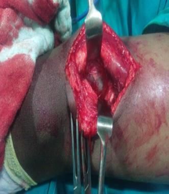
Singh R et al. Int J Res Orthop. 2021 Jan;7(1):168-170

looking overlying skin with no scars and discharging
sinuses. On palpation, there was no local raise of
temperature, swelling was non tender and non-pulsatile.
Skin over the swelling was pinchable and swelling was
mobile in one direction (along long axis of the thigh). It
was an oval shape swelling with dimensions around 5×4
cm. With uniform consistency and well-defined edges.
There was no neurovascular deficit noted in the affected
limb and knee range of motion (ROM) was 0-110 degree
in comparison to 0-130 degree on unaffected side. On
ultrasound evaluation, it was found to be ganglion cyst in
the muscle belly of quadriceps muscle.

Patient was operated under spinal anesthesia, with no                Figure 3: Intraoperative picture depicting cyst to be
tourniquet. About 7 cm midline incision along the long                arising from rectus femoris muscle belly and was
axis of thigh, centered over swelling was given. Skin and             incised along with the capsule without spillage of
subcutaneous tissue incised and retracted. Cyst was noted                                 contents.
to be arising from muscle belly and was incised along with
the capsule without spillage of contents.

Peri-operative period was uneventful. Suture removal
done on post-operative day 14 and was made to walk full
weight bearing on affected limb. Patient was followed up
for a period of one year and there were no complications
like recurrence.

                                                                     Figure 4: Cystic tumor approximately 4.1×2.5 cm in
                                                                                      size was identified.

    Figure 1: Sonographic image showing the non-
 lobulated mass of a cystic lesion (4.1×2.5 cm) within
 the rectus femoris of the quadriceps femoris muscle.

                                                                       Figure 5: Photomicrograph section shows a dense
                                                                     fibrotic cyst wall with no epithelial lining, suggestive
                                                                                        of ganglion cyst.

                                                                    DISCUSSION

                                                                    Ganglion cysts are benign soft tissue tumors with fluid
                                                                    filled cavity. The exact cause for its origin are unknown.
                                                                    However, it’s presumed to arise from synovium or tendon
                                                                    sheath.1 It is presumed that, as in baker’s cyst, the synovial
                                                                    tissue herniates and becomes loculated, which later
                                                                    transforms to form mesothelium lining membrane.2
Figure 2: Clinical picture depicting swelling was over
                                                                    Its clinical presentation varies from small asymptomatic
 distal 1/3rd of thigh with normal looking overlying
                                                                    swelling over dorsum of wrist to large swelling causing
     skin with no scars and discharging sinuses.

                              International Journal of Research in Orthopaedics | January-February 2021 | Vol 7 | Issue 1   Page 169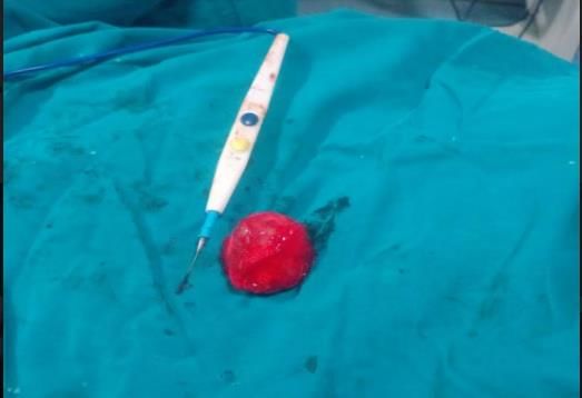
Singh R et al. Int J Res Orthop. 2021 Jan;7(1):168-170

pressure symptoms over adjacent neurovascular structures              managed by excision and biopsy with no post-operative
and soft tissue.1 Most common presentation is from                    complications and no recurrence after 1 year of follow up.
extensor digiti communis over dorsal aspect, followed by
space between flexor carpi radialis and abductor pollicis             Funding: No funding sources
longus tendon on volar aspect.3 Its source of origin varies           Conflict of interest: None declared
from tendon sheath, cartilage, cruciate ligament, nerves              Ethical approval: Not required
and muscles.4-7
                                                                      REFERENCES
Intramuscular ganglion cyst in quadriceps femoris is a rare
presentation and it usually present as palpable tender lump           1.    Brooks DM. Nerve compression by simple ganglia. J
without any accompanying symptoms. However, wasting                         Bone Joint Surg Br. 1952;34:391-400.
of associated limb could be noted, due to restricted use of           2.    Angelides AC, Wallace PF. The dorsal ganglion of
limb, secondary to pain.                                                    the wrist: its pathogenesis, gross and microscopic
                                                                            anatomy, and surgical treatment. J Hand Surg Am.
Plain radiographs of knee is unlikely to relieve any                        1976;1:228-35.
important diagnostic information. It is usually diagnosed             3.    Nelson CL, Sawmiller S, Phalen GS. Ganglions of
with ultra sound. However, the limitation with ultrasound                   the wrist and hand. J Bone Joint Surg Am.
are its inability to give definite information regarding cyst               1972;54:1459-64.
and its association with adjacent soft tissue. Magnetic               4.    Kang SY, Lee HJ, Lee SH. Intramuscular ganglion of
resonance imaging (MRI) is useful in such situations to                     the peroneus muscle mimicking peroneal
clearly mark the extent and its relation with adjacent soft                 compartment syndrome: a case report. J Korean
tissue. It is important to identify the extent of tumor, to                 Orthop Assoc. 2004;39:228-31.
avoid complications associated with incomplete excisions              5.    Lee YU, Kook SH, Chung EC, Youn EK, Park JY.
like recurrence.                                                            MRI of ganglion cysts in uncommon sites or with
                                                                            atypical appearance. J Korean Radiol Soc.
A study by Nelson et al had shown, cure rate of 94% after                   1999;41:393-9.
excision.3 Another study by Aydin et al on a case series of           6.    Yang SW, Teng HP, Tarng YW, Wong CY.
40 cases has shown recurrence rate of 22%.8 The common                      Intramuscular ganglion cyst of the quadriceps
surgical complications following excision include damage                    muscle: report of a case. Mid Taiwan J Med.
to adjacent neurovascular structure. As shown by Aydinet                    2002;7:193-7.
al in their study.8 The study showed 10% incidence of                 7.    Muckle DS, Monahan P. Intra-articular ganglion of
damage to palmar cutaneous nerve and 5% incidence of                        the knee: report of two cases. J Bone Joint Surg Br.
damage to radial nerve.                                                     1972;54:520-1.
                                                                      8.    Aydin A, Kabakas F, Erer M, Ozkan T, Tuncer S.
Intramuscular ganglion cyst in quadriceps femoris is                        Surgical treatment of volar wrist ganglia. Acta
unlikely presentation and there is limited literature on this               Orthop Traumatol Turc. 2003;37:309-12.
topic. Unlike ganglion cyst of hand, which is a common
form of presentation.
                                                                           Cite this article as: Singh R, Kumar PM, Gavaskar
                                                                           B, Praveena E. Uncommon presentation of a
CONCLUSION
                                                                           ganglionic cyst: a case study of intra muscular
                                                                           ganglion cyst of rectus femoris. Int J Res Orthop
In this case report, we present a 12 years old male child                  2021;7:168-70.
with intramuscular ganglion cyst in quadriceps tendon

                                International Journal of Research in Orthopaedics | January-February 2021 | Vol 7 | Issue 1   Page 170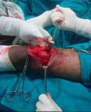
You can also read