Unusual Presentation of a Metastatic Lymph Node from Giant Phyllodes Tumour of Breast- A Case Report
←
→
Page content transcription
If your browser does not render page correctly, please read the page content below
Arch Clin Med Case Rep 2020; 4 (2): 253-258                                         DOI: 10.26502/acmcr.96550193

Case Report
    Unusual Presentation of a Metastatic Lymph Node from Giant
                   Phyllodes Tumour of Breast– A Case Report
Sherry Abraham*, Patricia Solomon, Rajesh B, Selvamani B

Department of Radiation Oncology Unit III, Christian Medical College, Vellore, Tamil Nadu, India

*
    Corresponding Author: Dr. Sherry Abraham, Senior Resident, Department of Radiation Oncology, Christian
Medical College, Vellore, Tamil Nadu, India, Tel: 9495529711; E-mail: drsherryabraham86@gmail.com

Received: 30 September 2019; Accepted: 02 March 2020; Published: 10 March 2020

Abstract
Background: Phyllodes tumours(PTs) of breast are rare fibroepithelial neoplasms which constitute 0.3% to 0.5% of
female breast tumours. Malignant PTs usually develop haematogenous spread and only
Arch Clin Med Case Rep 2020; 4 (2): 253-258 DOI: 10.26502/acmcr.96550193 Keywords: Malignant phyllodes tumour; Lymph node metastasis; Axillary lymphadenopathy 1. Introduction Phyllodes tumours(PTs) of breast are rare fibroepithelial neoplasms which constitute 0.3% to 0.5% of female breast tumours [1]. Their incidence is 2.1 per million and peaks from the age of 45 to 49 years [2]. World Health Organization classified PTs as benign, borderline and malignant based on histological findings [3]. The incidence of each type of PTs has been reported by the M.D. Anderson Cancer Center based on a large case series to be 58% in benign, 12% in borderline and 30% in malignant [4]. Local recurrence rate of up to 17% is seen in benign PTs and up to 27% in malignant tumours [5]. Malignant PTs usually develop haematogenous spread and only
Arch Clin Med Case Rep 2020; 4 (2): 253-258 DOI: 10.26502/acmcr.96550193
breast, replacing the normal parenchyma. Left breast was normal. Contrast enhanced computed tomography of
thorax revealed a large soft tissue density mass lesion of size 17 × 18 × 20 cm replacing the entire right breast
(Figure 1). Multiple cystic areas and linear branching vessels were noted within the lesion. Posteriorly, the pectoralis
muscles were not involved. Anteriorly, the lesion reached up to the skin with significant skin thickening in the upper
quadrant. There was significant right axillary lymphadenopathy, largest measuring upto 17 mm.
Trucut biopsy of the right breast lump showed compressed ducts lined by benign bilayered cuboidal epithelium with
stromal nuclear atypia and occasional mitotic activity suggestive of phyllodes tumour of aggressive behavior. Her
chest x-ray was normal. Ultrasound abdomen and pelvis did not show any focus of metastasis. She had modified
radical mastectomy of the right breast and level 1 and 2 axillary nodal clearance with latissimus dorsi flap
reconstruction. Operative findings were suggestive of a single large 5-kg tumor occupying the whole of the right
breast with dilated vessels over the surface, infiltrating into the pectoralis major muscle.
There were multiple large solid nodes at axillary levels 1 and 2. Histopathology of the specimen was suggestive of
malignant phyllodes tumour of maximum dimension of 22cm, with sarcomatous overgrowth, lymphovascular
invasion and poor margin clearance. Tumour was composed of sheets of round to short spindle cells with moderate
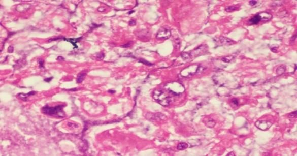
to marked nuclear pleomorphism and mitotic activity of >15/10 hpf (Figure 2 and 3). Focally proliferated epithelial
component forming occasional cleft like spaces and necrosis were also noted. Seven of 11 axillary lymph nodes
contained metastatic disease. The tumour cells were negative for ER, HER2, HMWCK, p63 and CK5/6 with
occasional foci showing faint staining for PR. The atypical proliferated stromal cells show patchy BCL2 positivity
and were negative for CD34. Background epithelial components were staining positively for HMWCK and Pan
Cytokeratin.
Figure 2: Stromal overgrowth, spindle shaped cells.
Archives of Clinical and Medical Case Reports 255Arch Clin Med Case Rep 2020; 4 (2): 253-258 DOI: 10.26502/acmcr.96550193
Figure 3: Pleomorphic atypical cells.
She received 4 cycles of adjuvant chemotherapy with single agent Doxorubicin. Post 4 cycles of chemotherapy, she
received radiotherapy to the chest wall. She was treated with 3D conformal radiotherapy using 6MV photons to a
dose of 60Gy in 30 fractions to the Planning Target Volume. She had been advised to be on follow up. Four months
later, she presented with complaints of cough and dyspnea on exertion for 2 weeks to emergency room. She was
found to have bilateral pleural effusion, pericardial effusion, tricuspid ball valve thrombus and acute submassive
pulmonary embolism. Though effusions were thought to be secondary to malignancy, cytology did not prove the
same. She succumbed to her illness within 72 hours of admission to emergency room.
3. Discussion
Malignant phyllodes tumour is a rare entity which mimics benign neoplasm clinically, but, is characterized by a
typical rapid growth. Microscopic findings is the basis of subclassification of PTs. Features consistent with a
malignant PT include high mitotic rate of >5/10hpf, stromal hypercellularity and pleomorphism, necrosis and
infiltrative tumour edge [6] – all of which were seen in histopathology of the specimen of our patient.
Malignant PTs behave like sarcomas with poor prognosis and haematogenous spread. Local recurrence rate upto
17% or distant metastasis upto 27% has been documented by Jhang Y and Kleer CG [5]. Pleomorphism, infiltrative
margins and stromal overgrowth are the poor histological findings related to recurrence in breast or distant
metastasis. Recurrence rate or metastasis is usually seen within the first two years after diagnosis, ranging from 1
month to 10 years with an average of 15 to 26 months [3]. Lung is the most common site of metastasis (66-84.5%)
followed by bone (28-39%) and rare sites include liver and brain [3]. Clinically palpable axillary nodes have been
identified in upto 10-15% of PTs; however, pathologically positive nodes have been present in onlyArch Clin Med Case Rep 2020; 4 (2): 253-258 DOI: 10.26502/acmcr.96550193
there is also a possibility of coexistent invasive carcinoma along with phyllodes tumor as demonstrated by Korula et
al.[9].
Wide local excision is the standard care for all histological types of phyllodes [10]. Hence, surgical excision with
greater emphasis on obtaining adequate surgical margin (atleast 1cm) is the mainstay of treatment of malignant PTs.
In terms of metastasis free survival and overall survival, studies have not shown significant difference between
mastectomy and breast conserving surgery. Mastectomy should be considered in larger tumours and recurrent
tumours with malignant histology. Role of axillary lymph node dissection in malignant PTs is controversial and
conflicting views are documented in the literature. Most of the time, axillary lymphadenopathy is due to reactive
hyperplasia. Majority of the investigators recommend against axillary lymph node dissection due to the low risk of
lymph node involvement, except in patients with proven metastatic nodes or in clinically palpable axillary nodes [7,
11]. Adjuvant radiotherapy is recommended to minimize local recurrence in malignant PTs, recurrent tumours and
tumours with close margins. Belkacemi et al found that radiotherapy was associated with superior 10 year local
control rate from 59% to 86% for malignant and borderline PTs [12]. Role of adjuvant chemotherapy in malignant
PTs is debatable. In an observational study by Morales-Vasquez in Mexico, 28 patients with malignant PT after
surgical resection, from January 1993 to December 2003, reported 58% recurrence free survival with adjuvant
chemotherapy with Doxorubicin and Dacarbazine as compared to 86% when in observation [13]. Turalba et al
demonstrated efficacy of Doxorubicin and Ifosfamide based chemotherapy in women with metastatic phyllodes
[14]. Our patient had recurrent giant PT of which, initial trucut biopsy showed PT of aggressive behavior with
highly suspicious hard axillary lymph nodes. Hence, modified radical mastectomy with axillary lymph node
dissection was done and the histopathology showed malignant PT with lymph node metastasis. She received
adjuvant radiation therapy and chemotherapy. She died due to pulmonary embolism at 13 months from the date of
diagnosis.
4. Conclusion
Malignant phyllodes tumour is a rare entity for which wide local excision with adequate margins is the mainstay of
treatment. Though the most common cause of axillary lymphadenopathy is reactive hyperplasia; rarely, the patients
may present with axillary node metastasis. Data regarding role of routine lymph node dissection remains
controversial; however, it is indicated in palpable axillary nodes.
References
1. Rowell MD, Perry RR, Hsiu JG, et al. Phyllodes tumors. Am J Surg 165 (1993): 376-379.
2. Mishra SP, Tiwary SK, Mishra M, et al. Phyllodes tumor of breast: a review article. ISRN Surg 2013
(2013): 361469.
3. Amir RA, Rabah RS, Sheikh SS. Malignant Phyllodes Tumor of the Breast with Metastasis to the Pancreas:
A Case Report and Review of Literature. Case Rep Oncol Med 2018 (2018): 1-5.
4. Chaney AW, Pollack A, McNeese MD, et al. Primary treatment of cystosarcoma phyllodes of the breast.
Cancer 89 (2000): 1502-1511.
Archives of Clinical and Medical Case Reports 257Arch Clin Med Case Rep 2020; 4 (2): 253-258 DOI: 10.26502/acmcr.96550193
5. Zhang Y, Kleer CG. Phyllodes Tumor of the Breast: Histopathologic Features, Differential Diagnosis, and
Molecular/Genetic Updates. Arch Pathol Lab Med 140 (2016): 665-671.
6. Tan BY, Acs G, Apple SK, et al. Phyllodes tumours of the breast: a consensus review. Histopathology 68
(2016): 5-21.
7. Chen W-H, Cheng S-P, Tzen C-Y, et al. Surgical treatment of phyllodes tumors of the breast: Retrospective
review of 172 cases. J Surg Oncol 91 (2005): 185-194.
8. Confavreux C, Lurkin A, Mitton N, et al. Sarcomas and malignant phyllodes tumours of the breast – A
retrospective study. Eur J Cancer 42 (2006): 2715-2721.
9. Korula A, Varghese J, Thomas M, et al. Malignant phyllodes tumour with intraductal and invasive
carcinoma and lymph node metastasis. Singapore Med J 49 (2008): e318-e321.
10. Verma S, Singh RK, Rai A, et al. Extent of surgery in the management of phyllodes tumor of the breast: a
retrospective multicenter study from India. J Cancer Res Ther 6 (2010): 511-515.
11. Ramakant P, Chakravarthy S, Cherian JA, et al. Challenges in management of phyllodes tumors of the
breast: A retrospective analysis of 150 patients. Indian J Cancer 50 (2013): 345.
12. Belkacémi Y, Bousquet G, Marsiglia H, et al. Phyllodes tumor of the breast. Int J Radiat Oncol Biol Phys
70 (2008): 492-500.
13. Morales‐ Vásquez F, Gonzalez‐ Angulo AM, Broglio K, et al. Adjuvant Chemotherapy with Doxorubicin
and Dacarbazine has No Effect in Recurrence-Free Survival of Malignant Phyllodes Tumors of the Breast.
Breast J 13 (2007): 551-556.
14. Turalba CI, el-Mahdi AM, Ladaga L. Fatal metastatic cystosarcoma phyllodes in an adolescent female:
case report and review of treatment approaches. J Surg Oncol 33 (1986):176-181.
Citation: Sherry Abraham, Patricia Solomon, Rajesh B, Selvamani B. Unusual Presentation of a Metastatic
Lymph Node from Giant Phyllodes Tumour of Breast– A Case Report. Archives of Clinical and Medical
Case Reports 4 (2020): 253-258.
This article is an open access article distributed under the terms and conditions of the
Creative Commons Attribution (CC-BY) license 4.0
Archives of Clinical and Medical Case Reports 258You can also read