Visualization of Gross Anatomy for Ultrasound Scanning Planes Using a Virtual Dissection Table
←
→
Page content transcription
If your browser does not render page correctly, please read the page content below
Int. J. Morphol.,
39(1):7-10, 2021.

                    Visualization of Gross Anatomy for Ultrasound
                   Scanning Planes Using a Virtual Dissection Table

                                 Visualización de la Anatomía Macroscópica para Planos de
                             Escaneo de Ultrasonido Utilizando una Mesa de Disección Virtual

                                                 Hyo-Min Cho1; Yongho Lee2 & Cheolpyo Hong2

CHO, H. M.; LEE, Y. & HONG, C. Visualization of gross anatomy for ultrasound scanning planes using a virtual dissection table. Int. J.
Morphol., 39(1):7-10, 2021.

          SUMMARY: There is a lack of visualization on gross anatomy planes for the non-orthogonal sections, such as subcostal and intercostal
oblique scanning planes of ultrasound imaging. The aim of the present study was to visualize the anatomical image of corresponding plane for the
oblique ultrasound scanning using a virtual dissection system. the oblique gross anatomy plane was constructed by appropriate segmentation
using a virtual dissection table. A suitable cutting of the body plane was accomplished by turning on and off the organ systems, particularly the
skeletal system, category, and structure. The right hepatic vein (RHV), middle hepatic vein (MHV), and left hepatic vein (LHV) for the right
subcostal oblique plane appeared in the single slice plane. The location of the liver, gallbladder, and kidneys differently appeared in the oblique
anatomical plane and body position. The results of this study suggest that using a virtual anatomy system contributes to improving the sonographer’s
ability to understand anatomy.

           KEY WORDS: Gross anatomy planes; Virtual dissection; Ultrasound scanning plane.

INTRODUCCIÓN

        Ultrasound imaging, also known as sonography or                        intercostal oblique plane (Fukuda, 1996; Mattoon et al.,
ultrasonography, employs mechanical pressure vibration to                      2014). Most gross anatomy information is provided with
generate images of the inside of the human body (Szabo,                        three standard orthogonal sections—namely, sagittal,
2004; Hoskins et al., 2010; Shung, 2015). Ultrasound                           coronal, and transverse planes.Transverse and longitudinal
imaging uses sound waves, which are typically non-ionizing                     planes are easily recognized by the three standard planes.
radiation, noninvasive, and safe (Barnett et al., 2000; Duck,
2008). Sonography is valuable for imaging soft tissues                                 However, there is a lack of visualization on gross
contrast of organs and tissues (Wells & Liang, 2011; Frulio                    anatomy planes for the subcostal and intercostal oblique
& Trillaud, 2013). Doppler ultrasonography is another                          scanning planes of ultrasound imaging.For instance, while
powerful tool to evaluate the blood velocity (Kremkau, 1990;                   the left, middle, and right hepatic vein branches can be
McDicken et al., 1992; Cohen et al., 2001; Grant et al.,                       routinely visualized using the right subcostal oblique
2003). Due to these characteristics, ultrasonography is widely                 scanning, such an image is not provided in gross anatomy
used in cardiology, endocrinology, orthopedics, obstetrics,                    planes. Physicians and technicians using ultrasound imaging
and gynecology (Ferrucci Jr., 1979; Kaminsky et al., 1997;                     system should only predict oblique imaging plane
Moore & Copel, 2011; Erçikti et al., 2017).                                    information through a formalized gross anatomy. The vir-
                                                                               tual dissection system based on the anatomy digital images
        In ultrasound imaging, scanning planes are                             allows a user to cutthe virtual body in any direction (Chung
established in the direction of the ultrasound beam lines and                  et al., 2015). Furthermore, an appropriate cutting of the
are determined by slightly moving the ultrasound transducer.                   section is accomplished by using the human organ systems
During the ultrasound examination, the transducer can be                       category of the virtual dissection system. In the present study,
moved to an arbitrary orientation, twisting, and rotating.                     we visualized the anatomical image of corresponding plane
Typically used ultrasound scan planes include the transverse                   for the oblique ultrasound scanning using a virtual dissection
plane, longitudinal plane, subcostal oblique plane, and                        system.

1
    Center for Medical Convergence Metrology, Korea Research Institute of Standards and Science (KRISS), Daejeon 34113, Rep. of Korea.
2
    Department of Radiological Science, Daegu Catholic University, Gyeongsan-si, Gyeongbuk, 38430, Rep. of Korea.

                                                                                                                                                 7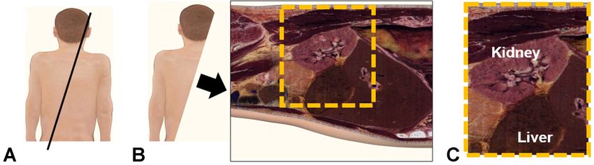
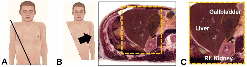
CHO, H. M.; LEE, Y. & HONG, C. Visualization of gross anatomy for ultrasound scanning planes using a virtual dissection table. Int. J. Morphol., 39(1):7-10, 2021.

MATERIAL AND METHOD

Virtual dissection system. Virtual dissection table produced                                   extended intercostal oblique planes were selected from the
by Anatomage company (San Jose, CA, United States) was                                         right superior to the left inferior directions (Fig. 3). The
used to generate the gross anatomy plane for imaging plane                                     appropriate cross section was chosen by the most similar
(Fig. 1a.). The Anatomage virtual dissection table was a                                       image plane to the right extended intercostal oblique
segmented 3D whole-body anatomy system providing                                               scanning of sonography. This cross section was selected by
various arbitrary planes and three orthogonal sections.                                        including the liver, gallbladder, and kidneys. The oblique
Digitized images of human anatomy were presented in an                                         plane on prone position was selected from the right superior
interactive manner on a life-sized touch screen.The oblique                                    to the left inferior direction (Fig. 4). The suitable oblique
and specialized gross anatomy planes were constructed by                                       plane was chosen by the most similar sonography scanning
appropriate segmentation. A suitable cutting of the body                                       image for the consideration of the liver and kidney location.
plane was accomplished by turning on and off the organ                                         Selected planes were obtained through repeated cutting.
systems, particularly the skeletal system, category, and
structure (Fig. 1b). The vascular and digestive system also
was used to appropriate cut the plane.                                                         RESULTS

Oblique anatomical images planes. The right costal margin
of the rib cage was designated by a skeletal model on the                                             The branches of hepatic veins were visualized as the
Anatomage table. The right subcostal oblique sectionsalong                                     corresponding anatomical images for the right subcostal
the oblique costal margin were selected from the left supe-                                    oblique plane (Fig. 2c). The right hepatic vein (RHV), middle
rior to the right inferior direction (Fig. 2). Various section                                 hepatic vein (MHV), and left hepatic vein (LHV) were
images were generated along the oblique planes, including                                      displayed on one section image. These veins appeared in
the intravenous branches in the liver. The optimal cross                                       the bottom right portion of the liver region in a sonography-
section was selected by the most similar image plane to the                                    like view. Figure 3c shows the anatomical image of the
right subcostal oblique scanning of sonography. The right                                      corresponding plane for the right extended intercostal oblique

Fig. 1. The Anatomage virtual dissection table images. Life-size dissection image of human anatomy on the coronal plane (a). The
skeletal system (b) and composite images of the skeletal, vascular, and digestive systems (c).

8
CHO, H. M.; LEE, Y. & HONG, C. Visualization of gross anatomy for ultrasound scanning planes using a virtual dissection table. Int. J. Morphol., 39(1):7-10, 2021.
sonography scanning. The gallbladder appeared in a higher supine position. The kidney appeared in a higher upper part
upper part than the kidney in a sonography-like view. The than the liver in the prone position in a sonography-like view
liver part was displayed on the right side of the sonography- (Fig. 4c). The organs were differently displayed along the
like view. Figures 2 and 3 were acquired on the virtual body cutting oblique plane and body position.
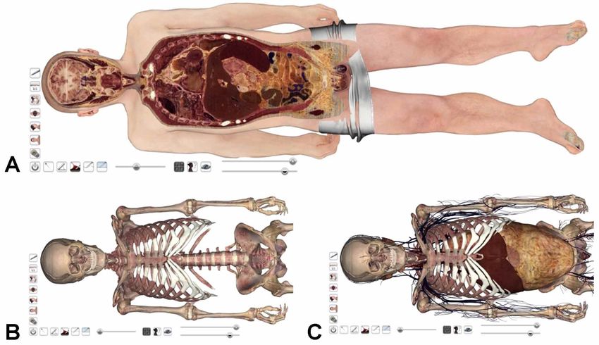
Fig. 2. The anatomical image of the corresponding plane for the right subcostal oblique scanning of sonography. The virtual anatomical
images (a) and the corresponding plane images (b and c). (The black line; cutting line, the yellow box: a sonography-like view, the right
hepatic vein, RHV; the middle hepatic vein, MHV; and the left hepatic vein, LHV).
Fig. 3. The anatomical image of the corresponding plane for the right extended intercostal oblique scanning of sonography. The virtual
anatomical images (a) and the corresponding plane images (b and c). The liver, gallbladder, and kidneys are represented in the identical
section, and the liver is shown above the kidney on the sonography-like view. (The black line; cutting line, the yellow box: a sonography-
like view).
Fig. 4. The anatomical image of the corresponding plane for the oblique scanning on prone position of sonography. The virtual anatomical
images (a) and the corresponding plane images (b and c). The liver and the right kidney are represented in the identical section, and the
kidney is shown above the liver on the sonography-like view. (The black line; cutting line, the yellow box: a sonography-like view).
9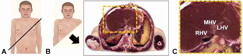
CHO, H. M.; LEE, Y. & HONG, C. Visualization of gross anatomy for ultrasound scanning planes using a virtual dissection table. Int. J. Morphol., 39(1):7-10, 2021.

DISCUSSION                                                                                        REFERENCES

         In this study, we visualized the oblique anatomical plane                                Barnett, S. B.; Ter Haar, G. R.; Ziskin, M. C.; Rott, H. D.; Duck, F. A. & Maeda, K.
                                                                                                      International recommendations and guidelines for the safe use of diagnostic
for the corresponding sonography-like image using a virtual                                           ultrasound in medicine. Ultrasound Med. Biol., 26(3):355-66, 2000.
dissection system. The RHV, MHV, and LHV appeared in sin-                                         Chung, B. S.; Shin, D. S.; Brown, P.; Choi, J. & Chung, M. S. Virtual dissection
gle slice plane (Fig. 2c). This image is similar to the one obtained                                  table including the visible korean images, complemented by free software of
                                                                                                      the same data. Int. J. Morphol., 33(2):440-5, 2015.
from the upper abdominal ultrasound scan. Such an image was                                       Cohen, L. S.; Escobar, P. F.; Scharm, C.; Glimco, B. & Fishman, D. A. Three-
not provided in gross anatomy images.Ultrasound probes are                                            dimensional power doppler ultrasound improves the diagnostic accuracy for
placed on the skin surface. The upper part of sonographic image                                       ovarian cancer prediction. Gynecol. Oncol., 82(1):40-8, 2001.
                                                                                                  Duck, F. A. Hazards, risks and safety of diagnostic ultrasound. Med. Eng. Phys.,
is a superficial region, while the lower part is a deep region of                                     30(10):1338-48, 2008.
the body position. In addition, the acquired ultrasound image                                     Erçikti, N.; Acer, N.; Apaydi, N.; Güven, I. & Zararsiz, G. Which method is gold
varies depending on the position of the patient in which the                                          standard for determination of thyroid volume? Int. J. Morphol., 35(2):452-8,
                                                                                                      2017.
probe is placed. The liver appeared in a higher upper part than                                   Ferrucci Jr., J. T. Body ultrasonography (first of two parts). N. Engl. J. Med.,
the kidney on the supine position in the upper abdominal                                              300(10):538-42, 1979.
ultrasound scan (Fig. 3c). Conversely, the kidney was                                             Frulio, N. & Trillaud, H. Ultrasound elastography in liver. Diagn. Interv. Imaging,
                                                                                                      94(5):515-34, 2013.
represented in a higher upper part than the liver in the patient                                  Fukuda, M. Ultrasonography of the Liver Anatomy, Procedure, Normal and
prone position (Fig. 4c). In addition, the sonography image has                                       Abnormal Findings in Diseased States. In: Fukuda, M.; Bergmann, H.; Padhy,
the limited scan field-of-view (FOV). The characteristics of                                          A. K. & Fukuhisa, K. (Eds.). Ultrasound and Radionuclide Images of Liver.
                                                                                                      An IAEA (CRP) Group Study. Mumbai, Himalaya Publishing House, 1996.
sonographic images mentioned above can be easily expressed                                        Grant, E. G.; Benson, C. B.; Moneta, G. L.; Alexandrov, A. V.; Baker, J. D.; Bluth,
by using the virtual anatomy dissection system.                                                       E. I.; Carroll, B. A.; Eliasziw, M.; Gocke, J.; Hertzberg, B. S.; et al. Carotid
                                                                                                      artery stenosis: gray-scale and Doppler US diagnosis--Society of Radiologists
                                                                                                      in Ultrasound Consensus Conference. Radiology, 229(2):340-6, 2003.
        Ultrasound imaging is affected by the patient's breathing,                                Hoskins, P. R.; Martin, K. & Thrush, A. Diagnostic Ultrasound: Physics and
movement, and probe handling status. Although the oblique                                             Equipment. Cambridge, Cambridge University Press, 2010.
plane extracted in the present study may not exactly match                                        Kaminsky, S.; Griffin, L.; Milsap, J. & Page, D. Is ultrasonography a reliable way
                                                                                                      to confirm the diagnosis of Morton's neuroma? Orthopedics, 20(1):37-9, 1997.
ultrasonic imaging anatomy, the virtual anatomy system helps                                      Kremkau, F. W. Doppler Ultrasound: Principles and Instruments. Chicago,
to better understand cross-sectional image anatomy.                                                   Saunders, 1990.
Furthermore, the quality of ultrasound images heavily depends                                     Mattoon, J. S.; Berry, C. R. & Nyland, T. G. Abdominal Ultrasound Scanning
                                                                                                      Techniques. Small Animal Diagnostic Ultrasound. 3rd ed. St Louis, Elsevier
on the operator. For an accurate diagnosis, operators should have                                     Saunders, 2014. pp.94-127.
a comprehensive understanding of oblique anatomy sections.                                        McDicken, W. N.; Sutherland, G. R.; Moran, C. M. & Gordon, L. N. Colour doppler
Consequently, the use of a virtual anatomy system contributes                                         velocity imaging of the myocardium. Ultrasound Med. Biol., 18(6-7):651-4,
                                                                                                      1992.
to improving the sonographer’s ability for diagnosis.                                             Moore, C. L. & Copel, J. A. Point-of-care ultrasonography. N. Engl. J. Med.,
                                                                                                      364:749-57, 2011.
                                                                                                  Shung, K. K. Diagnostic Ultrasound: Imaging and Blood Flow Measurements.
CHO, H. M.; LEE, Y. & HONG, C. Visualización de la anatomía                                           2nd ed. London, CRC Press, 2015.
macroscópica para planos de escaneo de ultrasonido utilizando una mesa                            Szabo, T. L. Diagnostic Ultrasound Imaging: Inside Out. New York, Academic
de disección virtual. Int. J. Morphol., 39(1):7-10, 2021.                                             Press, 2004.
                                                                                                  Wells, P. N. T. & Liang, H. D. Medical ultrasound: imaging of soft tissue strain
          RESUMEN: Existe una falta de visualización en los planos de                                 and elasticity. J. R. Soc. Interface, 8(64):1521-49, 2011.
anatomía macroscópica para las secciones no ortogonales, tal como los
planos de exploración oblicuos subcostales e intercostales en las imágenes
                                                                                                  Corresponding author:
de ultrasonido. El objetivo del presente estudio fue visualizar la imagen
anatómica del plano correspondiente para la ecografía oblicua mediante un
                                                                                                  Cheolpyo Hong, PhD
sistema de disección virtual. El plano de anatomía macroscópica oblicua se                        Assistant Professor
construyó mediante una adecuada segmentación utilizando una mesa de                               Department of Radiological Science
disección virtual. Se logró un corte correcto del plano corporal al encender                      Catholic University of Daegu
y apagar los sistemas de órganos, particularmente el sistema esquelético, la                      Hayang-ro 13-13, Hayang-eup
categoría y la estructura. La vena hepática derecha, la vena hepática media                       Gyeongsan-si
y la vena hepática izquierda para el plano oblicuo subcostal derecho apare-                       Gyeongbuk, 38430
cieron en el plano de corte único. La ubicación del hígado, la vesícula bi-                       REPUBLIC OF KOREA
liar y los riñones aparecieron de manera diferente en el plano anatómico
oblicuo y en la posición del cuerpo. Los resultados de este estudio sugieren
que el uso de un sistema de anatomía virtual ayuda a mejorar la capacidad                         Email: chong@cu.ac.kr
del ecografista para comprender la anatomía humana.
                                                                                                                                                             Received: 23-07-2020
        PALABRAS CLAVE: Planos de anatomía macroscópica; Di-
                                                                                                                                                             Accepted: 07-09-2020
sección virtual; Plano de exploración por ultrasonido.

10
You can also read