Role of conventional ultrasonography and color flow-doppler sonography in predicting malignancy in 'cold' thyroid nodules - European Journal of ...
←
→
Page content transcription
If your browser does not render page correctly, please read the page content below
European Journal of Endocrinology (1998) 138 41–46

Role of conventional ultrasonography and color flow-doppler
sonography in predicting malignancy in ‘cold’ thyroid nodules
T Rago, P Vitti, L Chiovato, S Mazzeo1, A De Liperi1, P Miccoli2, P Viacava3, F Bogazzi, E Martino and
A Pinchera
Istituto di Endocrinologia, 1Istituto di Radiologia, 2Cattedra di Anatomia Chirurgica, 3Istituto di Anatomia Patologica, University of Pisa, Pisa, Italy
(Correspondence should be addressed to P Vitti, Istituto di Endocrinologia, University of Pisa, Via Paradisa 2, 56124 Pisa, Italy)

                              Abstract
                              The aim of the present study was to establish the usefulness of conventional thyroid ultrasonography
                              (US) and color flow-doppler (CFD) sonography in the assessment of ‘cold’ thyroid nodules. One
                              hundred and four consecutive patients with thyroid nodules who were to undergo surgery were
                              examined by US and CFD before thyroidectomy. Conventional US evaluated the presence of a halo sign,
                              hypoechogenicity and microcalcifications. The vascular pattern on CFD was classified as follows: Type
                              I, absence of blood flow; Type II, perinodular blood flow; Type III, marked intranodular blood flow. On
                              histology, 30 nodules were diagnosed as malignant (carcinoma, CA) and 74 as benign nodules (BN).
                              On US, the echographic pattern most predictive for malignancy was absent halo sign, which was found
                              in 20/30 CA and in 17/72 BN (P ¼ 0.0001; specificity 77.0%; sensitivity 66.6%). The most specific
                              combination on US, absent halo sign/microcalcifications, was found in 8/30 CA and in 5/74 BN
                              (P < 0.005; specificity 93.2%, sensitivity 26.6%). The Type III pattern on CFD was found in 20/30 CA
                              and 38/74 BN (not statistically significant). The combination of absent halo sign on US with Type III
                              pattern on CFD was found in 15/30 CA and in 8/74 BN (P < 0.0001; specificity 89.0%, sensitivity
                              50.0%). The combination of absent halo sign/microcalcifications on US with Type III pattern on CFD
                              was the most specific combination of the two techniques, being found in 5/30 CA and in only 2/74 BN
                              (P < 0.01; specificity 97.2%, sensitivity 16.6%).
                                 In conclusion, findings on US and CFD become highly predictive for malignancy only when multiple
                              signs are simultaneously present in a thyroid nodule. Thus the predictive value of these techniques
                              increases at the expense of their sensitivity. Only in a small proportion of patients with thyroid
                              carcinoma is US and CFD information highly predictive of malignancy.

                              European Journal of Endocrinology 138 41–46

Introduction                                                                   flow. The usefulness of this technique in predicting
                                                                               malignancy of thyroid nodules is still controversial (14–
Thyroid nodules are a common finding in the general                            18). The aim of this study was to assess the ability of
population living in iodine sufficient areas; their                            conventional thyroid US and CFD sonography, either
prevalence dramatically increasing in areas of iodine                          alone or in combination, to predict malignancy of thyroid
deficiency (1, 2). The great majority of thyroid nodules                       ‘cold’ nodules.
are benign nodules (BN), less than 5% of them being
malignant (carcinomas, CA) (3–5). While cytological
examination of fine needle aspirates (FNAC), due to its
                                                                               Materials and methods
high sensitivity and specificity, is the best single test for
discriminating malignant thyroid nodules, (6–10)
                                                                               Patients
several studies have been performed to establish the                           The study included 104 consecutive patients, 70
ability of thyroid ultrasonography (US) to differentiate                       females (mean age 39.1 6 15 years, range 15–74
benign from malignant thyroid nodules. Indeed, com-                            years) and 34 males (mean age 48.8 6 13.9 years,
pared with FNAC, thyroid US has the advantage of being                         range 9–71 years), with a single thyroid nodule, either
a non-invasive procedure and giving immediate infor-                           in a normal thyroid (65 patients) or in a goiter (39
mation. Among several US patterns, hypoechogenicity                            patients), and who underwent surgery for compressive
of the nodule, microcalcifications and absence of halo                         symptoms or clinical suspicion of malignancy. All
sign were reported to be useful in predicting thyroid                          the nodules were ‘cold’ on 99mTc scintiscans and
malignancy (11–13).                                                            patients were euthyroid, as assessed by the measure-
   The availability of color flow-doppler (CFD) sonography                     ment of serum thyroid-stimulating hormone and free
allows the evaluation of nodular and perinodular blood                         thyroid hormones.

q 1998 Society of the European Journal of Endocrinology

                                                                                                               Downloaded from Bioscientifica.com at 01/16/2021 06:40:41PM
                                                                                                                                                             via free access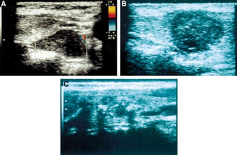
42    T Rago and others                                                                      EUROPEAN JOURNAL OF ENDOCRINOLOGY (1998) 138

Figure 1 Conventional echographic patterns. A: absence of halo sign in sporadic medullar carcinoma (longitudinal section).
B: hypoechogenicity in papillary thyroid carcinoma (longitudinal section). C: intranodular calcification, absence of halo sign and
hypoechogenicity are also present in papillary thyroid carcinoma (longitudinal section).

Conventional and CFD sonography                                        Results
Conventional US and CFD sonography were performed                      Histological examination demonstrated CA in 30
using a color doppler apparatus (AU 590 Asynchro-                      nodules: 18 papillary carcinomas, 6 medullary carci-
nous, Esaote Biomedica, Firenze, Italy), with a 7.5 MHz                nomas, 5 follicular carcinomas and 1 thyroid lym-
linear transducer. Data were collected blindly by two                  phoma. Seventy-four nodules were benign with the
independent examiners (T R and S M). When results of                   following histological pattern: 43 micro–macrofollicular,
the examiners were discordant, agreement was found                     18 microfollicular, 11 macrofollicular, and 2 Hurtle cell
by conjoint re-examination of the patient. Conven-                     adenoma (Table 1). The size of malignant nodules was
tional US evaluated: (i) the echogenicity of the nodule                4.2 6 5.7 ml (range 0.2–25); the size of BN was
with respect to the surrounding thyroid parenchyma;                    11.5 6 13.4 ml (range 0.2–60).
(ii) the presence of halo sign (transonic rim surround-
ing the lesion); and (iii) the presence of microcalcifica-
tions defined as hyperechoic spots less then 2 mm with                 Conventional US
acoustic shadowing. CFD evaluated the presence and                     US patterns considered were: (i) absence of halo sign;
the pattern of blood flow: Type I, absence of blood                    (ii) microcalcifications; and (iii) hypoechogenicity. The
flow; Type II, perinodular and absent or slight                        single US pattern that was most predictive of malig-
intranodular blood flow; and Type III, marked                          nancy (Table 2) was absent halo sign (P < 0.0001;
intranodular and absent or slight perinodular blood                    specificity 77.0%, sensitivity 66.6%). Absent halo sign/
flow (Figs 1 and 2).                                                   microcalcifications was the most specific double combi-
   Statistical evaluation was performed using the chi-                 nation of US patterns (P < 0.005; specificity 93.2%,
square test, and the predictivity test of Galen and                    sensitivity 26·6%). When all the three patterns were
Gambino (19).                                                          considered together, no gain in specificity was obtained.

                                                                                                    Downloaded from Bioscientifica.com at 01/16/2021 06:40:41PM
                                                                                                                                                  via free access
EUROPEAN JOURNAL OF ENDOCRINOLOGY (1998) 138 US and CFD sonography in ‘cold’ thyroid nodules 43
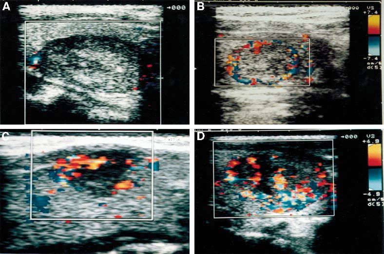
Figure 2 Color flow-doppler sonographic patterns. A: absence of blood flow (Type I). B: perinodular blood flow (Type II). C: slight
intranodular blood flow (Type III). D: marked intranodular blood flow (Type III).
CFD sonography sensitivity (50.0%) and specificity (75.6%) than the
absence of halo sign, similar to what has been observed
The CFD pattern was Type I in 23 nodules, Type II in
by other authors (12, 20–23). A hypoechoic aspect was
23, and Type III in 58 (Table 3). In particular, an
found in 66.6% CA and in 51.3% BN, in agreement
intranodular vascularization was found in 20/30 CA,
with the observations of Solbiati et al. (13) and
but also in 38/74 BN (not statistically significant).
Takashima et al. (12). Hypoechogenicity, absence of
halo sign and microcalcifications have already been
Combination of conventional US and CFD reported as single patterns suggestive of malignant
sonography thyroid nodules in previous studies (7, 23–26). A solid
nodule, with a hypoechoic aspect and irregular borders
For all US features, the combination with Type III was regarded as CA in most of the reports that considered
pattern on CFD increased the specificity but reduced the also combinations of several patterns (25–28). However,
sensitivity in predicting CA (Table 4). The most specific some of these studies were inhomogeneous for patient
double combination of US with Type III on CFD was selection since ‘hot’ thyroid nodules were also included.
absent halo sign/microcalcifications (P < 0.01; specifi-
city 97.2%, sensitivity 16.6%).
Table 1 Histological pattern in thyroid nodules.
Discussion Carcinoma (n ¼ 30) Benign nodule (n ¼ 74)
The absence of halo surrounding the nodule was the Type No. Type No.
pattern most predictive for malignancy on conventional
US. This sign was found in 66.6% CA and in 22.9% Papillary 18 Micro–macrofollicular 43
BN. The sensitivity (66.6%) and specificity (77.0%) of Medullary 6 Microfollicular adenoma 18
Follicular 5 Macrofollicular 11
this sign was higher than in other reports (12). The Lymphoma 1 Hurtle adenoma 2
finding of intranodular microcalcifications had lower
Downloaded from Bioscientifica.com at 01/16/2021 06:40:41PM
via free access44 T Rago and others EUROPEAN JOURNAL OF ENDOCRINOLOGY (1998) 138
Table 2 Conventional ultrasonographic patterns and histology in thyroid nodules.
Positive Negative
Benign Specificity Sensitivity predictive predictive
Echographic pattern Carcinoma nodules Significance (%) (%) value (%) value (%)
Absent halo sign 20/30 17/74 x2 ¼ 17:7 77.0 66.6 54.0 85.0
P < 0:0001
Microcalcifications 13/30 18/74 x2 ¼ 3:68 75.6 54.0 55.6 76.7
P < 0:05
Hypoechogenicity 20/30 38/74 x2 ¼ 2:00 48.6 66.6 34.4 78.2
P < 0:15
Absent halo sign þ 18/30 13/74 x2 ¼ 18:0 82.4 66.0 58.0 83.5
hypoechogenicity P < 0:0001
Absent halo sign þ 8/30 5/74 x2 ¼ 7:70 93.2 26.6 61.5 75.8
microcalcifications P < 0:005
Hypoechogenicity þ 9/30 15/74 x2 ¼ 1:13 79.7 30.0 26.4 73.7
microcalcifications P < 0:28
Table 3 Color flow-doppler (CFD) sonography and histological pattern in thyroid nodules.
Positive Negative
Benign Specificity Sensitivity predictive predictive
CFD pattern Carcinoma nodules Significance (%) (%) value (%) value (%)
Type I absence of blood flow 5/30 18/74 x2 ¼ 0:70 75.6 16.6 21.7 69.0
P ¼ 0:39
Type II perinodular blood flow 5/30 18/74 x2 ¼ 0:70 75.6 16.6 25.0 65.0
P ¼ 0:39
Type III intranodular blood flow 20/30 38/74 x2 ¼ 2:03 48.6 66.6 34.4 78.2
P ¼ 0:15
Table 4 Combination of conventional ultrasonography and color flow-doppler sonography (CFD) in thyroid nodules.
Positive Negative
Echographic pattern/ Benign Specificity Sensitivity predictive predictive
CFD Carcinoma nodules Significance (%) (%) value (%) value (%)
Absent halo sign/ 15/30 8/74 x2 ¼ 19:0 89.0 50.0 65.2 81.4
Type III P < 0:0001
Microcalcifications/ 7/30 10/74 x2 ¼ 1:50 86.4 23.3 41.1 73.5
Type III P < 0:20
Hypoechogenicity/ 14/30 22/74 x2 ¼ 2:70 70.2 44.0 38.8 76.4
Type III P ¼ 0:10
Absent halo sign þ 5/30 2/74 x2 ¼ 6:30 97.2 16.6 71.1 74.2
microcalcifications/ P < 0:01
Type III
Absent halo sign þ 13/30 6/74 x2 ¼ 17:7 91.8 43.3 68.4 80.0
hypoechogenicity/ P < 0:0001
Type III
Hypoechogenicity þ 6/30 8/74 x2 ¼ 1:50 89.1 20.0 42.8 73.3
microcalcifications/ P ¼ 0:20
Type III
Downloaded from Bioscientifica.com at 01/16/2021 06:40:41PM
via free accessEUROPEAN JOURNAL OF ENDOCRINOLOGY (1998) 138 US and CFD sonography in ‘cold’ thyroid nodules 45
In the present investigation only ‘cold’ thyroid nodules on 4 Mazzaferri EL & Ropfagha-Keyhani S. Solitary thyroid nodule:
scintiscans were included, and the predictive value of two diagnosis and management. Medical Clinics of North America 1988
72 1177–1211.
or more combined echographic patterns was evaluated. 5 Mazzaferri EL. Management of a solitary thyroid nodule. New
We found that the most predictive combination on England Journal of Medicine 1993 328 553–559.
conventional US was absence of halo sign plus micro- 6 Grant CS, Hay ID, Gough IR, McCarthy PM & Goeliner JR.
calcifications. This combination had a high specificity Long-term follow-up of patients with benign thyroid fine-
needle aspiration cytologic diagnoses. Surgery 1989 106 980–
(93.0%), but a low sensitivity (36.0%). The predictive 986.
value of other combinations was even lower. 7 Gharib H & Goellner JR. Fine needle aspiration biopsy of
Intranodular blood flow on CFD was found in 66.6% the thyroid: an appraisal. Annals of Internal Medicine 1993 118
of carcinomas and in 51.3% of BN. Thus the predictive 282–289.
value of CFD alone was poor, in agreement with data 8 Gharib H. Fine needle aspiration biopsy of thyroid nodules:
advantages and limitations and effect. Mayo Clinic Proceedings
reported by Fobbe et al. (15) and Solbiati et al. (25). 1994 69 44–49.
Other authors (29) suggested that carcinoma and 9 Walfish PG, Hazani E, Strawbridge HTG, Miskin M, Rosen B et al.
autonomous adenoma can be excluded in patients A prospective study of combined ultrasonography and needle
with nodular goiter when normal vascularization is aspiration biopsy in the assessment of the hypofunctioning
thyroid nodule. Surgery 1977 82 474–482.
demonstrated. In contrast, others (14, 30) did not find a 10 Miller JM, Hamburger JI & Kini S. Diagnosis of thyroid nodules:
correlation between the presence of specific color signals use of fine-needle aspiration and needle biopsy. Journal of the
and pathology, as the detection of color signals was American Medical Association 1979 241 481–486.
dependent on the size of the nodule rather than on its 11 Simeone JF, Daniels GH, Mueller PR, Maloof F, Van Sonnenberg E,
histology. On the other hand, Anguissola et al. (18) as Hall DA et al. High resolution real-time sonography of the thyroid.
Radiology 1982 145 431–435.
well as other authors (15–17, 28–34) reported that an 12 Takashima S, Fukuda H, Nomura N, Kishimoto H, Kim T &
increased nodular blood flow was associated with Kobayashi T. Thyroid nodules: re-evaluation with ultrasound.
thyroid carcinoma. In our series, we evaluated the Journal of Clinical Ultrasound 1995 23 179–184.
combination of each pattern on conventional US with 13 Solbiati T, Volterrani L, Rizzato G, Bazzocchi M, Busilacci P,
Canadiani F et al. The thyroid gland with low uptake lesions:
an intranodular blood flow on CFD. Absence of halo sign evaluation by ultrasound. Radiology 1985 155 187–191.
plus microcalcifications on US combined with intra- 14 Shimamoto K, Endo T, Ishigaki T, Sakuma S & Makino N. Thyroid
nodular blood flow on CFD was found to be highly nodules: evaluation with color doppler ultrasonography. Journal of
specific for malignancy, being seen in only 2/74 BN. Ultrasound Medicine 1993 12 673–678.
Unfortunately it was present in only 5/30 carcinomas. 15 Fobbe F, Finke R, Reichestein E, Schleusener H & Wolf KJ.
Appearance of thyroid disease using colour-coded duplex
Thus, the gain in specificity (97.2%) occurred at the sonography. European Journal of Radiology 1989 9 29–31.
expense of sensitivity. 16 Ralls PW, Mayekawa DS, Lee KP, Colletti PM, Radin DR,
In summary, we confirm that taken by itself no single Boswell WD et al. Color-flow doppler sonography in Graves’
pattern on US and CFD is highly predictive for disease: ‘thyroid inferno’. American Journal of Radiology 1988 150
781–784.
malignancy in ‘cold’ thyroid nodules. The combination 17 Lagalla R, Caruso G, Romano M, Midiri M, Novara V &
of patterns that are more frequently associated with Zappasodi F. Eco-color doppler nella patologia tiroidea. La
malignancy are the absence of halo sign and micro- Radiologica Medica 1993 85 109–113.
calcifications. An intranodular blood flow on CFD only 18 Anguissola R, Bozzini A, Campani R, Bottinelli O, Genovese E,
slightly increased the predictivity of signs observed on Guglielmoni B et al. Ruolo della Colour coded duplex sonography
nello studio della patologia tiroidea. La Radiologica Medica 1991
conventional US. 81 831–837.
In conclusion, findings on US and CFD become 19 Galen RS & Gambino SL. Beyond Normality: The Predictive Value
highly predictive for malignancy only when multiple and Efficiency of Medical Diagnosis. New York: Wiley and Son,
signs are simultaneously present in a thyroid nodule. 1980.
20 LiVolsi VA, Winfield MJ & Steinbach HL. Pathology of thyroid
However, the predictive value of these techniques tumors. In Comprehensive Management of Head and Neck Tumors, pp
increases at the expense of their sensitivity. Thus in 1599–1604. Eds SE Thawley & WR Panje. Philadelphia: W B
less than 20% of patients with CA can malignancy be Saunders, 1987.
predicted with high specificity by US and CFD 21 Margolin FR, Winfield J & Steinbach HL. Patterns of thyroid
sonography. calcification. Roentgenologic–histologic study of excised speci-
mens. Investigative Radiology 1967 2 208–214.
22 Barsakisn JG, Nishiyama RH & Rich CR. Microlithiasis (calco-
spherites) and carcinoma of the thyroid gland. Archives of
References Pathology and Laboratory Medicine 1960 69 493–498.
1 Belfiore A, Rosa GL, Giuffrida D, Regalbuto C, Lupo L, Fiumara A 23 Katz JF, Kane RA, Reyes J, Kam RA, Reyes J, Clarke MP et al.
et al. The management of thyroid nodules. Journal of Endocrino- Thyroid nodules: sonographic–pathologic correlation. Radiology
logical Investigation 1995 18 155–158. 1984 151 741–745.
2 Belfiore A, La Rosa GL, Padova G, Sava L, Ippolito O & Vigneri R. 24 Gorman B, Charboneau JW, James EM, Reading CC, Wold LE,
Prevalence of cold thyroid nodules and thyroid malignancies in Grant CS et al. Medullary thyroid carcinoma: role of high-
patients from an iodine deficient area. Cancer 1987 60 3096– resolution US. Radiology 1987 162 147–150.
3111. 25 Solbiati L, Ballarati E, Cioffi V, Poerio N, Croce F & Rizzatto G.
3 Trowbridge FL, Matovinovic J, McLaren GD & Nichaman MZ. Microcalcificazions: a clue in the diagnosis of thyroid
Iodine and goiter in children. Pediatrics 1975 56 82–90. malignancies. Radiology 1990 177 (Suppl) 140 (Abstract).
Downloaded from Bioscientifica.com at 01/16/2021 06:40:41PM
via free access46 T Rago and others EUROPEAN JOURNAL OF ENDOCRINOLOGY (1998) 138
26 Tan GH, Gharib H & Reading CC. Solitary thyroid nodule. using echo-color-doppler. Annales Italian Medicine Internal 1996 11
Comparison between palpation and ultrasonography. Archives of 175–179.
Internal Medicine 1995 155 2418–2423. 32 Spiezia S, Colao A, Assanti AP, Cerbone G, Picone GM, Merola B
27 Ross DS. Evaluation of the thyroid nodules. Journal of Nuclear et al. Usefulness of color echo doppler with power doppler in the
Medicine 1991 32 2181–2192. diagnosis of hypoechoic thyroid nodules: work in progress.
28 Cole-Beuglet C & Goldberg BB. New high-resolution ultrasound Radiological Medicine 1996 921 616–621.
evaluation of disease of the thyroid. Journal of the American Medical 33 Holden A. The role of color and duplex Doppler ultrasound in the
Association 1983 249 2941–2944. assessment of the thyroid nodules. Australasian Radiology 1995 35
29 Becker D, Bair HJ, Becker W, Gunter E, Lohner W, Lerch S et al. 343–349.
Thyroid autonomy with color-coded image-directed doppler 34 Schwaighofer F, Kurtaran A, Hubsch P, Fruhwald F, Barton P,
sonography internal hypervascularization for the recognition Trattnig S et al. Color-coded doppler sonography in thyroid gland
of autonomous adenoma. Journal of Clinical Ultrasound 1997 25 diagnosis: preliminary results. Rofo Fortschr gab Routgenstr
63–69. Nuklearmed 1988 149 310–313.
30 Argalia G, D’Ambrosio F, Lucarelli F, Mignosi U, Giuseppetti GM,
Passarini G et al. Echo doppler in the characterization of the
thyroid nodular goiter. Radiologica Medica 1995 89 651–657.
31 Urso M, Angelillis L & Ambrosio GB. Vascularization of single Received 26 May 1997
thyroid nodule as an indicator of malignant neoplasm: a study Accepted 19 August 1997
Downloaded from Bioscientifica.com at 01/16/2021 06:40:41PM
via free accessYou can also read