Dr Steve Colley
Queen Elizabeth Hospital Birmingham
                                      May 2014
←
→
Page content transcription
If your browser does not render page correctly, please read the page content below
Dr Steve Colley
Queen Elizabeth Hospital Birmingham
May 2014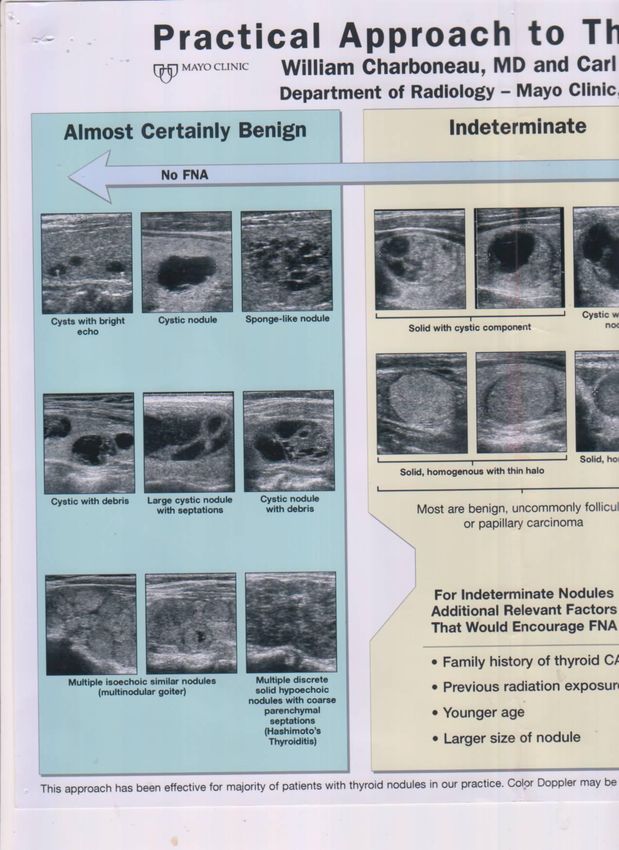
Thyroid cancer incidence Thyroid nodule appearances Scoring systems & Guidelines New BTA Guidelines Common misunderstandings Thyroid US standards
From: Increasing Incidence of Thyroid Cancer in the United States, 1973-2002

                                        Copyright © 2012 American Medical
                                          Association. All rights reserved.
From: Increasing Incidence of Thyroid Cancer in the United States, 1973-2002

.

                                        Copyright © 2012 American Medical
                                          Association. All rights reserved.
From: Increasing Incidence of Thyroid Cancer in the United States, 1973-2002

                                        Copyright © 2012 American Medical
                                          Association. All rights reserved.
Thyroid cancer: zealous imaging has increased detection and treatment of low risk tumours.
Brito et al. BMJ 2013; 347: 18 – 21.
Inappropriate use and reporting of imaging will result in an
epidemic of thyroid nodules, the majority of which will be benign.
Thyroid nodules are very com
                                                            50% of the adult population.
                                                            in approximately 25,000 pati
                                                            mon cause of benign thyroid
    Note: T his copy is for your personal non- commercial use only. To order presentation- ready
    copies for distr ibution to your colleagues or clients, contact us at www.rsna.org/rsnar ights.
                                                            7% of thyroid nodules are m
EDUCATION EXHIBIT                                           fied. The imaging modality               847
                                                                                                        o
                                                            resolution US. US is commo
                        US Featur es of Thyr oid            benign and malignant thyroid
                        M alignancy: Pear ls
                        and Pitfalls1
                         Abbreviation: FNA          fine-needle aspiration

                         RadioGraphics 2007; 27:847–865 ● Published online 10.1148/rg.2
                        Jenny K. Hoang, M BBS, FRANZCR ● Wai Kit Lee, M BBS, FRANZCR
 ONLINE-ONLY
     CME                M1From    the Departments
                           ichael Lee,  M BBS ● DarylofJohnson,
                                                         M edicalMImaging  (J.K.H.,
                                                                   BBS ● Stephen      W.K.L.,
                                                                                  Farrell,     M .L.),
                                                                                           M BBS,
                         sity of M elbourne, 41 Victoria Parade, Fitzroy 3065, Victoria, Australi
                        FRACS
   Seewww.rsna
   .org/education        RSNA Annual M eeting. Received M arch 22, 2006; revision requested
   /rg_cme.html.         relationships to disclose. Address correspondence to J.K.H. (e-mail
                        T hyroid nodules are common and occur in up to 50% of the adult
                        population;    however, less
                         See the commentary          than 7%following
                                                 by Langer    of thyroidthis
                                                                         nodules   are malignant.
                                                                             article.
   LEARNING
  OBJECTIVES            H igh-resolution ultrasonography (US) is commonly used to evaluate
                         ©
  After reading this
                        the  thyroid2007
                           RSNA,     gland, but US is frequently misperceived as unhelpful for
  article and taking    identifying features that distinguish benign from malignant nodules.
  thetest, thereader
    will beableto:      M icrocalcifications are one of the most specific US findings of a thy-
  D escribe common      roid malignancy. Other useful US features include a marked hypoecho-
US features of thy-
roid malignancy and     genicity, irregular margins, and the absence of a hypoechoic halo
the value and limita-   around the nodule. L ymphadenopathy and local invasion of adjacent
tions of each.
                        structures are highly specific features of thyroid malignancy but are less
  Correlate US fea-
tures with the patho-   commonly seen. T he number, size, and interval growth of nodules are
logic appearance and    nonspecific characteristics. Suspicious US features may be useful for
behavior of different
histologic types of     selecting patients for fine-needle aspiration biopsy when incidental
thyroid malignancy.     nodules are discovered and when multiple nodules are present. Com-
  Recognize atypical    mon interpretative pitfalls that may lead to failure to recognize a malig-
features of thyroid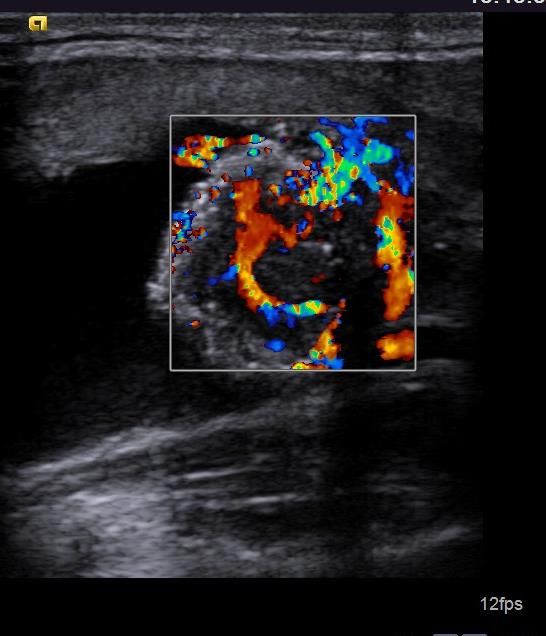
Sensitivity Specificity
Micro-calcifications 40% 90%
Absence of halo 66% 46%
Irregular margins 64% 84%
Hypo-echoic 83% 49%
Intra-nodular flow 70% 65%
MicroCa. & irreg m. 30% 95%
MicroCa. & hypoechoic 28% 95%
Solid & hypoechoic 73% 69%
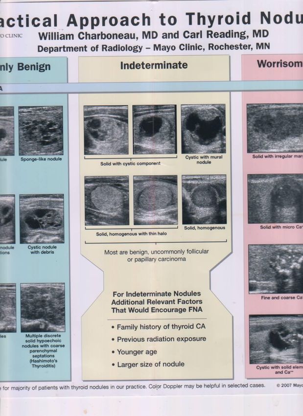
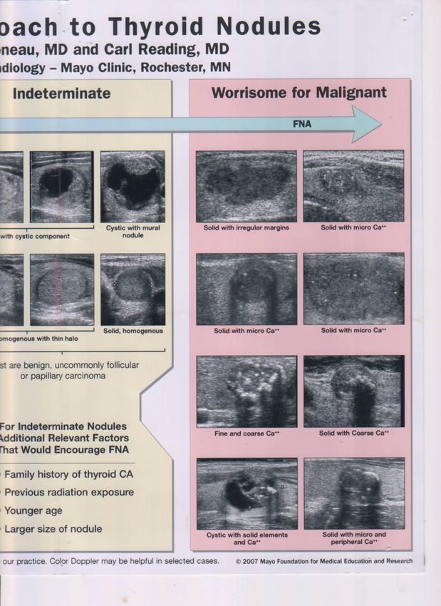
RSNA 2005 Mayo Clinic Thyroid US Chart TIRADS Scoring System U1 – U5 Classification
“This approach has been effective for the
majority of patients with thyroid nodules in our
practice. Colour doppler may be of use in
selected cases.”
2007 Mayo Clinic Foundation for Medical Education and Research Ultrasound is rarely of use in assessment of
thyroid nodules. It may help guide FNA. Separate chapter on thyroid nodule US
Rationalise thyroid US / FNA
Suggested standards for reports
Indications for FNA
Based upon US scoring system
U1 – U5
Follow up based upon US appearancesFIGURE 4. 1
Graphic compilation of the signs that can be used to differentiate benign from malignant nodules. These signs should be used to
guide the decision as to whether or not to carry out a FNAB based on the likelihood of malignancy in a nodule. The signs are
grouped to identify: normal thyroid gland (U1), benign nodule (U2), equivocal /indeterminate nodule (U3), suspicious (U4) and
malignant nodule (U5).
U1. Normal.
U2. Benign:
(a) halo, hyper- / iso-echoic
(b) cystic change +/- ring down sign (colloid)
(c) micro- cystic / spongiform
(d & e) peripheral egg shell calcification
(f) peripheral vascularity.
U3. Indeterminate/Equivocal:
(a) homogenous, hyper - echoic (markedly), solid, halo (follicular
lesion).
(b) ? hypo-echoic, equivocal echogenic foci, cystic change
(c) mixed/central vascularity.
U4. Suspicious:
(a) solid, hypo-echoic (cf thyroid)
(b) solid, very hypo-echoic (cf strap muscle)
(c) disrupted peripheral calcification, hypo-echoic
(d) lobulated outline
U5. Malignant
(a) solid, hypo-echoic, lobulated / irregular outline,
micro-calcification. (? Papillary carcinoma)
(b) solid, hypo-echoic, lobulated/irregular outline, globular
calcification (? Medullary carcinoma)
(c) intra-nodular vascularity
(d) shape (taller >wide)
(e) characteristic associated lymphadenopathyAggregation of micro-cystic spaces comprising >50% of a nodule 99.7% specificity, 100% if isoechoic nodule Moon W-J et al. Benign and Malignant Thyroid Nodules: US Differentiation, a multi-center retrospective study. Radiology 2008; 247(3): 762 – 770.
AP > TR in the transverse plane
FNA any indeterminate or suspicious /
malignant nodules
FNA of U3 – U5
U3 – most will be benign, but follicular lesions
are included, and the occasional cancer may
be present FNA of U3 – U5
U2
U2/3
U4 / 5Courtesy of Dr Andrew McQueen
AJR 2011;196: 655 - 660
Use of scoring systems is robust
Training possible
Good inter observer variability
Allows effective audit / follow Ultrasound based reporting system for thyroid
nodules improves patient management and
cost-effectiveness by reducing unnecessary
FNA
Horvath E et al. An ultrasonogram reporting system for thyroid nodules
stratifying cancer risk for clinical management. J Clin Endocrinol Metab 2009;
94: 1748 - 1751 American Thyroid Association (ATA)
American Association of Clinical
Endocrinologists (AACE)
Kim / Korean Society of Radiologists
Society of Radiologists in US (SRUS)
British Thyroid Association (BTA)
Mayo Clinic Thyroid US Chart Guidelines represent expert opinion based upon
a selection of retrospective studies
There is a lack of prospective randomised
control trials assessing effectiveness of
Guidelines in detecting thyroid cancer The majority of guidelines currently available
still recommend biopsy for the majority of
thyroid nodules
This leads to a massive cost implication, with
resultant surgery for often benign disease,
and all to exclude a cancer with excellent long
term survival ratesKim AACE SRUS 1398 nodules
FNA of any nodule with one of:
Markedly hypo-echoic
Micro-calcification
Irregular margins
Taller-than –wide shape
Kim EK, Park CS, Chung WY et al. New sonographic criteria for
recommending fine needle aspiration biopsy of nonpalpable solid nodules of
the thyroid. AJR 2002; 178: 687 – 691. FNA of any nodule with:
Marked hypo-echogenicity + one other:
Micro-calcification
Irregular margins
Taller-than-wide shape
Gharib H, Papini E, Valcavi R et al. American Association of Clinical
Endocrinologists and Associazone Medici Endocrinologi medical guidelines
for the diagnosis and management of thryoid nodules. Enodcr Pract 2006;
12: 63 – 102. FNA any nodule with:
> 10mm with micro-calcification
>15mm if solid
>15mm if coarse calcification
>20mm if solid and cystic
Frates MC, Benson CB, Charboneau JW et al. Management of thyroid
nodules detected at US: Society of Radiologists in Ultrasound consensus
conference statement. Radiology 2005; 237: 794 – 800. Kim Criteria
Sensitivity 92.7% Specificity 80.9%
NPV 97.3%
AACE Criteria
Sensitivity 74% Specificity 94.4%
NPV 95% SRUS
Uses size criteria
Sensitivity 35%
Specificity 54%
NPV 80% Nodule size Dominant Nodule FNA Follow up post benign FNA Nodule growth
Nodules > 4cm have been claimed as having
malignancy rates > 20%
Nodule size > 4cm increases neither the false
negative rate of FNA, nor the rate of
malignancy
Shrestha M, Crothers BA, Burch HB. The impact of thyroid nodule size on
the risk of malignancy and accuracy of fine-needle aspiration: a 10-year
study from a single institution. Thyroid. 2012;22:1251-6 661 nodules > 3cm diameter
US and FNA are accurate in nodules > 3cm.
US features are still predictive even with
larger nodule size
Yoon JH, Kwak JY, Moon HJ. The diagnostic accuracy of ultrasound guided
fine needle aspiration biopsy and the sonographic differences between
benign and malignant thyroid nodules 3cm or larger. Thyroid 2011: 21(9): 993
– 1000 FNA of a dominant nodule is a common but
mistaken practice
Decision to FNA should be based upon US
appearances.
Selecting nodules purely on size criteria
encourages lazy / incomplete assessmentRadiology 2010. 254 (1): 292 - 300 1343 nodules with US, FNA, pathological correlation Total: Benign 98.1% Malignant 1.9% Benign initial US + Thy 2 FNA: Benign 99.4% Malignant 0.6% Suspicious initial US + Thy 2 FNA: Benign 79.6% Malignant 20.4%
Clinico-radiologic-cytologic correlation
Benign US and benign FNAC does not need
repeating after 6 – 12 months
Low cost effectiveness
Suspicious US and Thy 2 must be repeated as
malignancy rates are significant Long term follow up of benign nodules
Associated with increased US studies
Associated with increased FNA rates
No improvement in malignancy detection rates
Lee S, Skelton TS, Zheng F. Biopsy proven benign thyroid nodule: is long
term follow up necessary. J Am Coll Surg 2013, 217(1): 81 – 8. Presence or absence of growth is not an
indicator of malignancy or benignity
Interval growth has low PPV for malignancy 294 / 330 Thy 2 nodules enlarged
Average 15% growth
74 nodules had significant growth (≈ 69%)
Re-FNA showed cancer in only 1 / 74.
Alexander EK et al. Natural History of Benign Solid & Cystic
Thyroid Nodules. Ann Int Medicine 2003; 138: 315 - 318
Growth of nodules is an expected finding in
benign thyroid disease Radiologist, sonographer, surgeon,
endocrinologist
Formal images, recorded on PACS, with
appropriate formal report on RIS system
Training in accordance with RCR Guidelines /
Non–radiologist US Training Document
Assessment of any indeterminate or suspicious
nodules Interest / regular practice of thyroid imaging
Participation at Thyroid MDT
US images / report stored on PACS system
Report attached to images
Assess the likelihood of cancer
Regular audit of cases / FNA results
MINIMUM STANDARD OF PRACTICE Massive cost to NHS of follow up / FNA
>£5 million per annum for UHB NHSFT for US + FNA
Stable mortality rates despite investigation
No CT feature reliably characterises nodules
Clinical evaluation (not US / FNA), but consider
US / FNA initially if:
Risk factors in history
Ill defined margins, young age, micro-calcification Focal FDG activity within thyroid Meta-analysis shows malignancy ≈ 35% US / FNA must be performed for focal uptake Soelberg KK, Bonnema SJ, Brix TH, Hegedus L. Risk of malignancy in thyroid incidentalomas detected by 18 FDG PET: a systematic review. Thyroid 2012; 22(9): 918 – 925.
US images stored on PACS system
Formal documented report
Report attached to images
Regular audit of cases / FNA results
Interest / regular practice of thyroid imaging
Participation at Thyroid MDT FNA of a dominant nodule in a MNG is a
common but mistaken practice
Use ultrasound to risk stratify nodules
Mayo Clinic
BTA U1-U5 scoring system
FNA indeterminate or suspicious / malignant
nodules Follow up post FNA should be based upon
nodule appearances on US
Close liaison with thyroid / endocrine team
Clinical history is important
Repeat sampling of Thy 2 but suspicious US
Incidental FDG avid nodules on PET CT need
definite follow up with US / FNACYou can also read