Visualizing Brain Activation during Planning: The Tower of London Test Adapted for Functional MR Imaging

Page created by Lorraine Wong
 
CONTINUE READING
Visualizing Brain Activation during Planning: The Tower of London Test Adapted for Functional MR Imaging
AJNR Am J Neuroradiol 21:1407–1414, September 2000

Visualizing Brain Activation during Planning: The Tower
  of London Test Adapted for Functional MR Imaging
   Richard H.C. Lazeron, Serge A.R.B. Rombouts, Willem C.M. Machielsen, Philip Scheltens, Menno P. Witter,
                                  Harry B.M. Uylings, and Frederik Barkhof

            BACKGROUND AND PURPOSE: Recent positron emission tomography and single-photon
         emission CT studies using the Tower of London test have shown that brain activation during
         planning activities primarily resides in the prefrontal cortex. In this study, we adapted the
         Tower of London test for functional MR imaging.
            METHODS: For use with functional MR imaging, a block design of the test was created, in
         which planning stages were contrasted with counting of colored balls. For nine healthy partic-
         ipants, multisection echo-planar functional MR imaging was performed to assess brain acti-
         vation based on changes in blood oxygen level. Activation maps for individual participants and
         a group average map were created.
            RESULTS: In the group average map, activation in the dorsolateral prefrontal cortex, the
         anterior part of the cingulate cortex, the cuneus and precuneus, the supramarginal and angular
         gyrus in the parietal lobe, and the frontal opercular area of the insula was seen. These findings
         are in agreement with grouped data of previous positron emission tomography results. Func-
         tional MR imaging enabled us to investigate brain activation during planning activities with
         high spatial (and temporal) resolution in individual patients, showing that the dorsolateral
         prefrontal cortex was activated in all participants studied.
            CONCLUSION: Presented is a working functional MR imaging version of the planning task.
         The high sensitivity of functional MR imaging may allow the use of this test for patients with
         possible (pre)frontal disorders.

Planning is defined as the ability to organize cog-                      processing abilities, it mainly depends on planning.
nitive behavior in time and space (1). It is neces-                      Patients with frontal lobe pathologic abnormalities
sary in situations in which a goal must be achieved                      (eg, frontal lobe dementia, multiple sclerosis) per-
through a series of intermediate steps, each of                          form worse than do healthy control participants.
which individually does not lead directly toward                            Neuropsychological studies have shown that le-
that goal. A well-known test to evaluate planning                        sions in the frontal lobe (mainly the prefrontal cor-
in neuropsychological research is the Tower of                           tex, which is located anterior to the motor part),
London test (2). For this test, the participant is in-                   might cause problems with planning (1–3). Recent
structed to move three different colored balls to                        positron emission tomography and single-photon
match a target configuration by using a minimum                          emission CT studies (4–7) that used the Tower of
number of moves. Although this test needs spatial                        London test confirmed that brain activity during
                                                                         planning is located mainly in the prefrontal area,
   Received November 10, 1999; accepted after revision February          particularly in the dorsolateral prefrontal cortex.
23, 2000.                                                                These data were based on averages of several par-
   From the Department of Radiology and the MS-MRI Centre                ticipants, because these techniques usually have too
(R.H.C.L., F.B.), the Department of Clinical Physics and In-             low a sensitivity to detect activation in individual
formatics (S.A.R.B.R.), and the Department of Neurology                  participants reliably.
(P.S.), Academic Hospital of the Vrije Universiteit, and the
Department of Anatomy and Embryology (W.C.M.M.,                             Functional MR imaging is a noninvasive tech-
M.P.W., H.B.M.U.), Faculty of Medicine, Vrije Universiteit,              nique with which to measure brain activity based
Amsterdam, The Netherlands.                                              on changes in the blood oxygen level (8, 9). Ad-
   This work was supported in part by grants 96-278 and 97-              ditional advantages of this technique compared
330 from Stichting Vrienden MS Research (to R.H.C.L.).                   with other functional brain imaging techniques are
   Address reprint requests to R.H.C. Lazeron, Academic Hos-
pital of the Vrije Universiteit, Department of Neurology, P.O.
                                                                         its high spatial and temporal resolution and the
Box 7057, 1007 MB Amsterdam, The Netherlands.                            ability to study individual subjects. Paradigms ap-
                                                                         plied in positron emission tomography studies,
q American Society of Neuroradiology                                     however, often cannot be used without major

                                                                  1407
Visualizing Brain Activation during Planning: The Tower of London Test Adapted for Functional MR Imaging
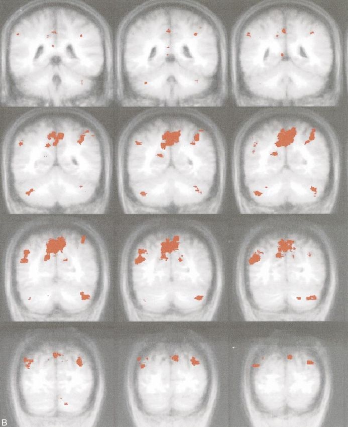
1408      LAZERON                                                                                                 AJNR: 21, September 2000

FIG 1. Example of the Tower of London screen.
   A, Sample screen of one of the configurations of a planning problem. Upper, baseline configuration; lower, target configuration. In
this example, the participant has been asked to move first the blue ball to the right rod, which is counterintuitive. Thereafter, the participant
has to place the yellow ball on top of the red ball, the blue ball at its destination, the yellow ball on top of the blue ball, the red ball on
the right rod, and, finally, the yellow ball at the target position (sixth move). Two alternatives are presented on each side of the screen,
from which the participant had to choose the correct answer. The participant was asked to respond by pressing the air bulb at the
corresponding side.
   B, Sample screen of the control configuration. The participant has to count the yellow and blue balls altogether. In this example, the
answer is six, which is indicated on the right.

changes in functional MR imaging, because there                             test was explained and practiced outside the procedure room
are important differences in the test situations of                         before MR imaging was performed.
the two techniques. In this study, the results of our
fMR imaging–adapted version of the planning task                                                       Participants
are presented.                                                                 Nine healthy students (five men and four women; mean age,
                                                                            22 years; age range, 20–27 years) were evaluated. The ethical
                                                                            review board of the Academic Hospital of the Vrije Univer-
                            Methods                                         siteit Amsterdam approved of the study, and all participants
                          Task Paradigm                                     provided informed consent.
   For application of the test with functional MR imaging, a
block design was created, in which an ‘‘active’’ condition con-                                      Data Acquisition
cerning planning and a ‘‘control’’ condition without planning
were alternated (36 s per block, including an instruction; nine                Imaging was performed on a 1.5-T MR system with a stan-
blocks in total). With the active condition, the participants are           dard circularly polarized head coil. Anatomic imaging was per-
presented a baseline and a target configuration on a single                 formed with a 3D gradient-echo T1-weighted sequence (15/7/1
screen (Fig 1) viewed through a mirror in the magnet bore.                  [TR/TE/excitations]; flip angle, 88; matrix, 256 3 256; field of
Both configurations consist of three balls of different colors              view, 220 3 220 mm; section thickness, 2 mm; number of sec-
(blue, yellow, and red) placed on three vertical rods, which are            tions, 82). The sections were planned in the coronal plane with
one, two, and three balls in height, respectively. The minimum              a rotation of approximately 308 of the cranial part in the anterior
number of necessary moves to reach the target has to be                     direction, to cover the whole brain in the least possible number
planned in mind. One ball can be moved at a time, and only                  of sections. For functional MR imaging, a whole-brain echo-
when there is no other ball on top. Sometimes counterintuitive              planar imaging sequence (4000/64/1; flip angle, 908; matrix, 64
moves are necessary to reach the target, one of the major as-               3 128 interpolated to 128 3 128 mm; field of view, 220 3
pects of planning. The participant holds two air bulbs and an-              220 cm; section thickness, 6 mm, intersection gap, 1.02 mm;
swers by pressing the one corresponding to the side where the               number of sections, 23) was used. The echo-planar imaging sec-
correct answer is shown; one of two possibilities displayed at              tions were planned parallel to the anatomic sections.
the bottom of the screen. With the control condition, partici-
pants simply have to count the yellow and blue balls together
and again choose the correct answer (total number of balls)                                           Data Analysis
from two possibilities. The display is almost the same as with                 The first step of postprocessing was correction of motion
the active condition, except that more balls are displayed, with            artifacts (10), with the consequence of corrupting the first and
every time another number of yellow and blue balls (Fig 1).                 last section of each volume, which were discarded from further
   Every condition block starts with an instruction of 4 s to               analysis. Next, the data were smoothed in-plane, resulting in
plan the moves (active condition) or count the balls (control               a full width at half maximum of 5 mm in plane. The following
condition). Easy (two to four moves) and difficult (five to sev-            steps were performed with AFNI software (11). Activation was
en moves) configurations are presented in separate blocks to                detected by correlating the time course of each voxel with a
make it possible to compare two different levels of planning                box car function representing the active and control blocks of
activity (easy and difficult) and a control situation without               the paradigm (Fig 2). Also, the two levels of planning diffi-
planning. The whole test is self-paced; a new trial (in the same            culty were correlated in the same way, without using the con-
block) is presented only after a response is obtained. No feed-             trol stage images. Voxels with a signal increase during the
back regarding the correctness of the answer is provided during             active condition had a positive correlation coefficient and were
the task. After 36 s, the next block starts with a new instruction          called positive activation. The opposite was true for voxels
(Fig 2), regardless of whether there was a response to the last             with a relatively reduced signal. The box car function was
trial. In total, 82 whole-brain volumes (nine blocks with nine              delayed 4 s (1 image) in time to account partly for the he-
volumes each and one volume preceding the start of the test)                modynamic response delay (12). The images that corresponded
were scanned (one scan was obtained every 4 s) (Fig 2). To                  with the time the instructions were displayed were not used in
ensure the participants were familiar with the procedure, the               the analysis (Fig 2).
Visualizing Brain Activation during Planning: The Tower of London Test Adapted for Functional MR Imaging
AJNR: 21, September 2000                                                                               BRAIN ACTIVATION              1409

FIG 2. Overview of the test, imaging, and data analysis. With this task paradigm, easy and difficult planning and counting are performed
in blocks. Every block lasts 36 s, including a 4-s instruction. A total of nine blocks were performed during the test. A total of 82 images
were obtained, including those of a dummy before the test started. To account for the hemodynamic response delay, a 4-s delay in the
analysis was used. The images obtained during the instruction (accounting for the hemodynamic response delay) were not used for
further calculations. The resulting 71 images were used for the analysis (24 obtained during the difficult planning problems, 24 obtained
during the easy planning problems, and 23 obtained during the control stage).

   The images were transformed into Talairach coordinate                the precuneus and cuneus and the left supramargin-
space (13) by defining reference line landmarks on the anatom-          al and angular gyrus (Fig 3B).
ic images. Individual activation maps were calculated and also
used to create a group average. Correction for multiple com-
                                                                           Most participants showed activation in the same
parisons was performed, accounting for the spatial extent of            gyri that were activated on the group average (Ta-
activation (14, 15). Only voxels with a P value of at least 1024        ble), and activation in the middle frontal gyrus (bi-
were considered active, and 3D clusters of at least 104 mm3             laterally) especially occurred in all participants. We
(ie, five connected active voxels) were included, resulting in a        did not find any significant differences in the acti-
mean activation map of all participants with an overall P value         vation when comparing the two levels of difficulty
, .05. In all individual and group images, the macroscopic              (easy and difficult planning).
position of significant activations was defined based on the
visible gyral-sulcal pattern to specify the location in more an-           The areas observed with a higher signal with the
atomic detail.                                                          control condition were the middle part of the cin-
                                                                        gulate gyrus and the middle part of the insula on
                                                                        both sides, the fusiform gyrus and the pre- and
                        Results                                         postcentral gyrus. The majority of cases (67–100%)
   All nine participants were studied successfully.                     showed activation in the same gyri that were acti-
Because we used a self-paced paradigm, the num-                         vated on the group average. An exception was the
ber of answers varied, with a mean of 13.7 answers                      activation in the fusiform gyrus, which had a higher
(range, 9–17 answers) during both planning con-                         signal with the control condition compared with the
ditions together, 78.7% of which were correct                           planning condition, in only 44% of the participants
(66.7–100%). For the easy configurations, a mean                        (Fig 3 and Table).
of 8.5 answers (6–11 answers) was provided, 82%
of which (57–100%) were correct; for the difficult
configurations, the results were 5.2 answers (3–8                                            Discussion
answers), 71% of which (50–100%) were correct.                             A planning task, such as the Tower of London,
With the control condition (only counting), the                         has proven to be sensitive to prefrontal lesions (3,
number of correct answers was 98% (89–100%),                            5, 16). A new version of the planning task, adapted
and the participants gave 36.4 answers (29–48                           for functional MR imaging, is presented. The group
answers).                                                               analysis showed activation during the planning
   Activation in the group average images during                        stages in the dorsolateral prefrontal cortex, the an-
the active condition (easy and difficult configura-                     terior part of the cingulate cortex, the cuneus and
tions combined) was seen on both sides in the fron-                     precuneus, the supramarginal and angular gyrus in
tal and parietal lobes, the cerebellum, and the insula                  the parietal lobe, and the frontal opercular area of
(Fig 3 and Table). The frontal area showed acti-                        the insula. These findings are in full agreement
vation bilaterally in the middle frontal gyrus and                      with grouped data of previous positron emission
the adjacent part of the inferior frontal sulcus (with                  tomography results (5–7). In addition, functional
some preference for the right hemisphere) and in                        MR imaging enabled us to investigate brain acti-
the anterior part of the cingulate gyrus (Fig 3A).                      vation during planning activities with high spatial
The parietal and occipital regions involved were                        (and temporal) resolution in individual participants,
1410      LAZERON                                                                                            AJNR: 21, September 2000

FIG 3. Activated areas during the active condition of the Tower of London task. During the active condition (planning stage) of the task,
activation (red) on the group average map (shown in Talairach format with coronal orientation) is shown. In the brain area from 23
anterior to 149 posterior, no activation was seen.
  A, Frontal regions. Coronal sections of coordinates 244 to 23 (anterior part of the brain). Activity was noted in the dorsolateral
prefrontal cortex, the anterior part of the cingulate cortex, a part of the precentral cortex, and the frontal opercular area of the insula
during planning. The right side shows slightly more activation than the left side. cont’d →
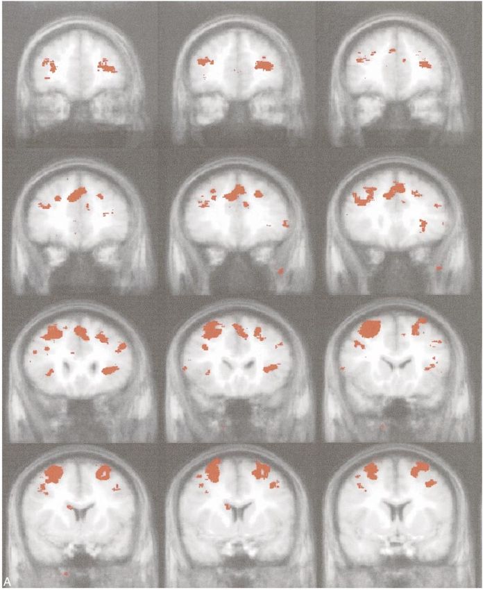
AJNR: 21, September 2000                                                                           BRAIN ACTIVATION              1411

  B, Parietal/occipital regions. Coronal sections of coordinates 149 to 180 (posterior part of the brain). Activation was noted in the
cuneus and precuneus region, the marginal and angular gyrus in the parietal lobe, and the cerebellum during the active condition.
1412                                                                                                        LAZERON                                                                                                                                                                                                                                                                                                            AJNR: 21, September 2000

                                                                                                                                                                                                                                                                                                                                                                                                 showing that the dorsolateral prefrontal cortex was

                                                                                                                                                        9
                                                                                                                                                        9
                                                                                                                                                        9

                                                                                                                                                                                   9
                                                                                                                                                                                   9
                                                                                                                                                                                   9
                                                                                                                                                                                   9

                                                                                                                                                                                                                                                              9
                                                                                                                                                                                                                                                                                                                                                                                                 activated in all individual participants studied.
                                                                                                                                                                                                                                                                                                                                                                                                    Previous studies have indicated that activation
                                                                                                                                                                                                                                                                                                                                                                                                 associated with this task performance occurs es-

                                                                                                                                                        8
                                                                                                                                                        8

                                                                                                                                                                                   8
                                                                                                                                                                                   8
                                                                                                                                                                                   8
                                                                                                                                                                                   8

                                                                                                                                                                                                                    8
                                                                                                                                                                                                                    8
                                                                                                                                                                                                                                                                                                                                                                                                 pecially in the prefrontal cortex (1, 2, 4–7). Three
                                                                                                                                                                                                                                                                                                                                                                                                 positron emission tomography studies that evalu-
                                                                                                                                                                                                                                                                                                                                                                                                 ated the Tower of London test with healthy control
                                                                                                                                                        7
                                                                                                                                                        7
                                                                                                                                                        7

                                                                                                                                                                                   7
                                                                                                                                                                                   7
                                                                                                                                                                                   7
                                                                                                                                                                                   7
                                                                                                                                                                                   7
                                                                                                                                                                                   7
                                                                                                                                                                                   7
                                                                                                                                                                                                                                                                                                                                                                                                 participants (5–7) are herein discussed in compar-
                                                                                                                                                                                                                                                                                                                                                                                                 ison with our results.
                                                                                                                                                                                                                                                                                                                                                                                                    The positron emission tomography studies con-
                                                                                                                                                        6
                                                                                                                                                        6
                                                                                                                                                        6

                                                                                                                                                                                   6
                                                                                                                                                                                   6
                                                                                                                                                                                   6

                                                                                                                                                                                                                6
                                                                                                                                                                                                                6
                                                                                                                                                                                                                6
                                                                                                                                                                                                                                                                                                                                                                                                 ducted by Owen et al (5), Baker et al (6), and Dagh-
                                                                                                             Subjects Active** day/yr

                                                                                                                                                                                                                                                                                                                                                                                                 er et al (7) used a block paradigm in which the par-
                                                                                                                                          Individuals

                                                                                                                                                                                                                                                                                                                                                                                                 ticipants had to plan the moves and press on a touch
                                                                                                                                                        5
                                                                                                                                                        5
                                                                                                                                                        5

                                                                                                                                                                                                        5
                                                                                                                                                                                                                                                                                                                                                                                                 screen the number of moves (6) or perform each
                                                                                                                                                                                                                                                                                                                                                                                                 move separately by pressing on a touch screen the
                                                                                                                                                                                                                                                                                                                                                                                                 ball that has to be moved and thereafter the place to
                                                                                                                                                                                   4
                                                                                                                                                                                   4
                                                                                                                                                        4
                                                                                                                                                        4

                                                                                                                                                                                                        4
                                                                                                                                                                                                        4
                                                                                                                                                                                                        4
                                                                                                                                                                                                        4

                                                                                                                                                                                                                                                                                                                                                                                                 which it had to be moved (5, 7). With the control
                                                                                                                                                                                                                                                                                                                                                                                                 condition, the participants did not need planning but
                                                                                                                                                                                                                                                                                                                                                                                                 only had to view the subsequent moves (6) or press
                                                                                                                                                                                   3
                                                                                                                                                                                   3
                                                                                                                                                        3
                                                                                                                                                        3
                                                                                                                                                        3

                                                                                                                                                                                                                3

                                                                                                                                                                                                                                                                                                                                                                                                 the touch screen at the highlighted locations corre-
                                                                                                                                                                                                                                                                                                                                                                                                 sponding with locations pressed during the planning
                                                                                                                                                                                                                                                                                                                                                                                                 condition (5, 7). Dagher et al not only analyzed the
                                                                                                                                                                                   2
                                                                                                                                                                                   2
                                                                                                                                                        2
                                                                                                                                                        2
                                                                                                                                                        2

                                                                                                                                                                                   2
                                                                                                                                                                                   2

                                                                                                                                                                                                                    2
                                                                                                                                                                                                                    2

                                                                                                                                                                                                                                                                                                                                                                                                 activation during planning, but the planning stages
                                                                                                                                                                                                                                                                                                                                                                                                 were analyzed also in a parametric way, based on
                                                                                                                                                                                                                                                                                                                                                                                                 task complexity (7).
                                                                                                                                                                                                                                                                  * Direction in Talairach space (from negative to positive) RL 5 right/left; AP 5 anterior/posterior; IS 5 inferior/superior.
                                                                                                                                                        1
                                                                                                                                                        1

                                                                                                                                                                                                 1
                                                                                                                                                                                                 1

                                                                                                                                                                                                                                                              1

                                                                                                                                                                                                                                                                                                                                                                                                    The three aforementioned positron emission to-
                                                                                                                                                                                                                                                                                                                                                                                                 mography studies (group analysis) showed frontal
                                                                                                                                          Number

                                                                                                                                                                                                                                                                                                                                                                                                 lobe activation, in the dorsolateral prefrontal cortex
                                                                                                                                                                                   7
                                                                                                                                                                                   7
                                                                                                                                                        9
                                                                                                                                                        9
                                                                                                                                                        6

                                                                                                                                                                                   5
                                                                                                                                                                                   7
                                                                                                                                                                                   4
                                                                                                                                                                                   5
                                                                                                                                                                                   7

                                                                                                                                                                                                                                                                                                                                                                                                 bilaterally (predominantly right hemisphere), the
                                                                                                                                                                                                                                                                  ** Days of number of subjects who showed activity in the neighborhood of the center of the peak activation.

                                                                                                                                                                                                                                                                                                                                                                                                 anterior cingulate gyrus bilaterally, and some fron-
       Areas of fMR activation during the Tower of London task; planning condition (mean of all subjects)

                                                                                                                                                                                                                                                                                                                                                                                                 tal lobe motor areas. Activation was also noted in
                                                                                                                               Volume

                                                                                                                                                        7254
                                                                                                                                                        8741
                                                                                                                                                         183

                                                                                                                                                                                   10 142
                                                                                                                                                                                    1141
                                                                                                                                                                                    1027
                                                                                                                                                                                    1164
                                                                                                                                                                                      452
                                                                                                                                                                                      672
                                                                                                                                                                                    2684
                                                                                                                               (mm 3 )

                                                                                                                                                                                                                                                                                                                                                                                                 other frontal areas but not with complete consisten-
                                                                                                                                                                                                                                                                                                                                                                                                 cy across the three studies; Owen et al (5) activated
                                                                                                                                                                                                                                                                                                                                                                                                 the left medial frontal cortex, and Baker et al (6)
                                                                                                                                                                                                                                                                                                                                                                                                 activated the right rostrolateral prefrontal cortex.
                                                                                                                                                                                                                                                                                                                                                                                                 All three studies also showed activation in the cau-
                                                                                                                                                        59
                                                                                                                                                        54
                                                                                                                                                        22

                                                                                                                                                                                    33
                                                                                                                                                                                    50
                                                                                                                                                                                     7
                                                                                                                                                                                   233
                                                                                                                                                                                   237
                                                                                                                                                                                    27
                                                                                                                                                                                    20
                                                                                                                                          IS
                                                                                                             Center of Peak Activation*

                                                                                                                                                                                                                                                                                                                                                                                                 date nuclei (the left one in the study conducted by
                                                                                                                                                                                                                                                                                                                                                                                                 Baker et al and the right one in the other two stud-
                                                                                                                                                                                                                                                                                                                                                                                                 ies). In addition to the frontally located activated
                                                                                                                                                                                                                                                                                                                                                                                                 areas, activation was also seen in the right anterior
                                                                                                                                          AP
                                                                                                                                                        211
                                                                                                                                                        216
                                                                                                                                                        238

                                                                                                                                                                                    70
                                                                                                                                                                                    62
                                                                                                                                                                                   223
                                                                                                                                                                                    66
                                                                                                                                                                                    56
                                                                                                                                                                                    80
                                                                                                                                                                                    67

                                                                                                                                                                                                                                                                                                                                                                                                 insula (frontal opercular area), the medial parietal
                                                                                                                                                                                                                                                                                                                                                                                                 cortex (precuneus) bilaterally, the left inferior pa-
                                                                                                                                                                                                                                                                                                                                                                                                 rietal cortex, the right superior parietal cortex bi-
                                                                                                                                                                                                                                                                                                                                                                                                 laterally, the lateral occipital cortex, and the left
                                                                                                                                          RL
                                                                                                                                                         28
                                                                                                                                                        228
                                                                                                                                                          9

                                                                                                                                                                                     1
                                                                                                                                                                                    38
                                                                                                                                                                                    29
                                                                                                                                                                                    39
                                                                                                                                                                                   239
                                                                                                                                                                                    31
                                                                                                                                                                                   245

                                                                                                                                                                                                                                                                                                                                                                                                 cerebellum and vermis (5–7). Those areas are prob-
                                                                                                                                                                                                                                                                                                                                                                                                 ably activated not only by the planning process it-
                                                                                                                                                                                                                                                                                                                                                                                                 self but also by the motor and visual processes
                                                                                                                                                        Supramarginal and angular gyrus (parietal lobe), left
                                                                                                                                                        Cuneus and precuneus, around parieto-occipital sul-
                                                                                                                                                        Middle frontal gyrus and adjacent part of inferior

                                                                                                                                                                                                                                                                                                                                                                                                 needed to perform this planning.
                                                                                                                                                                                                                                                                                                                                                                                                    In the study conducted by Dagher et al (7), the
                                                                                                                                                                                                                                                                                                                                                                                                 activated areas during planning could be divided,
                                                                                                                                                                                                                                                                                                                                                                                                 as a result of the parametric analysis, into those that
                                                                                                                                                        Cingulate gyrus, anterodorsal part, left

                                                                                                                                                                                                                    Lateral occipital gyrus, left and right

                                                                                                                                                                                                                                                                                                                                                                                                 did not correlate with the task complexity, such as
                                                                                                                                                                                                                                                                                                                                                                                                 the areas belonging to the dorsal stream of visual
                                                                                                                                          Active Area

                                                                                                                                                          frontal sulcus, left and right

                                                                                                                                                                                                                                                                                                                                                                                                 input (visual and posterior parietal cortical areas)
                                                                                                                                                                                                                                                                                                                                                                                                 and the execution of arm movements (frontal lobe
                                                                                                                                                        Cerebellum, left and right
                                                                                                                                                        Insula, anterior part, left

                                                                                                                                                                                                                                                                                                                                                                                                 motor areas) and those that correlated with the task
                                                                                                                                                          cus, left and right

                                                                                                                                                                                                                                                                                                                                                                                                 complexity, such as lateral premotor cortex, rostral
                                                                                                                                                                                                                                                                                                                                                                                                 anterior cingulate cortex, dorsolateral prefrontal
                                                                                                                                                                                                                                                                                                                                                                                                 cortex bilaterally, and the right dorsal caudate
                                                                                                                                                                                                                                                                                                                                                                                                 nucleus.
                                                                                                                                                                                                                                                                                                                                                                                                    In our functional MR imaging study, we found
                                                                                                                                                                                                                                                                                                                                                                                                 globally the same activated areas as in the afore-
AJNR: 21, September 2000                                                                  BRAIN ACTIVATION               1413

mentioned positron emission tomography studies             lying brain areas activated, so the test score, and
(Table). Concerning the frontal areas, we observed         thereby its within-participants reliability, was less
significant activation in the middle frontal gyrus         important for our goal. The high intersubject con-
and the adjacent part of the inferior frontal sulcus,      cordance for prefrontal activation may relate to the
the precentral cortex, and the anterior part of the        fact that for this test, it is not the number of correct
cingulate gyri. As in the positron emission tomog-         answers but the process of planning (resulting in a
raphy studies, activation was also noted in the cau-       correct answer or not) that is the most important
date nuclei, but the volume of this activation was         determinant.
below our cluster size limit. We found only one               One of the main ideas regarding the use of func-
main difference with the positron emission tomog-          tional MR imaging was to study participants indi-
raphy studies. In our experiment, the occipital lobe       vidually (Table). All nine participants tested
and the primary motor areas were more active dur-          showed significant activation in the dorsolateral
ing the control condition. This could be explained         prefrontal cortex when analyzed individually. The
in that with the control condition, the total number       other areas that showed activation in the group
of configurations processed was higher.                    analysis were also seen in most participants. Future
   One of the main areas activated during this plan-       research with functional MR imaging will enable
ning task is the dorsolateral prefrontal cortex. Ac-       us to correlate those findings with individual
tivation in this area is thought to be associated with     parameters.
active processing of both spatial and nonspatial in-
formation. Left-right differences probably exist, but
there is no consensus regarding the nature of those                            Conclusion
differences. Baker et al (6) refer to literature on           The Tower of London test was successfully
positron emission tomography in which the spatial          adapted for functional MR imaging, and the acti-
information is predominantly represented in the            vated areas found were consistent with those of
dorsolateral prefrontal cortex of the right hemi-          previous positron emission tomography studies, es-
sphere, whereas nonspatial working memory                  pecially in the prefrontal cortex. Also, functional
should be positioned predominantly at the left side.       MR imaging allowed us to show significant acti-
In our study, we noted bilateral frontal activation        vation in individual participants. The dorsolateral
with a slightly larger activated area on the right         prefrontal cortex was active in all individual par-
side. This may be taken to indicate that spatial in-       ticipants. The benefits of the functional MR imag-
formation processing is a prominent feature of the         ing procedure could enable us to use this adapted
Tower of London paradigm. Such a suggestion                version of this test to evaluate individual patients
seems logical in view of the test; moreover, acti-         with presumed prefrontal dysfunctions.
vation of the precuneus and inferior parietal lobe
has been associated with spatial processes and is
correlated with prefrontal activity (6, 7).                                     Acknowledgments
   In contrast with the study conducted by Dagher             The Dutch MR Centre for MS Research is supported by the
et al (7), who found complexity-related activation         Stichting Vrienden MS Research, the University Hospital Vrije
when performing a parametric analysis on five dif-         Universiteit, and the Medical Faculty of the Vrije Universiteit.
ficulty levels, including one-move problems (which
require almost no planning), no differences in ac-
tivation were found in our study when comparing                                      References
the easy and difficult conditions. This could be ex-        1. Owen AM. Cognitive planning in humans: neuropsychologi-
                                                               calal, neuroanatomical and neuropharmacological perspec-
plained by the decreased amount of data, our de-               tives. Prog Neurobiol 1997;53:431–450
sign of only two levels, and too small a difference         2. Shallice T. Specific impairments of planning. Philos Trans R
of the two levels of planning. Another possible ex-            Soc Lond B Biol Sci 1982;298:199–209
                                                            3. Shallice T, Burgess PW. Deficits in strategy application follow-
planation is the difference in the response to be              ing frontal lobe damage in man. Brain 1991;114:727–741
provided: in our test, one number, and in the study         4. Morris RG, Ahmed S, Syed GM, Toone BK. Neural correlates
conducted by Dagher et al, the whole planning se-              of planning ability: frontal lobe activation during the Tower
quence, the last of which probably requires more               of London test. Neuropsychologia 1993;31:1367–1378
                                                            5. Owen AM, Doyon J, Petrides M, Evans AC. Planning and spatial
planning activities.                                           working memory: a positron emission tomography study in
   Regarding the interpretation of the test score, for         humans. Eur J Neurosci 1996;8:353–364
all except one participant, the test score was clearly      6. Baker SC, Rogers RD, Owen AM, et al. Neural systems engaged
                                                               by planning: a PET study of the Tower of London task. Neu-
beyond chance expectations (ie, more than 50%                  ropsychologia 1996;34:515–526
correct answers). The test score is of assistance           7. Dagher A, Owen AM, Boecker H, Brooks DJ. Mapping the net-
only in determining whether the participant has                work for planning: a correlation PET activation study with
                                                               the Tower of London task. Brain 1999;122:1973–1987
performed the test or when no activation or acti-           8. Ogawa S, Lee TM, Nayak AS, Glynn P. Oxygenation-sensitive
vation in unexpected areas is seen, which was not              contrast in magnetic resonance image of rodent brain at high
the case for our participants. With the non-imaging            magnetic fields. Magn Reson Med 1990;14:68–78
versions of the test, the reliability of the test scores    9. Kwong KK, Belliveau JW, Chesler DA et al. Dynamic magnetic
                                                               resonance imaging of human brain activity during primary
within individual participants is sometimes criti-             sensory stimulation. Proc Natl Acad Sci U S A 1992;89:5675–
cized. Our study tried only to localize the under-             5679
1414      LAZERON                                                                                         AJNR: 21, September 2000

10. Woods RP, Cherry SR, Mazziotta JC. Rapid automated algo-          14. Cox RW, Jesmanowicz A, Hyde JS. Real-time functional mag-
    rithm for aligning and reslicing PET images. J Comput Assist          netic resonance imaging. Magn Reson Med 1995;33:230–236
    Tomogr 1992;16:620–633                                            15. Forman SD, Cohen JD, Fitzgerald M, Eddy WF, Mintun MA, Noll
11. Cox RW. AFNI: software for analysis and visualization of func-        DC. Improved assessment of significant activation in function-
    tional magnetic resonance neuroimages. Comput Biomed Res              al magnetic resonance imaging (fMRI): use of a cluster-size
    1996;29:162–173                                                       threshold. Magn Reson Med 1995;33:636–647
12. Kim SG, Ugurbil K. Functional magnetic resonance imaging of       16. Rezai K, Andreasen NC, Alliger R, Cohen G, Swayze V, O’Leary
    the human brain. J Neurosci Methods 1997;74:229–243                   DS. The neuropsychology of the prefrontal cortex. Arch Neurol
13. Talairach J, Tournoux P. Co-Planar Stereotaxic Atlas of the Hu-       1993;50:636–642
    man Brain. Stuttgart: Thieme Verlag, 1988
You can also read