Scientific Background The Brain's Navigational Place and Grid Cell System
←
→
Page content transcription
If your browser does not render page correctly, please read the page content below
Scientific Background
                   The Brain’s Navigational Place and Grid Cell System

The 2014 Nobel Prize in Physiology or           Introduction
Medicine is awarded to Dr. John M. O’Keefe,     The sense of place and the ability to navigate
Dr. May-Britt Moser and Dr. Edvard I.           are some of the most fundamental brain
Moser for their discoveries of nerve cells in   functions. The sense of place gives a
the brain that enable a sense of place and      perception of the position of the body in the
navigation. These discoveries are ground        environment and in relation to surrounding
breaking and provide insights into how          objects. During navigation, it is interlinked
mental functions are represented in the         with a sense of distance and direction that is
brain and how the brain can compute             based on the integration of motion and
complex cognitive functions and behaviour.      knowledge of previous positions. We
An internal map of the environment and a        depend on these spatial functions for
sense of place are needed for recognizing
                                                recognizing     and     remembering        the
and remembering our environment and for
                                                environment to find our way.
navigation. This navigational ability, which
requires integration of multi-modal sensory
information, movement execution and             Questions about these fundamental brain
memory capacities, is one of the most           functions have engaged philosophers and
complex of brain functions. The work of the     scientists for a long time. During the 18th
2014 Laureates has radically altered our        century the German philosopher Immanuel
understanding of these functions. John          Kant (1724-1804) argued that some mental
O’Keefe discovered place cells in the           abilities exist independent of experience. He
hippocampus that signal position and            considered perception of place as one of
provide the brain with spatial memory           these innate abilities through which the
capacity. May-Britt Moser and Edvard I.         external world had to be organized and
Moser discovered in the medial entorhinal       perceived.
cortex, a region of the brain next to
hippocampus, grid cells that provide the        A concept of a map-like representation of
brain with an internal coordinate system        place in the brain was advocated for by the
essential for navigation. Together, the         American experimental psychologist Edward
hippocampal place cells and the entorhinal      Tolman, who studied how animals learn to
grid cells form interconnected nerve cell       navigate (Tolman, 1948). He proposed that
networks that are critical for the              animals could experience relationships
computation of spatial maps and                 between places and events and that the
navigational tasks. The work by John            exploration of the environment gradually
O’Keefe, May-Britt Moser and Edvard Moser       resulted in the formation of a cognitive map
has       dramatically      changed       our   that enabled animals to navigate and find
understanding of how fundamental
                                                the optimal path through the environment.
cognitive functions are performed by neural
                                                In this view, cognitive maps represent the
circuits in the brain and shed new light onto
                                                environment as a gestalt that allows the
how spatial memory might be created.
                                                subject to experience the room and navigate.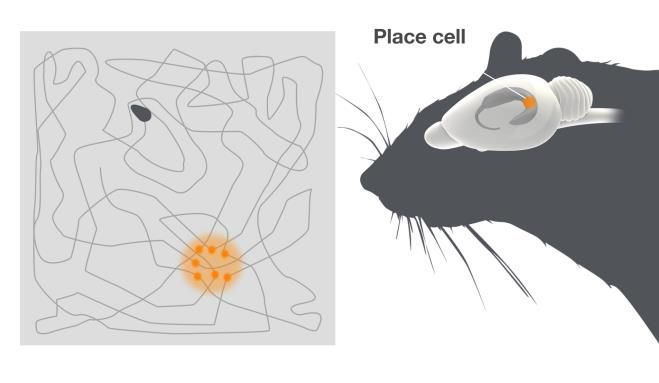
Tolman’s theory opposed the prevailing view The firing pattern of these cells was
among behaviourists that complex completely unexpected. Place cells were
behaviours are achieved by chains of active in a way that had not been seen for
sensory-motor response relationships. But it any cells in the brain before. Individual place
did not address where in the brain these cells were only active when the animal was
functions may be localized and how the in a particular place in the environment,
brain computes such complex behaviours. namely their place field. By systematically
The advent of techniques to record from changing the environment and testing
cells in the brain of animals that were freely different theoretical possibilities for the
moving in the environment, using chronically creation of the place fields O’Keefe showed
implanted micro wires (Sturmwasser, 1958), that place cell firing did not merely reflect
made it possible to approach these activity in sensory neurons, but that it
questions. represented a complex gestalt of the
environment.
Finding the place cells
John O’Keefe had a background in Different place cells could be active in
physiological psychology, working with different places and the combination of
Ronald Melzack at McGill University before activity in many place cells created an
he moved to the laboratory of the pain internal neural map representing a particular
researcher Patrick Wall at University College environment (O'Keefe, 1976; O'Keefe and
in London, where he started his work on Conway, 1978). O’Keefe concluded together
behaving animals in the late 1960s. There he with Nadel that place cells provide the brain
discovered the place cells, when recording with a spatial reference map system, or a
from neurons in the dorsal partition of sense of place (O'Keefe and Nadel, 1978). He
hippocampus, called CA1, together with showed that the hippocampus can contain
Dostrovsky, in rats moving freely in a multiple maps represented by combinations
bounded area (O'Keefe and Dostrovsky, of activity in different place cells that were
1971) (Figure 1). active at different times in different
environments. A specific serial combination
of active place cells may therefore represent
a unique environment, while other
combinations represent other environments.
Through O’Keefe’s discoveries, the cognitive
map theory had found its representation in
the brain.
A prerequisite for O’Keefe’s experiments
was the development of appropriate
recording techniques to be used in freely
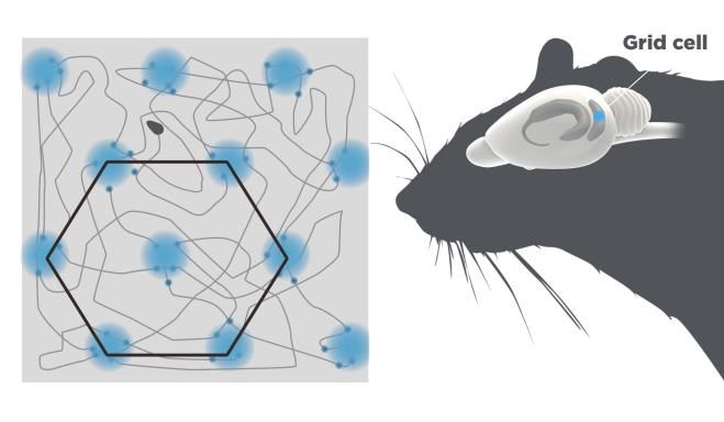
Figure 1. Place cells. To the right is a schematic of the
moving animals. Although O’Keefe was an
rat. The hippocampus, where the place cells are
located is highlighted. The grey square depicts the early user of these techniques, he was not
open field the rat is moving over. Place cells fire when the first to record from hippocampal or
the animal reaches a particular location in the other nerve cells in intact animals (see
environment. The dots indicate the rat’s location in O’Keefe and Nadel 1978). However,
the arena when the place cell is active. Different place
researchers mostly used restricted
cells in the hippocampus fire at different places in the
arena. behavioural task or strict stimulus-response
protocols. In contrast, O’Keefe recorded the
2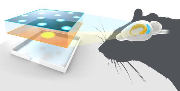
cellular activity during natural behaviour,          May-Britt Moser and Edvard Moser, who
which allowed him to observe the unique              were studying the hippocampus, both during
place fields and relate the neural activity in       their PhD work in Per Andersen’s laboratory
the place cells to represent the sense of            in Oslo and afterwards both as visiting
place.                                               scientists in Richard Morris’ laboratory in
                                                     Edinburgh and John O’Keefe’s laboratory in
In subsequent experiments, O’Keefe showed            London, asked whether the place cell firing
that the place cells might have memory               can be generated from activity outside
functions (O'Keefe and Conway, 1978;                 hippocampus. The major input to the
O'Keefe and Speakman, 1987). The                     hippocampus comes from a structure on the
simultaneous rearrangement in many place             dorsal edge of the rat’s brain, the entorhinal
cells in different environments was called           cortex. A large part of the output from the
remapping and O’Keefe showed that                    entorhinal cortex projects to the dentate
remapping is learned, and once it is                 gurus in hippocampus, which in turn connect
established, it can be stable over time (Lever       to the region in the hippocampus called CA3,
et al., 2002). The place cells may therefore         and further to CA1 in the dorsal
provide a cellular substrate for memory              hippocampus. Interestingly, this is the same
processes, where a memory of an                      the part of the brain in which John O’Keefe
environment can be stored as specific                first found the place cells. In 2002, the
combinations of place cells.                         Mosers found that disconnecting projections
                                                     from the entorhinal cortex through CA3 did
At first, the proposition that the                   not abolish the CA1 place fields (Brun et al.,
hippocampus was involved in spatial                  2002). These findings, and the knowledge
navigation was met with some scepticism.             that medial entorhinal cortex is also directly
However, it was later appreciated that the           and reciprocally connected to the CA1
discovery of place cells, the meticulous             region, prompted May-Britt Moser and
demonstration that these cells represent a           Edvard Moser to look in the medial
mental map far from primary sensory input,           entorhinal cortex for place coding cells. In a
and the proposal that hippocampus contains           first study they established, similar to what
an inner map that can store information              others had shown, that the medial
about the environment, were seminal.                 entorhinal cortex contained cells that shared
O’Keefe’s discovery sparked a large number           characteristics with the place cells in
of experimental and theoretical studies on           hippocampus (Fyhn et al., 2004). However,
how place cells are engaged in generating            in a later study using larger encounters for
spatial information and in spatial memory            the animals to move in, they discovered a
processes. The general notion from these             novel cell type, the grid cells, that had
studies is that the key function of the place        unusual properties, (Hafting et al., 2005).
cells are to create a map of the environment,
although they may also be involved in                The grid cells showed an astonishing firing
measuring        distance     under     some         pattern. They were active in multiple places
circumstances (Ravassard et al., 2013).              in the open box that together formed nodes
                                                     of an extended hexagonal grid (Figure 2),
From hippocampus to grid cells in the                similar to the hexagonal arrangements of
                                                     holes in a beehive.
entorhinal cortex
Through the 1980s and 1990s the prevailing
                                                     Grid cells in the same area of the medial
theory was that the formation of place fields
                                                     enthorinal cortex fire with the same spacing
originated within the hippocampus itself.

                                                 3
and orientation of the grid, but different al. 2008). The existence of border cells was
phasing, so that together they cover every predicted by theoretical modelling by
point in the environment. O’Keefe and colleagues (Hartley, et al. 2000).
The Mosers showed that the grid cells, the
head direction cells, and the border cells,
projected to hippocampal place cells (Zhang
et al. 2013). Using recordings from multiple
grid cells in different parts of the entorhinal
cortex, the Mosers also showed that the grid
cells are organized in functional modules
with different grid spacing ranging in
distance from a few centimetres to meters,
thereby covering small to large
Figure 2. Grid cells. The grid cells are located in the environments.
entorhinal cortex depicted in blue. A single grid cell
fires when the animal reaches particular locations in The Mosers further explored the
the arena. These locations are arranged in a relationship between grid cells and place
hexagonal pattern. cells in theoretical models (Solstad et al.,
2006), lesion experiments (Bonnevie et al.,
The Mosers found that the distance of the 2013; Hafting et al., 2008), and in remapping
grid fields varies in the medial entorhinal experiments (Fyhn et al. 2007). These and
cortex with the largest fields in the ventral other studies by Mosers and O’Keefe, as well
part of the cortex. They also showed that the as by others, have shown that there is a
grid formation did not arise out of a simple reciprocal influence between grid cells in the
transformation of sensory or motor signals, medial entorhinal cortex and place cells in
but out of complex network activity. the hippocampus and that other spatially-
tuned cells in the entorhinal cortex, in
The grid pattern had not been seen in any particular the border cells (Figure 3), may
brain cells before! The Mosers concluded contribute in the generation of the firing
that the grid cells were part of a navigation pattern of the place cells (Brandon et al.,
or path integration system. The grid system 2011; Koenig et al., 2011; Bush, Berry and
provided a solution to measuring movement Burgess, 2014, Bjerkness et al. 2014).
distances and added a metric to the spatial
maps in hippocampus.
The Mosers further showed that grid cells
were embedded in a network in the medial
entorhinal cortex of head direction cells and
border cells, and in many cases, cells with a
combined function (Solstad et al., 2008).
Head-direction cells were first described by
James Ranck (1985) in another part of the
brain, the subiculum. They act like a
compass and are active when the head of an Figure 3. A schematic showing grid cells (blue) and
animal points in a certain direction. Border place cells (yellow) in the entorhinal cortex and
cells are active in reference to walls that the hippocampus, respectively.
animal encounters when moving in a closed
environment (Solstad et al., 2008; Savelli, et
4The Mosers’ discovery of the grid cells, a The importance of the discovery of
spatial metric coordination system, and their place cells and grid cells for research
identification of the medial entorhinal cortex
in cognitive neuroscience
as a computational centre for spatial
It is an emergent theme that place-coding
representation, is a break-through that
cells in the hippocampal structures are
opens up new avenues to advance the
involved in storing and/or retrieving spatial
understanding of the neural mechanisms
memories. In the 1950s Scoville and Milner
underlying spatial cognitive functions.
(1957) published their report on the patient
Henry Molaison (HM), who had his two
The grid and place cell systems are hippocampi surgically removed for
found in many mammalian species treatment of epilepsy. The loss of
including humans hippocampi caused severe memory deficits,
Since the initial description of place and grid as evident by the clinical observation that
cells in rat and mice, these cell types have HM was unable to encode new memories,
also been found in other mammals (Killian et while he could still retrieve old memories.
al., 2012; Ulanovsky et al., 2007; Yartsev et HM had lost what has later been named
al., 2011, 2013;). Humans have large episodic memory (Tulving and Markowitch
hippocampal-entorhinal brain structures and 1998), referring to our ability to remember
these structures have long been implicated self-experienced events. There is no direct
in spatial learning and episodic memory evidence that place cells are coding episodic
(Squire, 2004). A number of studies support memory. However, place cells can encode
the idea that the human brain has a spatial- not only for the current spatial location, but
coding system that is similar to that found in also where the animal has just been and
non-human mammals. Thus, researchers where it is going next (Ferbinteanu and
have found place-like cells in the Shapiro, 2003). The past and present may
hippocampus (Ekstrom et al., 2003; Jacobs also be overlapping in time in place cells
et al., 2010) and grid-like cells in the when animals are rapidly tele-transported
entorhinal cortex (Jacobs et al., 2013) when between two physical different
directly recording from nerve cells in the environments (Jezek et al., 2011). An
human brain of patients with epilepsy encoding of places in the past and present
undergoing pre-surgical investigation. Using might allow the brain to remember
functional imaging (fMRI). Doeller et al. temporally ordered representations of
(2010) have also provided support for the events, like in the episodic memory.
existence of grid cells in the human
entorhinal cortex. After a memory has been encoded, the
memory undergoes further consolidation,
The similarity of the hippocampal-entorhinal e.g. during sleep. Ensemble recording with
structure in all mammals and the presence multi-electrodes in sleeping animals has
of hippocampal-like structures in non- made possible the study of how memories
mammalian vertebrates with navigational of spatial routes achieved during active
capacity suggest that the grid-place cells navigation are consolidated. Groups of place
system is a functional and robust system cells that are activated in a particular
that may be conserved in vertebrate sequence during the behaviour display the
evolution. same sequence of activation in episodes
during the subsequent sleep (Wilson and
McNaughton, 1994). This replay of place cell
activity during sleep may be a memory
5consolidation mechanism, where the workers have showed in a mouse model of
memory is eventually stored in cortical Alzheimer’s disease that the degradation of
structures. place fields correlated with the deterioration
of the animals’ spatial memory (Cacucci et
Together the activity of place cells may be al., 2008). There is no immediate translation
used both to define the position in the of such results to clinical research or practice.
environment at any given time, and also to However, the hippocampal formation is one
remember past experiences of the of the first structures to be affected in
environment. Maybe related to this notion is Alzheimer’s disease and knowledge about
the findings that the hippocampus of London the brain’s navigational system might help
taxi drivers, which undergoes extensive understand the cognitive decline seen in
training to learn how to navigate between patients with this diseases.
thousands of places in the city without a
map, grew during the year long training Conclusions
period and that the taxi drivers after this The discoveries of place and grid cells by
training had significantly larger hippocampal John O’Keefe, May-Britt Moser and Edvard I.
volume than control subjects (Magurie et al. Moser present a paradigm shift in our
2000, Woollett and Maguire, 2011). understanding of how ensembles of
specialized cells work together to execute
Relevance for humans and medicine higher cognitive functions. The discoveries
Brain disorders are the most common cause have profoundly promoted new research
of disability and despite the major impact on with grid and place cell systems now found
people’s life and on the society, there is no in many mammals, including humans.
effective way to prevent or cure most of Studies of the navigation system have
these disorders. The episodic memory is opened new avenues for studying how
affected in several brain disorders, including cognitive processes are computed in the
dementia and Alzheimer’s disease. A better brain.
understanding of neural mechanisms
underlying spatial memory is therefore
important, and the discoveries of place and
Ole Kiehn and Hans Forssberg
grid cells have been a major leap forward to
Karolinska Institutet
advance this endeavour. O’Keefe and co-
Ole Kiehn, MD, PhD
Professor of Neuroscience, Karolinska Institutet
Member of the Nobel Committee
Member of the Nobel Assembly
Hans Forssberg, MD, PhD
Professor of Neuroscience , Karolinska Institutet
Adjunct Member of the Nobel Committee
Member of the Nobel Assembly
Illustrations: Mattias Karlen
6Cited literature
Bjerknes, T.L., Moser, E.I. and Moser, M.B. (2014). Representation of geometric borders in the
developing rat. Neuron, 82(1), 71-78.
Bonnevie, T., Dunn, B., Fyhn, M., Hafting, T., Derdikman, D., Kubie, J.L., Roudi, Y., Moser, E.I.,
and Moser, M.B. (2013). Grid cells require excitatory drive from the hippocampus. Nature
Neuroscience 16, 309-317.
Brandon, M.P., Bogaard, A.R., Libby, C.P., Connerney, M.A., Gupta, K., and Hasselmo, M.E.
(2011). Reduction of theta rhythm dissociates grid cell spatial periodicity from directional
tuning. Science 332, 595-599.
Brun, V.H., Otnass, M.K., Molden, S., Steffenach, H.A., Witter, M.P., Moser, M.B., and Moser,
E.I. (2002). Place cells and place recognition maintained by direct entorhinal-hippocampal
circuitry. Science 296, 2243-2246.
Bush. D., Barry, C., Burgess, N. (2014). What do grid cells contribute to place cell firing? Trends
in Neuroscience, 37(3), 136-145
Cacucci, F., Yi, M., Wills, T.J., Chapman, P. and O´Keefe, J. (2008) Place cell firing correlates
with memory deficits and amyloid plaque burden in Tg2576 Alzheimer mouse model. PNAS,
105, 7863-7868.
De Hoz, L., and Wood, E.R. (2006). Dissociating the past from the present in the activity of
place cells. Hippocampus, 16, 704-715.
Doeller, C.F., Barry, C., and Burgess, N. (2010). Evidence for grid cells in a human memory
network. Nature 463, 657-661.
Ekstrom, A.D., Kahana, M.J., Caplan, J.B., Fields, T.A., Isham, E.A., Newman, E.L., and Fried, I.
(2003). Cellular networks underlying human spatial navigation. Nature 425, 184-188.
Ferbinteanu, J., and Shapiro, M.L. (2003). Prospective and retrospective memory coding in
the hippocampus. Neuron, 40, 1227-1239.
Fyhn, M., Hafting, T., Treves, A., Moser, M.B., and Moser, E.I. (2007). Hippocampal remapping
and grid realignment in entorhinal cortex. Nature 446, 190-194.
Fyhn, M., Molden, S., Witter, M.P., Moser, E.I., and Moser, M.B. (2004). Spatial representation
in the entorhinal cortex. Science 305, 1258-1264.
Hafting, T., Fyhn, M., Bonnevie, T., Moser, M.B., and Moser, E.I. (2008). Hippocampus-
independent phase precession in entorhinal grid cells. Nature 453, 1248-1252.
Hafting, T., Fyhn, M., Molden, S., Moser, M.B., and Moser, E.I. (2005). Microstructure of a
spatial map in the entorhinal cortex. Nature 436, 801-806.
Hartley, T., Burgess, N., Lever, C., Cacucci, F. and O'Keefe, J. (2000). Modeling place fields in
terms of the cortical inputs to the hippocampus. Hippocampus, 10(4), 369-379.
Jacobs, J., Kahana, M.J., Ekstrom, A.D., Mollison, M.V., and Fried, I. (2010). A sense of direction
in human entorhinal cortex. PNAS 107, 6487-6492.
Jacobs, J., Weidemann, C.T., Miller, J.F., Solway, A., Burke, J.F., Wei, X.X., Suthana, N., Sperling,
M.R., Sharan, A.D., Fried, I., and Kahana, M.J. (2013). Direct recordings of grid-like neuronal
activity in human spatial navigation. Nature Neuroscience, 6, 1188-1190.
Jezek, K., Henriksen, E.J., Treves, A., Moser, E.I., and Moser, M.B. (2011). Theta-paced
flickering between place-cell maps in the hippocampus. Nature, 478, 246-249.
Killian, N.J., Jutras, M.J., and Buffalo, E.A. (2012). A map of visual space in the primate
entorhinal cortex. Nature 491, 761-764.
7Langston, R.F., Ainge, J.A., Couey, J.J., Canto, C.B., Bjerknes, T.L., Witter, M.P., Moser, E.I., and
Moser, M.B. (2010). Development of the spatial representation system in the rat. Science
328, 1576-1580.
Lever, C., Wills, T., Cacucci, F., Burgess, N., and O'Keefe, J. (2002). Long-term plasticity in
hippocampal place-cell representation of environmental geometry. Nature 416, 90-94.
Maguire, E.A., Gadian, D.G., Johnsrude, I.S., Good, C.D., Ashburner, J., Frackowiak, R.S. and
Frith C.D. (2000). Navigation-related structural change in the hippocampi of taxi drivers.
PNAS, 97(8), 4398-4403.
O'Keefe, J. (1976). Place units in the hippocampus of the freely moving rat. Experimental
neurology 51, 78-109.
O'Keefe, J., and Conway, D.H. (1978). Hippocampal place units in the freely moving rat: why
they fire where they fire. Experimental brain research 31, 573-590.
O'Keefe, J., and Dostrovsky, J. (1971). The hippocampus as a spatial map. Preliminary evidence
from unit activity in the freely-moving rat. Brain research 34, 171-175.
O'Keefe, J., and Nadel, L. (1978). The Hippocampus as a Cognitive Map (Oxford Univeristy
Press ).
O'Keefe, J., and Speakman, A. (1987). Single unit activity in the rat hippocampus during a
spatial memory task. Experimental brainresearch 68, 1-27.
Ranck, J.B. (1985). Head direction cells in the deep cell layer of dorsal presubiculum in freely
moving rats. In Electrical Activity of the Archicortex, C.V. G. Buzsáki, ed. (Budapest:
Akademiai Kiado), pp. 217-220.
Ravassard, P., Kees. A., Willers, B., Ho, D., Aharoni, D., Cushman, J., Aghajan, Z.M. and Mehta
M.R. (2013) Multisensory control of hippocampal spatiotemporal selectivity. Science,
340(6138), 1342-1346.
Sargolini, F., Fyhn, M., Hafting, T., McNaughton, B.L., Witter, M.P., Moser, M.B., and Moser, E.I.
(2006). Conjunctive representation of position, direction, and velocity in entorhinal cortex.
Science 312, 758-762.
Savelli, F., Yoganarasimha, D and Knierim, J.J (2008). Influence of boundary removal on the
spatial representations of the medial enthorinal cortex. Hippocampus, 18, 1270-1282.
Scoville, W.B., and Miller, B. (1957). Loss of recent memory after bilateral hippocampal lesions.
Journal of Neurology Neurosurgery and Psychiatry, 20, 11-21.
Solstad, T., Boccara, C.N., Kropff, E., Moser, M.B., and Moser, E.I. (2008). Representation of
geometric borders in the entorhinal cortex. Science 322, 1865-1868.
Solstad, T., Moser, E.I., and Einevoll, G.T. (2006). From grid cells to place cells: a mathematical
model. Hippocampus 16, 1026-1031.
Squire, L.R. (2004). Memory systems of the brain: a brief history and current perspective.
Neurobiology of learning and memory 82, 171-177.
Stensola, H., Stensola, T., Solstad, T., Froland, K., Moser, M.B., and Moser, E.I. (2012). The
entorhinal grid map is discretized. Nature 492, 72-78.
Strumwasser, F. (1958). Long-term recording from single neurons in brain of unrestrained
mammals. Science, 127, 469-670.
Tolman, E.C. (1948). Cognitive maps in rats and men. Psychological Review, 55, 189-208.
Tulving, E. and Markowitsch, H.J. (1998). Episodic and declarative memory: role of the
hippocampus. Hippocampus, 8, 198-204.
8Ulanovsky, N., and Moss, C.F. (2007). Hippocampal cellular and network activity in freely
moving echolocating bats. Nat Neurosci 10, 224-233.
Wilson, M.A., and McNaughton, B.L. (1994). Reactivation of hippocampal ensemble memories
during sleep. Science 265, 676-679.
Woollett K. and Maguire E.A. (2011). Acquiring "the Knowledge" of London's layout drives
structural brain changes. Current. Biology, 21(24), 2109-2114
Yartsev, M.M., and Ulanovsky, N. (2013). Representation of three-dimensional space in the
hippocampus of flying bats. Science 340, 367-372.
Yartsev, M.M., Witter, M.P., and Ulanovsky, N. (2011). Grid cells without theta oscillations in
the entorhinal cortex of bats. Nature 479, 103-107.
Zhang, S.J., Ye, J., Miao, C., Tsao, A., Cerniauskas, I., Ledergerber, D., Moser, M.B., and Moser,
E.I. (2013). Optogenetic dissection of entorhinal-hippocampal functional connectivity.
Science 340, (6128)232627.
9You can also read