Vol. 3 No. 1 April 2020 - eISSN 2635-5280 - Journal of Neurointensive Care
←
→
Page content transcription
If your browser does not render page correctly, please read the page content below
eISSN 2635-5280

Vol. 3 · No. 1 · April 2020
eISSN 2635-5280
                                                                                                                  http://www.e-jnic.org

Vol. 3 · No. 1 · April 2020

Aims and Scope
Journal of Neurointensive Care (J Neurointensive Care, JNIC) is the official journal of the Korean Neurointensive
Care Society and is published biannually (the last day of April and October). It is a peer reviewed, open access
journal aimed at publishing all aspects of neurointensive care medicine, such as stroke, brain and spine trauma,
perioperative neurosurgical intensive care, neuro-pediatric severe anormaly, CNS infection, seizure, myelitis and
etc. It is intended for all neurointensive care providers as neurosurgeons, neurologists, anesthesiologists, emergency
physicians, and critical care nurses treating patients with urgent neurologic disorders.

Open Access
This is an open-access article distributed under the terms of the Creative Commons Attribution Non- Commercial
License (http://creativecommons.org/licenses/by-nc/4.0) which permits unrestricted noncommercial use,
distribution, and reproduction in any medium, provided the original work is properly cited.

Subscription information
The Korean Neurointensive Care Society will send J Neurointensive Care for free to some important individuals
and institutions. Full text PDF files are also available at the official website (http://www.e-jnic.org). To order a
subscription to J Neurointensive Care, please contact out editorial office.

Publisher: Jeong-Taik Kwon

Editors-in-Chief: Dong-Hyuk Park

Editorial Office
Department of Neurosurgery, Korea University College of Medicine
73, Inchon-ro, Seongbuk-gu, Seoul 02841, Korea
Tel: +82-2-920-6833 Fax: +82-2-929-0629 E-mail: jnic.editor@gmail.com

Printing Office M2community Co.
8th FL, DreamTower, 66 Seongsui-ro, Seongdong-gu, Seoul 04784, Korea
Tel: +82-2-2190-7300 Fax: +82-2-2190-7333 E-mail: journal@m2community.co.kr

Copyright © 2020 by Korean Neurointensive Care Society
∞This paper meets the requirements of KS X ISO 9706, ISO 9706-1994 and ANSI/NISO Z39.48-1992 (Permanence of Paper)
eISSN 2635-5280
http://www.e-jnic.org
Editorial Board Editors-in-Chief
Dong-Hyuk Park Korea University, Korea
Associate Editor
Jun Seok W. Hur Korea University, Korea
Editorial Board
Jin Hwan Cheong Hanyang University, Korea
Won-Sang Cho Seoul National University, Korea
Kyu-Sun Choi Hanyang University, Korea
Joon ho Chung Yonsei University, Korea
Eun Jin Ha Seoul National University, Korea
Kyung Sool Jang The Catholic University of Korea, Korea
Ju Ho Jeong Dongguk University, Korea
Kwang Wook Jo The Catholic University of Korea, Korea
Sung Pil Joo Chonnam National University, Korea
Chang-Hyun Kim Keimyung University, Korea
Tae Gon Kim CHA University, Korea
Young Jin Kim Dankook University, Korea
Young Woo Kim The Catholic University of Korea, Korea
Young Zoon Kim Sungkyunkwan University, Korea
Doo-Sik Kong Sungkyunkwan University, Korea
Hyon-Jo Kwon Chungnam National University, Korea
Soon Chan Kwon Ulsan University, Korea
Jong-Young Lee Hallym University, Korea
Sang Weon Lee Pusan National University, Korea
Sung Ho Lee Kyung-Hee University, Korea
Taek Kyun Nam Chung-Ang University, Korea
Cheol Wan Park Gachon University, Korea
Jun Bum Park Ulsan University, Korea
Keun Young Park Yonsei University, Korea
Young Seok Park Chonbuk National University, Korea
Jeong-Am Ryu Sungkyunkwan University, Korea
Sang Woo Song Konkuk University, Korea
Chan Jong Yoo Gachon University, KoreaVol. 3 · No. 1 · April 2020
CONTENTS
Review Article
1 Management of central nervous system metastases
Sang-Hoon Lee, Kyung-Jae Park, Dong-Hyuk Park, Jang-Bo Lee, Shin-Hyuk Kang, Tai-Hyoung Cho, Jung-Yul Park, Yong-Gu Jung,
Junseok W Hur
Original Reports
6 Prognostic Value of Early Hyperglycemia in Neurocritically Ill Patients
Junghoon Han, Yun Im Lee, Jeong-Am Ryu
12 Role of Cancer Emboli as a Metastatic Core on the Growth of Brain Metastasis in Patients with Non-Small Cell
Lung Cancer
Jinuk Kim, Gyuseo Jung, Hoon Gi Kim, Jeong Yeon Kim, Geun Young Yang, Young Zoon Kim
20 Prognostic Factors of Clinical Outcome after Aneurysmal Clipping in the Aged Patients with Unruptured
Intracranial Aneurysms
Min-Woo Son, Jin-Woo Park, Kyung-Jae Park, Shin-Hyuk Kang, Yong-Gu Jung, Jung-Yul Park, Dong-Hyuk Park
Case Report
26 Superior Mesenteric Infarction During Management of Patient with Cerebral Infarction
Min-Woo Son, Sang-Hoon Lee, Dong-Hyuk ParkeISSN 2635-5280
Review Article
J Neurointensive Care 2020;3(1):1-5
https://doi.org/10.32587/jnic.2020.00262
Management of central nervous system metastases
Sang-Hoon Lee, Kyung-Jae Park, Dong-Hyuk Park, Jang-Bo Lee, Shin-Hyuk Kang, Tai-Hyoung Cho,
Jung-Yul Park, Yong-Gu Jung, Junseok W Hur
Department of Neurosurgery, College of Medicine, Korea University, Seoul, Korea
Received: April 10, 2020
Central nervous system (CNS) metastases are divided into brain metastasis and intramedullary
Accepted: April 17, 2020
spinal cord metastasis (ISCM). Although the blood-brain barrier (BBB) and blood-spinal barrier
Corresponding Author: (BSB) protect the brain and spinal cord, metastases occur when these barriers break under ab-
Junseok W Hur M.D., Ph.D. normal conditions. Brain metastasis accounts for the largest number of brain tumors, however,
Korea University Anam Hospital, ISCM rarely occurs. For brain metastasis, whole brain radiotherapy (WBRT), stereotactic radio-
73, Goryeodae-ro, Seongbuk-gu, surgery (SRS), surgery, and chemotherapy can be considered, and for ISCM, radiotherapy (RT),
Seoul, 02841, Korea surgery, Cyberknife SRS, and chemotherapy can be considered. As treatment options vary de-
Tel: +82-2-920-5729 pending on the patient’s life expectancy, performance status (PF), extent and number of metas-
Fax: +82-2-929-0629 tases, and the type of primary cancer, careful patient evaluation should be performed prior to
E-mail: hurjune@gmail.com treatment of CNS metastases.
Keywords: Brain; Central nervous system; metastases; Intramedullary spinal cord
INTRODUCTION multiple brain metastases is confirmed. However, nowadays,
surgery has an important role in management for carefully se-
Among central nervous system (CNS) metastases, brain me- lected cases. Surgery provides definite and accurate diagnosis,
tastasis accounts majority cases compare to intramedullary spi- reduces intracranial mass effect, improves neurological symp-
nal cord metastases (ISCM). Brain metastases remain the com- toms, and may also improve overall survival (OS), all with low
monest type of brain tumor, being four times more common morbidity and mortality rates29). It is generally accepted that to
than primary brain tumors23). The management of patients with benefit from surgery, a patient with brain metastases must have
multiple brain metastases remains a difficult challenge for neu- reasonable medical fitness, with a systemic disease process
rosurgeons. Treatment strategies for brain metastases depend on amenable to benefit from local tumor control30). Unlike brain
several factors. Some patients may be candidates for whole brain metastasis, the role of surgical treatment in ISCM is controver-
radiotherapy (WBRT), while others may require surgical resec- sial. Chemotherapy is usually considered by the type of primary
tion followed by WBRT or local radiation therapy. Stereotactic cancer and applied for systemic control, however, the efficacy
radiosurgery (SRS) has added another dimension to the man- for the ISCM is unclear. The RT is generally considered as a pal-
agement of these lesions. The patient with metastatic brain tu- liative treatment for ISCM treatment16,20).
mor has a poor prognosis, and historically, treatment has gener-
ally consisted of administering WBRT after the diagnosis of
Copyright © 2020 The Korean Neurointensive Care Society
This is an Open Access article distributed under the terms of the Creative Commons Attribution Non-Commercial License (http://creativecommons.org/licenses/by-nc/4.0/)
which permits unrestricted non-commercial use, distribution, and reproduction in any medium, provided the original work is properly cited.
www.e-jnic.org 1Management of CNS metastasis Sang-Hoon Lee et al.
EPIDEMIOLOGY chemotherapy. Many patients are treated with a combination of
these, and treatment decisions must take concern about factors
The majority of brain metastases originate from one of three such as patient age, functional status, primary tumor type, extent
primary malignancies; lung cancer (40%–50%), breast cancer of extracranial disease, prior therapies, and number of intracrani-
(15%–25%), and melanoma (5%–20%). Among these, melano- al lesions. Most ISCM patients are diagnosed with a neurological
ma has the highest propensity to metastasize to the brain, with a deficit. Ninety-three % of the patients showed motor dysfunc-
50% rate of brain involvement reported in patients dying of mel- tion, 78% of the patients showed sensory abnormalities, and 62%
anoma2). The frequency of metastatic brain tumors appears to of the patients showed urinogenital dysfunction16). For patients
be rising as a result of superior imaging modalities and earlier who previously diagnosed with primary cancer, ISCM can be
detection as well as longer survival after a primary cancer diag- considered, but if an intramedullary spinal cord tumor is diag-
nosis because of more effective treatment of systemic disease11). nosed without knowing the primary cancer, it is highly likely to
In the case of ISCM, lung cancer metastases (54%) is the most be mistaken for a primary tumor. While the surgery is the treat-
common cause, followed by breast cancer metastases (11%)19). ment of choice for primary intramedullary tumor, in contrast,
ISCM is a rare systemic cancer that autopsy studies have con- surgery is performed in highly selective cases for ISCM. Careful
firmed that ISCM is present in 0.9% to 2.1% of cancer patients. diagnosis is needed prior to the treatment.
However, almost 95% of these lesions are clinically silent and re-
main undiagnosed before death, so they are actually less fre- TREATMENT MODALITIES – BRAIN
quently encountered in practice7,9,10,18,31,36). METASTASIS
PATHOPHYSIOLOGY Systemic chemotherapy
Chemotherapy has traditionally played a limited role in the
Under physiologic condition, CNS metastases hardly occur treatment of brain metastases, and has been reserved for patients
because of the firm blood-brain barrier (BBB) and blood-spinal who have failed other treatment modalities or for diseases known
barrier (BSB). However, in pathologic condition, inflammatory to be “chemo-sensitive,” such as lymphoma, small-cell lung can-
mediators cause increased permeability of the BBB/BSB. In this cer, germ-cell tumor and breast cancer11). Incredulous stance re-
environment, cancer cells can attach to microvascular endothe- garding the usefulness of chemotherapy for brain metastases
lial cells and invade the BBB/BSB, cause CNS metastases1,14). A arises from the reason that most agents cannot cross the BBB, be-
typical CNS metastases route is hematogeneous spreading. In cause of their large molecular weight or hydrophilic property.
brain metastasis, arterial spreading is known as a major cause, The degree to which a given agent is believed to penetrate the
and venous spreading through Batson plexus is also considered BBB is usually based on pharmacokinetic animal and/or human
as an important route for ISCM10,13,17,18,31,34). Leptomeningeal studies comparing plasma with CSF drug concentrations after
dissemination by the cerebrospinal fluid (CSF) is also an im- intravenous or oral administration. This method may underesti-
portant metastasis mechanism. In particular, it explains why the mate the concentration of drug delivered to the tumor, however,
brain and ISCM often appear simultaneously9,31). Direct inva- because brain metastases are known to have local BBB break-
sion is also known as a case of metastasis, however, this is mainly down (demonstrated on magnetic resonance imaging (MRI) by
limited in the case of ISCM9,37). contrast enhancement and peritumoral edema). This is corrobo-
rated by studies showing roughly equivalent intracranial and ex-
CLINICAL ASSESSMENTS tracranial response rates to chemotherapeutic agents assumed to
have little BBB penetration, particularly when first-line agents for
The management of brain metastases can be divided into the systemic cancer are chosen4,6,33). The success of an agent may
symptomatic and therapeutic strategies. Symptomatic therapy therefore rest more heavily upon its inherent activity against the
often includes corticosteroids to reduce peritumoral edema and systemic tumor than its putative ability to cross the BBB.
anticonvulsants to prevent recurrent seizures. In addition, there
is accumulating data to suggest that medications such as methyl- Radiotherapy
phenidate and donepezil can improve cognition, mood, and The mainstay of treatment for brain metastases over the past
quality of life in patients with brain tumors26,35). Therapeutic ap- five decades has been corticosteroids and WBRT. Nonrandom-
proaches to brain metastases include surgery, WBRT, SRS, and ized studies suggest that WBRT increases the median survival
2 www.e-jnic.orgManagement of CNS metastasis Sang-Hoon Lee et al.
time by 3–4 months over approximately 1 month without treat- TREATMENT MODALITIES – ISCM
ment and 2 months with corticosteroids alone. Although reports
of the response rate after WBRT alone vary, complete responses Radiotherapy
(CRs) or partial responses (PRs) have been documented in ap- The RT is considered as standard therapy for palliative treat-
proximately 60% of patients in randomized controlled studies ment for ISCM8,10,13,18,38). However, the efficacy is limited to ra-
conducted by the radiation-therapy oncology group21). Stasis or diosensitive tumor as small cell carcinoma, breast carcinoma,
Improvement of neurologic symptoms occurs nearly the same or lymphoma8,13,15,28,38). Furthermore, radiation myelitis due to
proportion, even though symptom response defined separately radiotoxicity should be considered.
in studies5).
Surgery
SRS Surgery should be performed in highly selective patients. They
Although there is controversy exists, particularly those with a should have good performance status, single CNS metastasis,
limited number of brain metastases, can be treated effectively and long enough life expectancy. In surgical technical aspect, as
with SRS alone11). The assumed rationale for exclusion of WBRT the microscopic surgical skill has been advanced and neurophysi-
is to spare patients the risk for late neurotoxicity from WBRT. Pa- ologic intraoperative monitoring (IOM) has been developed,
tients who were not treated upfront WBRT are typically moni- surgical outcome gradually improved. Some groups claim that
tored closely with serial MRI scans and treated with WBRT or the ISCM shows fair borderline that normal neural structure is
additional SRS at recurrence11). well preserved along surgery, however, some groups assert the
opposite12,25). There is little evidence that surgical resection could
Surgery improve OS, however, neurologic improvement has been
Management of patients with brain metastases has been evolv- achieved in some reports12,40).
ing over time, with a general tendency towards a more aggressive
treatment approach22). Benefits of surgical resection include the Chemotherapy
provision of an accurate and definite diagnosis, immediate relief As the BSB block the chemical, chemotherapy has little effect
of neurological symptoms caused by extensive perilesional ede- for ISCM treatment20). However, if the primary cancer is suit-
ma or mass effect, and local control of disease. Advances in surgi- able for specific chemotherapy, it could be applied as adjuvant
cal technique have led to lower rates of morbidity and mortality3). therapy for RT or surgery19).
Muacevic et al., demonstrate in their retrospective review of
management of solitary metastasis of less than 3.5 cm of diame- Steroid
ter concluded that result of surgery with WBRT is comparable to Steroid can reduce spinal cord edema and stabilize BSB, which
SRS in local tumor control rate27). helps relieving pain and delay neurologic deterioration. Even
Table 1. Management recommendation for brain metastases (adopted from Lin et al.)24)
Consider systemic therapy Consider WBRT Consider SRS Consider surgical resection No treatment is reasonable
BM from highly chemotherapy- CNS and systemic progression OM (1-3) or multiple BMs, Uncertain diagnosis of Systemic progression of disease,
sensitive primary tumor of disease, with few systemic especially if PT is known to CNS lesion(s) with few treatment options
treatment options and poor be radiotherapy resistant and poor PS
PS
BM found on screening MRI Multiple (> 3-10) BMs, Postsurgical resection of a single 1-2 BMs, especially when
with planned systemic especially if PT known to be BM, especially if ≥ 3 cm and in associated with extensive
treatment radiotherapy sensitive the posterior fossa cerebral edema
BM from primary tumor Postsurgical resection of a Local relapse after surgical Dominant BM in a critical
with identified molecular dominant BM with multiple resection of a single BM location
alteration amenable to (> 3-10) remaining BMs
targeted therapy
Other therapeutic options have Salvage therapy for recurrent Salvage therapy for recurrent
been exhausted and there is a BM after SRS or WBRT OM (1-3) after WBRT
reasonable drug available failure
BM: brain metastases; CNS: central nervous system; MRI: Magnetic-resonance imaging; OM: oligometastases; PS: performance status; SRS: stereotactic
radiosurgery; WBRT: whole brain radiotherapy.
www.e-jnic.org 3Management of CNS metastasis Sang-Hoon Lee et al.
though steroid cannot prolong survival, it is commonly used 496.
with other treatment modalities19,20). 6. Boogerd W, Dalesio O, Bais EM, van der Sande JJ. Response of
brain metastases from breast cancer to systemic chemotherapy.
Cyberknife Stereotactic Radiosurgery (SRS) Cancer 1992;69:972–980.
Some groups reported ISCM were treated safely with Cy- 7. Chason J, Walker FB, Landers JW. Metastatic carcinoma in the
berknife SRS without severe complication32,39). However, the to- central nervous system and dorsal root ganglia. A prospective
tal population is too small for the conclusion. Additional studies autopsy study. Cancer 1963;16:781–787.
are essential to build stronger evidence. 8. Choucair A. Myelopathies in the cancer patient: incidence,
presentation, diagnosis and management. Oncology
CONCLUSION 1991;5:25–31; discussion 35-27.
9. Costigan DA, Winkelman MD. Intramedullary spinal cord me-
Recommendation of clinical decision making in treatment of tastasis: a clinicopathological study of 13 cases. J Neurosurg
brain metastasis is below (Table 1)24). Many factors such as che- 1985;62:227–233.
mosensitivity of primary tumor, number and size of brain metas- 10. Edelson RN, Deck MD, Posner JB. Intramedullary spinal cord
tases (BM), clinical course such as local relapse or recurrence of metastases: clinical and radiographic findings in nine cases.
BM, and patient factors such as KPS should be considered. For Neurology 1972;22:1222–1222.
ISCM, golden standard is still controversial. RT is generally per- 11. Eichler AF, Loeffler JS. Multidisciplinary management of brain
formed and surgery is applied in selective patients. Cyberknife metastases. Oncologist 2007;12:884–898.
SRS could be considered as well. Due to the variety of treatment 12. Goyal A, Yolcu Y, Kerezoudis P, Alvi MA, Krauss WE, Bydon
options, meticulous clinical assessment of patients and disease is M. Intramedullary spinal cord metastases: an institutional re-
mandatory. view of survival and outcomes. J Neurooncol 2019;142:347–
354.
NOTES 13. Grem JL, Burgess J, Trump DL. Clinical features and natural
history of intramedullary spinal cord metastasis. Cancer 1985;
Conflict of interest 56:2305–2314.
No potential conflict of interest relevant to this article was re- 14. Hawkins BT, Davis TP. The blood-brain barrier/neurovascular
ported. unit in health and disease. Pharmacol Rev 2005;57:173–185.
15. Holoye P, Libnoch J, Cox J, Kun L, Byhardt R, Almagro U, et al.
REFERENCES Spinal cord metastasis in small cell carcinoma of the lung. Int J
Radiat Oncol Biol Phys 1984;10:349–356.
1. Abbott NJ, Rönnbäck L, Hansson E. Astrocyte-endothelial in- 16. Hrabalek L. Intramedullary spinal cord metastases: review of
teractions at the blood-brain barrier. Nat Rev Neurosci 2006; the literature. Biomed Pap Med Fac Univ Palacky Olomouc
7:41. Czech Repub 2010;154:117–122.
2. Amer MH, Al-Sarraf M, Baker LH, Vaitkevicius VK. Malignant 17. Hur JW, Lee S, Lee JB, Cho TH, Park JY. What are MRI find-
melanoma and central nervous system metastases: incidence, ings of Spine Benign Metastasizing Leiomyoma? Case report
diagnosis, treatment and survival. Cancer 1978;42:660–668. with literature review. Eur Spine J 2015;24 Suppl 4:S600–605.
3. Barker FG 2nd. Craniotomy for the resection of metastatic 18. Jellinger K, Kothbauer P, Sunder-Plassmann E, Weiss R. Intra-
brain tumors in the U.S., 1988-2000: decreasing mortality and medullary spinal cord metastases. J Neurol 1979;220:31–41.
the effect of provider caseload. Cancer 2004;100:999–1007. 19. Kalaycı M, Cağavi F, Gül S, Yenidünya S, Açıkgöz B. Intramed-
4. Bernardo G, Cuzzoni Q, Strada MR, Bernardo A, Brunetti G, ullary spinal cord metastases: diagnosis and treatment-an illus-
Jedrychowska I, et al. First-line chemotherapy with vinorel- trated review. Acta neurochirurgica 2004;146:1347–1354.
bine, gemcitabine, and carboplatin in the treatment of brain 20. Kalita O. Current insights into surgery for intramedullary spinal
metastases from non-small-cell lung cancer: a phase II study. cord metastases: a literature review. Int J Surg Oncol
Cancer Invest 2002;20:293–302. 2011;2011.
5. Bezjak A, Adam J, Barton R, Panzarella T, Laperriere N, Wong 21. Khuntia D, Brown P, Li J, Mehta MP. Whole-brain radiothera-
CS, et al. Symptom response after palliative radiotherapy for py in the management of brain metastasis. J Clin Oncol 2006;
patients with brain metastases. Eur J Cancer 2002;38:487– 24:1295–1304.
4 www.e-jnic.orgManagement of CNS metastasis Sang-Hoon Lee et al.
22. Lagerwaard FJ, Levendag PC. Prognostic factors in patients 33. Robinet G, Thomas P, Breton JL, Lena H, Gouva S, Dabouis G,
with brain metastases. Forum (Genova) 2001;11:27–46. et al. Results of a phase III study of early versus delayed whole
23. Laghari AA, Ahmed SI, Shamim MS. Role of surgery in brain brain radiotherapy with concurrent cisplatin and vinorelbine
metastases. J Pak Med Assoc 2017;67:1299–1300. combination in inoperable brain metastasis of non- small-cell
24. Lin X, DeAngelis LM. Treatment of Brain Metastases. J Clin lung cancer: Groupe Francais de Pneumo-Cancerologie
Oncol 2015;33:3475–3484. (GFPC) Protocol 95-1. Ann Oncol 2001;12:59–67.
25. Lv J, Liu B, Quan X, Li C, Dong L, Liu M. Intramedullary spi- 34. Schiff D, O'Neill BP. Intramedullary spinal cord metastases:
nal cord metastasis in malignancies: an institutional analysis clinical features and treatment outcome. Neurology 1996;
and review. OncoTargets and therapy 2019;12:4741. 47:906–912.
26. Meyers CA, Weitzner MA, Valentine AD, Levin VA. Methyl- 35. Shaw EG, Rosdhal R, D'Agostino RB Jr, Lovato J, Naughton
phenidate therapy improves cognition, mood, and function of MJ, Robbins ME, et al. Phase II study of donepezil in irradiated
brain tumor patients. J Clin Oncol 1998;16:2522–2527. brain tumor patients: effect on cognitive function, mood, and
27. Muacevic A, Kreth FW, Horstmann GA, Schmid-Elsaesser R, quality of lif. J Clin Oncol 2006;24:1415–1420.
Wowra B, Steiger HJ, et al. Surgery and radiotherapy compared 36. Smaltino F, Bernini F, Santoro S. Computerized tomography in
with gamma knife radiosurgery in the treatment of solitary ce- the diagnosis of intramedullary metastases. Acta neurochirur-
rebral metastases of small diameter. J Neurosurg 1999;91:35– gica 1980;52:299–303.
43. 37. Sutter B, Arthur A, Laurent J, Chadduck J, Friehs G, Clarici G,
28. Murphy K, Feld R, Evans W, Shepherd F, Perrin R, Sima A, et et al. Treatment options and time course for intramedullary
al. Intramedullary spinal cord metastases from small cell carci- spinal cord metastasis Report of three cases and review of the
noma of the lung. J Clin Oncol 1983;1:99–106. literature. Neurosurg Focus 1998;4:E5.
29. Mut M. Surgical treatment of brain metastasis: a review. Clin 38. Tognetti F, Lanzino G, Calbucci F. Metastases of the spinal
Neurol Neurosurg 2012;114:1–8. cord from remote neoplasms: study of five cases. Surg Neurol
30. Narita Y, Shibui S. Strategy of surgery and radiation therapy for 1988;30:220–227.
brain metastases. Int J Clin Oncol 2009;14:275–280. 39. Veeravagu A, Lieberson RE, Mener A, Chen Y-R, Soltys SG,
31. Okamoto H, Shinkai T, Matsuno Y, Saijo N. Intradural paren- Gibbs IC, et al. CyberKnife stereotactic radiosurgery for the
chymal involvement in the spinal subarachnoid space associat- treatment of intramedullary spinal cord metastases. J Clin Neu-
ed with primary lung cancer. Cancer 1993;72:2583–2588. rosci 2012;19:1273–1277.
32. Parikh S, Heron DE. Fractionated radiosurgical management 40. Wilson DA, Fusco DJ, Uschold TD, Spetzler RF, Chang SW.
of intramedullary spinal cord metastasis: a case report and re- Survival and functional outcome after surgical resection of in-
view of the literature. Clin Neurol Neurosurg 2009;111:858– tramedullary spinal cord metastases. World Neurosurg 2012;
861. 77:370–374.
www.e-jnic.org 5eISSN 2635-5280
Original Article
J Neurointensive Care 2020;3(1):6-11
https://doi.org/10.32587/jnic.2020.00255
Prognostic Value of Early Hyperglycemia in Neurocritically
Ill Patients
Junghoon Han1, Yun Im Lee2, Jeong-Am Ryu1,2
1
Department of Neurosurgery, Samsung Medical Center, Sungkyunkwan University School of Medicine, Seoul, Korea
2
Department of Critical Care Medicine, Samsung Medical Center, Sungkyunkwan University School of Medicine, Seoul, Korea
Received: March 3, 2020
Accepted: March 24, 2020 Objective
To evaluate the relationship of early hyperglycemia and neurological prognosis in neurocritically
Corresponding Author: ill patients.
Jeong-Am Ryu, M.D., Ph.D. Methods
Department of Critical Care This was a retrospective study of adult patients admitted to the neurosurgical intensive care unit
Medicine and Department of (ICU) from January 2010 to July 2019. Primary outcome was neurological status at 6-month fol-
Neurosurgery, Samsung Medical low-up assessed with the Glasgow Outcome Scale (GOS, 1 to 5).
Center, Sungkyunkwan University
Results
School of Medicine, 81 Irwon-ro,
A total of 202 patients were analyzed in this study. Of them, 70 (34.7%) patients had early hyper-
Gangnam-gu, Seoul 06351, Korea
glycemia (≥200 mg/dL within 48 hours after ICU admission). Brain tumor (39.6%) and sub-
Tel: +82-2-3410-6399
Fax: +82-2-2148-7088 arachnoid hemorrhage (17.8%) were the most common reasons for ICU admission. Nine-
E-mail: lamyud.ryu@samsung.com ty-three (46.0%) patients had favorable neurological outcomes (GOS of 4 or 5). Poor neurologi-
cal outcome was more common in the early hyperglycemia group than in the non-hypoglycemia
group (71.4% vs. 44.7%, pEarly hyperglycemia and prognosis Junghoon Han et al.
INTRODUCTION were excluded if they were admitted to departments other than
neurosurgery.
Hyperglycemia is common in critically ill patients. It could be
associated with poor prognosis of these patients2,7). Similarly, hy- Definitions and outcomes
perglycemia is frequently accompanied in neurocritically ill pa- We retrospectively reviewed all neurocritcally ill patients who
tients6,7). Hyperglycemia is a significantly prognostic marker of were hospitalized in the neurosurgical ICU for more than 7 days.
poor neurological outcome of patients with ischemic stroke or in- Serum glucose levels were measured after the neurosurgical ICU
tracerebral hemorrhage1,5,13,16). However, there are limited reports admission. Baseline glucose levels was defined as peak level within
of neurological prognosis according to hyperglycemia in neurocri- 48 hours. Minimum glucose level was determined as minimal lev-
tially ill patients12,17). el within 48 hours. Fluctuation of glucose level was expressed as
Acute stress-related hyperglycemia can be develop in patients the difference between baseline glucose level and minimal glucose
with stroke or myocardial infarction1,11,15). Hyperglycemia may be level. Subjects were classified into three groups based on their
caused by a complex interplay between counteracting regulatory baseline glucose levels (96–150 mg/dL, 151–199 mg/dL and ≥
hormones such as cortisol, glucagon, growth hormone, and cyto- 200 mg/dL). Early hyperglycemia was defined as ≥ 200 mg/dL
kines in these patients17). Especially, early hyperglycemia is associ- of baseline glucose level. The primary endpoint was poor neuro-
ated with mortality in patients with ST-segment elevation myo- logical outcome at six months after the admission. This neurolog-
cardial infarction only without diabetes mellitus (DM)17). Howev- ical status was accessed with the Glasgow Outcome Scale (GOS,
er, it is unclear whether early hyperglycemia is closely linked to 1 to 5)14). In this study, GOSs of 4 and 5 were classified as good
prognosis in patients in neurocritically ill patients only without neurological outcomes whereas GOSs of 1, 2, and 3 were consid-
DM6). ered as poor neurological outcomes. Medical records of patients
Glycerin and corticosteroid are commonly used in neurocriti- were thoroughly reviewed. Two independent intensivists (YIL
cally ill patients with severe brain edema12). These medications and JAR) measured patients’ GOSs. If the GOS did not match
can raise serum glucose levels. However, the relationship between between these two intensivists, an agreement was reached
neurological prognosis and medication-related hyperglycemia has through their discussion.
not been reported yet. Therefore, the purpose of this study was to
investigate the relationship of early hyperglycemia and neurologi- Statistical analyses
cal prognosis in neurocritically ill patients. Whether their progno- Our center has constructed the “Clinical Data Warehouse Dar-
sis was related to hyperglycemia itself or whether it depended on win-C” designed for investigators to search and retrieve de-identi-
causes of hyperglycemia such as comorbidities, stress, and drugs fied medical records from the electronic archive system. After fi-
was also investigated. nalizing the patient list for this study, clinical data and laboratory
data were extracted from the Clinical Data Warehouse Darwin-C.
METHODS All data are presented as means ± standard deviations (SD) for
continuous variables and numbers (percentages) for categorical
Study population and design variables. Data were compared using one-way analysis of variance
This was a retrospective, single-center, observational study of with Tukey’s honestly significant difference post-hoc test for con-
adult patients admitted to the neurosurgical intensive care unit tinuous variables and Chi-square test or Fisher’s exact test for cat-
(ICU) at Samsung Medical Center from January 2010 to July egorical variables. Variables with P-values of less than 0.05 in uni-
2019. This study was approved by the Institutional Review Board variate analyses and clinically relevant variables were subjected to
of Samsung Medical Center (approval number: SMC 2020-02- a stepwise multiple logistic regression model to obtain statistically
113). The requirement for informed consent was waived due to meaningful predictor variables. These clinically relevant variables
its retrospective nature. We included adult patients admitted to were age, gender, DM, hypertension, early hyperglycemia, Acute
the neurosurgical ICU during the study period. Of those who Physiology and Chronic Health Evaluation (APACHE) II score
were hospitalized in the neurosurgical ICU for more than 7 days, on ICU admission, and the use of mannitol, glycerin or dexa-
we excluded patients under age 18, those who did not have brain methasone. Adequacy of the prediction model was also deter-
injury or spinal injury, those who did not have serum glucose lev- mined using the Hosmer-Lemeshow test. The Kaplan-Meier
els within 48 hours after the neurosurgical ICU admission, and method was used to generate survival curves, which were com-
those who had insufficient medical records. Additionally, patients pared using log-rank test. All tests were two-sided and P-values of
www.e-jnic.org 7Early hyperglycemia and prognosis Junghoon Han et al.
less than 0.05 were considered statistically significant. Data were mission among baseline characteristics (Table 1).
analyzed using IBM SPSS statistics version 20 (IBM, Armonk,
NY, USA). Clinical outcomes
Among 202 neurocritically ill patients, 180 (89.1%) patients
RESULTS survived until discharge from the hospital and 172 (85.1%) pa-
tients survived until 6 months. Of these 172 survivors, 93 (46.0%)
Baseline characteristics patients had favorable neurological outcomes (GOS of 4 or 5).
A total of 202 patients were analyzed in this study. Among these The entire distribution of GOS is shown in Fig. 1. Poor neurologi-
patients, 70 patients (34.7%) had early hyperglycemia. Mean age cal outcome was more common in the early hyperglycemia group
of all patients was 57.7 ± 16.0 years. There were 97 (48.0%) male than that in the non-hypoglycemia group (71.4% vs. 44.7%, p <
patients. Malignancy (56.4%) and hypertension (45.5%) were the 0.001). In addition, in-hospital mortality was higher in the early
most common comorbidities. Brain tumor (39.6%) and subarach- hyperglycemia group than that in the non-hypoglycemia group
noid hemorrhage (17.8%) were the most common reasons for (18.6% vs. 6.8%, p= 0.011). However, in patients with early hyper-
ICU admission. There was no significant difference in gender, co- glycemia, poor neurological outcome was not significantly differ-
morbidities, reason for admission, use of mannitol, or Glasgow ent according to the presence of DM (31.4% vs. 40.0%, p = 0.649).
Coma Scale among the three groups except for age, DM, use of In addition, in patients with early hyperglycemia, in-hospital mor-
glycerin and dexamethasone, and APACHE II score on ICU ad- tality was not significantly different according to the presence of
Table 1. Baseline characteristics
Baseline glucose level (mg/dL)
Variables
96-150 (n = 61) 151-199 (n = 71) ≥ 200 (n = 70) p-value
Age (yr) — mean ± SD 51.0 ± 18.0 59.1 ± 14.7* 62.0 ± 13.5*Early hyperglycemia and prognosis Junghoon Han et al.
DM (4.3% vs. 14.3%, p= 0.069, Table 2). 1.020–1.066), APACHE II score on ICU admission (adjusted
Minimum glucose level was higher in patients with poor neuro- OR: 1.06, 95% CI: 1.003–1.113), and early hyperglycemia (ad-
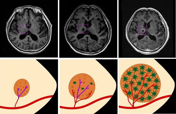
logical outcome than those with good neurological outcome justed OR: 2.32, 95% CI: 1.188–4.528) were significantly associ-
(154.7 ± 46.0 mg/dL vs. 139.5 ± 30.6 mg/dL, p = 0.006). Fluctua- ated with poor neurological outcomes in neurocritically ill pa-
tion of glucose level did not show significant difference between tients (Hosmer-Lemeshow Chi-squared = 5.43, df = 8, p = 0.711,
patients with poor neurological outcome and those with good Table 3). In addition, the 90-day mortality rate was significantly
neurological outcome (56.1 ± 70.8 mg/dL vs. 39.8 ± 46.3 mg/dL, lower in patients without early hyperglycemia compared to those
p = 0.052). with early hyperglycemia (8.3% vs 17.1%, log-rank test, p = 0.011,
Multivariable logistic regression analysis revealed that age (ad- Fig. 2).
justed odd ratio [OR]: 1.04, 95% confidence interval (CI):
DISCUSSION
Neurolgical outcomes In this study, we investigated the relationship of early hypergly-
80 cemia and neurological prognosis in neurocritically ill patients.
■ Non-DM
■ DM This study had the following major findings. First, poor neurolog-
ical outcome was more common in the early hyperglycemia group
21
60 than that in the non-hypoglycemia group. In-hospital mortality
Number of patients
9
40 46 5
41 Table 3. Multivariable logistic regression for the relationship of
38 clinically relevant variables associated with poor neurological outcome
5
Variables B Adjusted OR (95% CI) p-value
20 25 Age 0.042 1.04 (1.020–1.066)Early hyperglycemia and prognosis Junghoon Han et al.
100 sis of critically ill patients. A few studies have shown that the rela-
tionship between hyperglycemia and poor prognosis is unclear in
Survival probability (%)
90 DM patients17). Although stress-related hyperglycemia could be
important to predict the prognosis of critically ill patients, it might
80 be confused with DM-related hyperglycemia. In this study, regard-
less of DM or use of drugs, early hyperglycemia itself was associat-
70
Non-hyperglycemia ed with poor neurological outcome in neurocritically ill patients.
Early hyperglycemia This relationship might be due to a neurotoxic effect of hypergly-
60
cemia itself in patients with brain injuries.
In this study, early hyperglycemia occurred more frequently in
50
0 20 40 60 80 100
patients who used glycerine or dexamethasone than in those who
Day did not. Corticosteroid and multiple osmotic agents might be used
in patients with severe brain damages. Therefore, hyperglycemia
Fig. 2. Kaplan-Meier 90-day survival analysis comparing early might arise from these medications in more sick patients. However,
hyperglycemia and nonhyperglycemia groups of neurocritically ill
patients. Red solid line, non-hyperglycemia; blue dotted line, early in multivariable variable analysis, the use of glycerine or dexameth-
hyperglycemia. p=0.011, based on log-rank tests. asone was not associated with poor neurological outcomes. Neu-
rological prognosis was more associated with early hyperglycemia
itself compared to the use of glycerine or dexamethasone in this
was also higher in the early hyperglycemia group than that in the study. Therefore, it is necessary to consider drug-related hypergly-
non-hypoglycemia group. Second, in patients with early hypergly- cemia when treating neurocritically ill patients.
cemia, clinical prognoses were similar between DM group and In this study, early hyperglycemia associated with poor neuro-
non-DM group. Third, multivariable logistic regression analysis logical outcome in neurocritically ill patients. Especially, early hy-
revealed that age, APACHE II score on ICU admission, and early perglycemia itself could elevate risk of poor neurological outcome
hyperglycemia were significantly associated with poor neurologi- in these patients. Therefore, aggressively hyperglycemic control
cal outcomes in neurocritically ill patients. Fourth, early hypergly- could be important to protect their brain from hyperglycemia in-
cemia itself could be a significant predictor for poor neurological ducing brain damage in patients with early hyperglycemia. In addi-
outcome in neurocritically ill patients regardless of comorbidities tion, management of these patients needs to be aware of drugs that
or drugs. are at risk for inducing hyperglycemia, such as glycerin and dexa-
Hyperglycemia is associated with poor prognosis in various ICU methasone.
patients2,3,8,10). Hyperglycemia is a predictor associated with poor This study has several limitations. First, it was a retrospective re-
neurological prognosis in patients with neurological diseases such view of medical records. Second, administration of corticosteroid
as ischemic stroke, intracerebral hemorrhage, and aneurysmal sub- or multiple osmotic agents was determined by a neurointensivist
arachnoid hemorrhage5,9,13,16). Indeed, hyperglycemia itself could or a neurosurgeon. It was not protocol-based. Therefore, the
be harmful to the brain7). Complex factors such as free radical for- non-randomized nature of registry data might have resulted in se-
mation and oxidative injury, activation of N-methyl-D-aspartate lection bias. Third, in a few patients, causes of hyperglycemia were
receptors, raised intracellular calcium, triggering of inflammatory difficult to distinguish between hyperglycemia caused by stress or
and apoptotic pathways, and alterations in lactate metabolism are DM if DM was not diagnosed on admission. Finally, our study had
associated with primary toxic-ischemic injury and secondary1,4,7). limited statistical power due to its small sample size. Although this
In addition, ischemic penumbra could be injured due to direct study provides valuable insight, prospective large-scale studies are
neurotoxic effect arising from hyperglycemia1). needed to evaluate the relationship of early hyperglycemia and
In critically ill patients, hyperglycemia could be caused by vari- neurological prognosis in neurocritically ill patients to obtain evi-
ous factors2,7). Especially, stress-related hyperglycemia mainly re- dence-based conclusions.
sults from the release of stress hormones caused by acute illness6).
In addition, stress-related hyperglycemia might be associated with CONCLUSION
the extent and severity of ischemic damage in patients with stroke
or ST-segment elevation myocardial infarction1,17). Therefore, In this study, poor neurological outcome in neurocritcally ill pa-
stress-related hyperglycemia might be associated with the progno- tients was associated with early hyperglycemia. In addition, DM,
10 www.e-jnic.orgEarly hyperglycemia and prognosis Junghoon Han et al.
the use of corticosteroid, or the use of multiple osmotic agents was in neurocritical care patients: a systematic review and meta-anal-
not associated with neurological prognosis. Therefore, early hyper- ysis. Crit Care 2012;16:R203.
glycemia itself could be a significant predictor associated with neu- 8. Krinsley JS. Association between hyperglycemia and increased
rological outcome in neurocritically ill patients. hospital mortality in a heterogeneous population of critically ill
patients. Mayo Clin Proc 2003;78:1471–1478.
NOTES 9. Kruyt ND, Biessels GJ, DeVries JH, Luitse MJ, Vermeulen M,
Rinkel GJ, et al. Hyperglycemia in aneurysmal subarachnoid
Conflict of interest hemorrhage: a potentially modifiable risk factor for poor out-
We declare that we have no conflict of interest. come. J Cereb Blood Flow Metab 2010;30:1577–1587.
10. Malmberg K, Ryden L, Wedel H, Birkeland K, Bootsma A,
Informed consent Dickstein K, et al. Intense metabolic control by means of insulin
Informed consent was obtained from each participant included in patients with diabetes mellitus and acute myocardial infarc-
in this study. tion (DIGAMI 2): effects on mortality and morbidity. Eur
Heart J 2005;26:650–661.
Acknowledgements 11. Melamed E. Reactive hyperglycaemia in patients with acute
We would like to thank Hye Jung Kim, nursing director of the stroke. J Neurol Sci 1976;29:267–275.
neurosurgical intensive care unit, for providing excellent advice 12. Ryu JA, Jung W, Jung YJ, Kwon DY, Kang K, Choi H, et al. Early
and engaging in fruitful discussions. We would also like to thank all prediction of neurological outcome after barbiturate coma ther-
nurses of the neurosurgical intensive care unit at Samsung Medical apy in patients undergoing brain tumor surgery. PLoS One
Center. 2019;14:e0215280.
13. Saxena A, Anderson CS, Wang X, Sato S, Arima H, Chan E, et
REFERENCES al. Prognostic Significance of Hyperglycemia in Acute Intracere-
bral Hemorrhage: The INTERACT2 Study. Stroke 2016;
1. Capes SE, Hunt D, Malmberg K, Pathak P, Gerstein HC. Stress 47:682–688.
hyperglycemia and prognosis of stroke in nondiabetic and dia- 14. Sekhon MS, Griesdale DE, Robba C, McGlashan N, Needham
betic patients: a systematic overview. Stroke 2001;32:2426– E, Walland K, et al. Optic nerve sheath diameter on computed
2432. tomography is correlated with simultaneously measured intra-
2. Finfer S, Chittock DR, Su SY, Blair D, Foster D, Dhingra V, et al. cranial pressure in patients with severe traumatic brain injury.
Intensive versus conventional glucose control in critically ill pa- Intensive Care Med 2014;40:1267–1274.
tients. N Engl J Med 2009;360:1283–1297. 15. Sewdarsen M, Jialal I, Vythilingum S, Govender G, Rajput MC.
3. Gale SC, Sicoutris C, Reilly PM, Schwab CW, Gracias VH. Poor Stress hyperglycaemia is a predictor of abnormal glucose toler-
glycemic control is associated with increased mortality in criti- ance in Indian patients with acute myocardial infarction. Diabe-
cally ill trauma patients. Am Surg 2007;73:454–460. tes Res 1987;6:47–49.
4. Godoy DA, Di Napoli M, Rabinstein AA. Treating hyperglyce- 16. Tsivgoulis G, Katsanos AH, Mavridis D, Lambadiari V, Roffe C,
mia in neurocritical patients: benefits and perils. Neurocrit Care Macleod MJ, et al. Association of Baseline Hyperglycemia With
2010;13:425–438. Outcomes of Patients With and Without Diabetes With Acute
5. Gofir A, Mulyono B, Sutarni S. Hyperglycemia as a prognosis Ischemic Stroke Treated With Intravenous Thrombolysis: A
predictor of length of stay and functional outcomes in patients Propensity Score-Matched Analysis From the SITS-ISTR Reg-
with acute ischemic stroke. Int J Neurosci 2017;127:923–929. istry. Diabetes 2019;68:1861–1869.
6. Guo YJ, Zhou Y, Zhang SY, Wei Q, Huang Y, Xia WQ, et al. Op- 17. Yang JH, Song PS, Song YB, Hahn JY, Choi SH, Choi JH, et al.
timal target range for blood glucose in hyperglycaemic patients Prognostic value of admission blood glucose level in patients
in a neurocritical care unit. Diab Vasc Dis Res 2014;11:352– with and without diabetes mellitus who sustain ST segment ele-
358. vation myocardial infarction complicated by cardiogenic shock.
7. Kramer AH, Roberts DJ, Zygun DA. Optimal glycemic control Crit Care 2013;17:R218.
www.e-jnic.org 11eISSN 2635-5280
Original Article
J Neurointensive Care 2020;3(1):12-19
https://doi.org/10.32587/jnic.2020.00227
Role of Cancer Emboli as a Metastatic Core on the Growth
of Brain Metastasis in Patients with Non-Small Cell Lung
Cancer
Jinuk Kim1, Gyuseo Jung1, Hoon Gi Kim1, Jeong Yeon Kim2, Geun Young Yang2, Young Zoon Kim1
1
Division of Neuro Oncology and Department of Neurosurgery, Samsung Changwon Hospital, Sungkyunkwan University School of Medicine, Changwon, Korea
2
Department of Anesthesiology and Pain Medicine, Samsung Changwon Hospital, Sungkyunkwan University School of Medicine, Changwon, Korea
Received: January 18, 2020
Accepted: February 3, 2020 Objective
Brain metastasis (BM) was a common complication of patient with non-small cell lung cancer
Corresponding Author: (NSCLC) and associated with a poor prognosis. The study was to evaluate the effect of cerebral
Young Zoon Kim, M.D., Ph.D. infarction (CI) which was originated from cancer emboli on the risk of BM in NSCLC for preven-
Division of Neuro Oncology and tive therapy strategy.
Department of Neurosurgery, Methods
Samsung Changwon Hospital, Three hundred seven patients with newly diagnosed NSCLC in our institute from July 2013 to
Sungkyunkwan University School July 2018 were retrospectively analyzed. The diagnostic criteria of CI refereed to Updated Criteria
of Medicine, 158 Paryong-ro, for Population-Based Stroke and Transient Ischemic Attack Incidence Studies for the 21st Centu-
Masanhoewon-gu, Changwon ry. Depending on magnetic resonance imaging (MRI), the patients were divided into the BM
51353, Korea group and control group (without BM). Then, the prevalence of CI and baseline clinicopatholog-
Tel: +82-55-233-5241 ical parameters were evaluated and compared between the two groups.
Fax: +82-55-233-8040 Results
E-mail: yzkim@skku.edu Of the 307 patients, 204 patients (66.4%) had CI and 52 patients (16.9%) had BM. Especially, the
prevalence of CI in the NSCLC patients with BM was 84.6%, which was significantly higher than
that of 62.7% in the NSCLC patients without BM (p = 0.002). Following univariate logistic re-
gression analysis and the multivariate model, the results demonstrated that CI was a significant in-
dependent risk factor for BM in NSCLC (odds rate, 3.303; 95% confidence interval, 1.437-7.593;
p = 0.005). What’s more, CI contributed to a worse prognosis in NSCLC patients with BM. Dy-
namical trace confirmed CI could promote BM in NSCLC patient.
Conclusions
CI could be associated with a metastatic tropism to the brain and then with an increased risk of
BM in NSCLC patient. Therefore, targeted intervention of the metastatic core of CI could offer
promising approach for the prevention, prognostic evaluation, and therapy of BM in NSCLC pa-
tients for better clinical outcome.
Keywords: Cancer emboli; Metastatic core; Non-small cell lung cancer; Brain metastasis; Cere-
bral infarction
Copyright © 2020 The Korean Neurointensive Care Society
This is an Open Access article distributed under the terms of the Creative Commons Attribution Non-Commercial License (http://creativecommons.org/licenses/by-nc/4.0/)
which permits unrestricted non-commercial use, distribution, and reproduction in any medium, provided the original work is properly cited.
12 www.e-jnic.orgMetastatic core from cancer emboli Jinuk Kim et al.
INTRODUCTION CLC patients who were continuously admitted to Sungkyunkwan
University Samsung Changwon Hospital from July 2013 to July
Lung cancer remains one of the most frequently diagnosed can- 2018. The inclusion criteria were as follows: 1) all patients with
cers as well as the leading cause of cancer-related mortality world- histologically confirmed NSCLC, 2) did not receive any surgery,
wide, in which non-small cell lung cancer (NSCLC) constitutes chemotherapy, radiotherapy, molecular targeted therapy, or immu-
85% cases2,20). Unfortunately, in nearly 20% of NSCLC patients, notherapy in other hospitals before admission, 3) for the purpose
brain metastasis (BM) is already present at diagnosis, with up to of detecting distant organ metastasis, all patients accepted brain,
50% of patients developing BM throughout the disease course12,28). chest, abdominal and pelvic imaging, such as ultrasound, comput-
BM in NSCLC complicates the clinical picture and portends a ed tomography (CT), magnetic resonance imaging (MRI) and
poor prognosis with median survival of 3-7 months12,28). However, positron emission computed tomography/computed tomography
risk factors and underlying mechanisms that the most common (PET/CT), 4) complete medical records. The exclusion criteria
site of distant metastasis of lung cancer is the brain remain the mys- were as follows: 1) NSCLC patients with synchronous distant me-
tery and have not been well addressed. Thus, to best improve the tastases except for brain, 2) patients with NSCLC accompanied by
overall survival (OS) and quality of life for NSCLC patients, it is malignant tumors in other parts of the body, 3) patients with CI
highly significant to illustrate the clinical risk factors of target BM caused by other pathogenic factors. In total, 307 patients were en-
in NSCLC for prevention strategies and specific therapies. rolled in this study.
The tumor metastases are formed by a complex interaction be-
tween cancer cells and microenvironment, which is the "seed-soil" Data collection
hypothesis26,29). The "seed-soil" hypothesis sets forth the concept Data collected included age, gender, body mass index (BMI),
that a conducive microenvironment, or metastatic core, is neces- histological type, and primary tumor size and location. In addition,
sary for disseminating cancer cells to engraft distant sites17,23). The laboratory tests were also covered, including D-Dimer (D-D) and
common functions of metastatic core include anchorage, survival the tumor markers of carcinoembryonic antigen (CEA) and car-
support, protection from external insults, licensing proliferation bohydrate antigen 125 (CA125). The survival time from the onset
and outgrowth3). Long-term since, much attention has focused on of BM were evaluated. The follow-up duration lasted until the
the molecular and genetic factors of cancer cells as “seed” endowed death or July 31, 2019. OS from diagnosis of BM was evaluated.
metastatic advantage. Meanwhile, the metastatic core creating a The Institutional Review Board (IRB) of our hospital approved
fertile “soil” for cancer cell to lodge and grow has been largely ne- the study protocol (IRB number: SCMC 2019-02-015). All stud-
glected. Therefore, the microenvironment of organs as the risk fac- ies were conducted according to guidelines of the Declaration of
tors to determine metastatic colonization are particularly import- Helsinki for biomedical research. Informed consent was waived
ant for exploration, which might be most amenable to therapeutic due to its retrospective nature.
interventions.
A very interesting one appears in several studies which demon- The diagnosis of cerebral infarction
strated that patients with lung cancer were prone to induce cerebral The diagnostic criteria of CI refer to Updated Criteria for Popu-
infarction (CI) when compared with non-cancer control4). What’s lation-Based Stroke and Transient Ischemic Attack Incidence Stud-
more, CI occurrence could worse patient’s prognosis in advanced or ies for the 21st Century8), which points out that CI can be diag-
post-operative recurrent NSCLC14). These achievements inspire us nosed regardless of the duration of nervous symptoms/ signs
from the perspective of “soil” to draw attention to the effect of CI as when there are neuroimaging findings of responsible ischemic le-
metastatic core on the risk of target-specific BM in NSCLC patients sion. However, when no evidence of imaging evidence of responsi-
in the study, which may lead to detect therapeutic strategies and ble lesion can be obtained, the duration of symptoms and signs ex-
prevent metastasis at its earliest inception. The aim of this study is ceeding 24h is still the time limit for diagnosis of ischemic stroke8).
to evaluate the effect of CI which was originated from cancer embo-
li on the risk of BM in NSCLC for preventive therapy strategy. Imaging evaluation of BM in NSCLC
The patients had undergone a brain computed tomography
PATIENTS AND METHODS (CT) scan. When brain lesion(s) could not be excluded, patients
would undergo a magnetic resonance imaging (MRI). The diag-
Study population nosis of BM on MRI was determined by two different neuroradiol-
We conducted a retrospective study of all newly diagnosed NS- ogists (YM Kim and MO Sunwoo) who were blinded to the clini-
www.e-jnic.org 13Metastatic core from cancer emboli Jinuk Kim et al.
cal and pathological findings. Depending on the imaging examina- MRI without any brain symptom, while other patients took the ex-
tion results of CI and BM, NSCLC patients were divided into the amination due to some symptoms, such as headache, hemiplegia
BM group and control group (without BM). Then, the prevalence and psychiatrical disorder. The summary of patients’ characteris-
of CI and baseline clinicopathological parameters were evaluated tics of BM group and control group are shown in Table 1. The re-
and compared between the two groups. sult demonstrated that patients with NSCLC are prone to induce
CI with the prevalence of 66.45 %. Especially, the prevalence of CI
Statistical analysis in the NSCLC patients with BM was 84.6%, which was significant-
The prevalence of CI in the two groups was compared by the chi ly higher than that of 62.7% in the NSCLC patients without BM (p
square test, other patient characteristics were compared either the = 0.002, χ 2 test). In addition, there were significantly differences
chi square test, Fisher's exact test, or Wilcoxon two-sample test. in primary tumor location (lobe), histology (type), D-D, CEA,
Univariate and multivariate analyses were performed using logistic and CA125 between BM group and control group. By contrast,
regression to assess the risk factors for BM. OS was plotted using there were no significant differences in age, BMI, sex, and primary
the Kaplan Meier method. Differences in OS were analyzed using tumor size between the two groups Table 1.
the log-rank test. p < 0.05 was considered to be statistically signifi-
cant. All statistical analyses were performed using SPSS version Risk factors for BM in NSCLC patient
25.0 software. Following univariate logistic regression analysis, CI, primary tu-
mor location (lobe), histology (type), D-D, CEA, and CA125
RESULTS were chosen as risk factors for BM in NSCLC. The results illustrat-
ed that BM was significantly associated with CI (odds rate [OR],
Characteristics of NSCLC patient 3.266; 95% confidence interval [CoI], 1.475-7.231; p = 0.004).
A total of 307 NSCLC patients (185 men and 122 women) with Besides, adenocarcinoma (p = 0.011), middle region tumor loca-
a median age of 64 years (range of 25–83 years) were confirmed tion (p = 0.005), increased CEA level (p = 0.002), increased
for the analysis in our study. Among them, 52 patients (16.9%) had CA125 level (p < 0.001) and higher D-D level (p = 0.027) were
BM, and they were then divided into BM group. NSCLC patients also linked with increased risks of BM in NSCLC Table 2.
without BM were regarded as the control group. In the 52 NSCLC Next, we assessed the significance of CI with respect to BM by
patients with BM, 22 patients (42.3%) underwent a brain CT or using the multivariate model according to the factors that are prov-
Table 1. Baseline characteristics of NSCLC patients with/without BM
BM (-) BM (+)
p-value
(N=255) (N=52)
Age (year) Median (range) 62 (25-83) 62.5 (43-78) 0.920
BMI kg/m² Median (range) 23.18(16-34) 22.85 (14-30) 0.088
Sex, Male (%) 154 (60.4) 31 (59.6) 0.920
Primary tumor size (cm) Median (range) 3 (0.4-14) 3.6 (0.8-11) 0.912
Primary tumor location 0.020
Upper lobe (%) 152 (59.6) 26 (50.0)
Lower lobe (%) 89 (34.9) 17 (32.7)
Middle (%) 14 (5.5) 9 (17.3)
Histological type 0.028
Adenocarcinoma (%) 180 (70.6) 46 (88.5)
Squamous carcinoma (%) 58 (22.7) 5 (9.6)
Other (%) 17 (6.7) 1 (1.9)
Presence of CI (%) 160 (62.7) 44 (84.6) 0.002
D-D (ng/ml >500) 182 (0.11-14612) 304.5 (52-7640) 5 4.11 (0.1-746.7) 9.675 (1.28-1068) 35 19.44 (3.1-2846) 49.15 (6.61-796.7)You can also read