2022 Calendar - Revolutionizing structural biology with 3DEM - RCSB PDB
←
→
Page content transcription
If your browser does not render page correctly, please read the page content below

Revolutionizing
                                 structural biology
                                       with 3DEM

2022 Calendar
      RCSB.ORG
      A Living Digital Data
      Resource that Enables
      Scientific Breakthroughs
                                       Learn more: rcsb.org | pdb101.rcsb.org
3D electron microscopy (3DEM) is revolutionizing the field                                            Experimental
of structural biology. This technique has become the                                                  Structure
method of choice for determining structures of large
macromolecular complexes. Thanks to recent                                                            Determination
                                                                                                    ATOMIC STRUCTURE
advances in sample preservation,                                                                      with 3DEM
                                                                                              DETERMINATION WITH 3DEM
microscopy, and computing, it’s
also increasingly used for
smaller structures.
This calendar features twelve
molecular assemblies studied                                               1. Sample Preparation

by 3DEM. They further                                                      Sample preparation starts with isolating the protein of interest from cells
                                                                           or organisms. To stabilize and ensure that proteins are in a native-like
our understanding of                                                       conformation for the experiment, some molecular modifications can
biology and serve as a                                                     be applied, like placing the proteins in nanodiscs or adding stabilizing
gateway to advances                                                        nanobodies (see illustrations for February, March, and August). A tiny
                                                                           amount of the sample is placed on a small grid and spread in a single
in technology and                                                          layer. Then the sample is frozen in liquid ethane.
medicine.
                                                             2. Data Collection
                                                             The sample is placed inside an electron microscope and is exposed to a beam of
                                                             accelerated electrons. The electrons scatter as they hit the sample and thousands of
                                                             images are captured.
                                          3. Data Analysis
                                          The data obtained are 2D projection images showing the macromolecules in many different orientations.
                                          The images are grouped by orientation and then combined computationally to reconstruct a 3DEM map
                                          of the molecule.
4. The EM Map and Atomic Model
                                                                                                                     The 3DEM Resolution:
                                   The EM map shows the overall shape of the protein structure (shown in             Small numbers are better than big
                                   cyan on the left). Using the map and advanced computing techniques
                                   along with existing sequence and experimental data, atoms are positioned          When we talk about resolution in atomic
                                   within the map to create an atomic structure model (shown in gold).               structure, we use the Ångstrom unit. One
                                                                                                                     Ångstrom is approximately the width of one
                                           5. Data Access                                                            hydrogen atom. In both X-ray crystallography
                                                                                                                     and 3DEM, a resolution of 1 Ångstrom is
                                           3DEM maps are freely available in a public repository, the Electron
                                                                                                                     highly desirable. A protein structure at this
                                           Microscopy Data Bank (EMDB). Atomic structure models obtained
                                                                                                                     resolution has hydrogen atoms visible and all
                                            from 3DEM data are stored in the Protein Data Bank (PDB).
                                                                                                                     other atoms mapped accurately. 3DEM maps
                                              Unique IDs are assigned to these maps (e.g., EMD-11657)
                                                                                                                     at greater Ångstrom resolution are typically
                                                and models (e.g., PDB ID 2rte).
                                                                                                                     not as detailed.
                                                 To explore in 3D, vist ebi.ac.uk/emdb (maps) and
                                                                                                                     The 3DEM structure determined at the
                                                  RCSB.org (atomic models).
                                                                                                                     highest resolution is currently Apoferritin
                                                                                                                     solved at ~1.2 Å (see January illustration).
                                                                                                                     Shown below are examples of the 3DEM
                                                                                                                     map-to-atomic model fit for each type of
                                                                                                                     amino acid at this resolution.

           The PDB released its first 3DEM entry, the
         ground-breaking structure of bacteriorhodopsin,
                                                                                              PDB Structure 1BRD
    in 1991 (shown right). Since then, more than 9,000                       R. Henderson et al. (1990) Model for
3DEM structures have been made available in the PDB                             the structure of bacteriorhodopsin
                                                                           based on high-resolution electron cryo-
archive, most of them in the last few years.                                microscopy. J Mol Biol 213: 899–929.

In 2017, Jacques Dubochet, Joachim Frank, and Richard Henderson were recognized
with the Nobel Prize in Chemistry for their contributions to experimental determination
of atomic structures using single-particle cryo-electron microscopy.                                                       Learn more: rcsb.org | pdb101.rcsb.org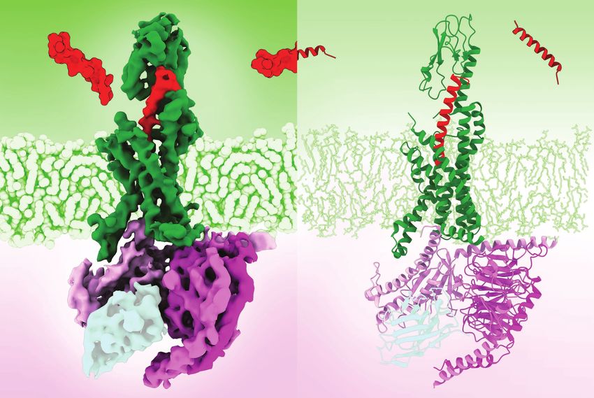
Learn more: rcsb.org | pdb101.rcsb.org

JANUARY 2022                                                                        Ferritin
                                                                                    Ferritin helps balance iron levels within
Sunday   Monday          Tuesday   Wednesday   Thursday   Friday   Saturday         living organisms. It forms a spherical shell
                                                                                    composed of 24 subunits (shown in shades
26       27              28        29          30         31       1                of blue). Reactive and potentially toxic iron
                                                                                    atoms (red) enter the shell through the
                                                                                    3-fold pores (shown on the right) where
                                                                   New Year’s Day   they form non-reactive small crystallites.
                                                                                    The iron mineral is released as needed.
2        3               4         5           6          7        8                Ferritin that is not combined with iron
                                                                                    is called apoferritin.
                                                                                    As a structure of extreme stability, ferritin
                                                                                    is one of the most widely used test
                                                                                    samples in 3DEM. The map of apoferritin
9        10              11        12          13         14       15               at approximately 1.2 Å (shown on the
                                                                                    right) is currently the EM structure with
                                                                                    the highest resolution. It is precise enough
                                                                                    to unambiguously identify the position of
                                                                                    individual atoms in the protein.
16       17              18        19          20         21       22
                                                                                    PDB Structure 6Z6U
         Martin Luther                                                              3DEM Map EMD-11103
         King Jr. Day
                                                                                    K.M. Yip et al. (2020) Atomic-resolution
                                                                                    protein structure determination by cryo-EM.
23       24              25        26          27         28       29               Nature 587: 157–161.

30       31              1         2           3          4        5

                                                                                    Learn more: rcsb.org | pdb101.rcsb.org
Learn more: rcsb.org | pdb101.rcsb.org
FEBRUARY 2022                                                                          GABAA receptor
                                                                                       GABAA receptors reside in neurons in the
Sunday   Monday            Tuesday          Wednesday   Thursday   Friday   Saturday   cell membrane. One protein chain (shown
                                                                                       in orange with EM map overlay) is repeated
30       31                1                2           3          4        5          five times radially to form the pentameric
                                                                                       channel. Upon binding of the GABA
                                                                                       neurotransmitter, the channel opens and
                           Lunar New Year                                              allows negatively charged ions to enter the
                                                                                       neuron, offsetting the positive charges of
6        7                 8                9           10         11       12         the ions driving the neuronal signal.
                                                                                       This activity marks the channel as
                                                                                       an important drug target for many
                                                                                       anti-stimulatory agents.
                                                                                       The structure shown here has a nanobody
13       14                15               16          17         18       19         (light blue, encircling the receptor) bound
                                                                                       at the GABA binding site. This helps to
                                                                                       stabilize and orient the channel during
         Valentine’s Day                                                               the experiment. At 1.7 Å resolution, this
                                                                                       structure is the most detailed portrait of
20       21                22               23          24         25       26         a GABAA receptor to date and serves as
                                                                                       an invaluable aid for current and future
                                                                                       structure-based drug discovery efforts.
         Presidents’ Day

                                                                                       PDB Structure 7A5V
27       28                1                2           3          4        5          3DEM Map EMD-11657
                                                                                       T. Nakane et al. (2020) Single-particle cryo-EM
                                                                                       at atomic resolution. Nature 587: 152–156.

6        7                 8                9           10         11       12

                                                                                       Learn more: rcsb.org | pdb101.rcsb.org
Learn more: rcsb.org | pdb101.rcsb.org
MARCH 2022                                                                             GLP-1 Receptor
                                                                                       Glucagon-like peptide 1 (GLP-1, red) is
Sunday                   Monday   Tuesday   Wednesday   Thursday   Friday   Saturday   a hormone regulating insulin secretion,
                                                                                       carbohydrate metabolism, and appetite.
27                       28       1         2           3          4        5          When GLP-1 binds to its receptor (green),
                                                                                       it activates the G protein (purple) inside the
                                                                                       cell, starting a cascade of responses in the
                                                                                       insulin-regulating pathway. The receptor is
                                                                                       a target for many type 2 diabetes drugs.
6                        7        8         9           10         11       12         3DEM has provided the first clear picture of
                                                                                       the full-length activated receptor in complex
                                                                                       with G protein, highlighting key interactions
                                                                                       not visible in the X-ray structures of the
                                                                                       individual components, and revealing how
13                       14       15        16          17         18       19         the different domains cooperate to transmit
                                                                                       signals upon binding to the GLP-1 hormone.
Daylight Saving Time                                                                   The structure has a nanobody bound to
starts (North America)   Pi Day                                                        the G protein (shown in light blue) that
                                                                                       helped the researchers to capture the
20                       21       22        23          24         25       26         active conformation of the receptor.

                                                                                       PDB Structure 5VAI
                                                                                       3DEM Map EMD-8653
                                                                                       Y. Zhang et al. (2017) Cryo-EM structure of the
27                       28       29        30          31         1        2          activated GLP-1 receptor in complex with a
                                                                                       G protein. Nature 546: 248–253.

3                        4        5         6           7          8        9

                                                                                       Learn more: rcsb.org | pdb101.rcsb.org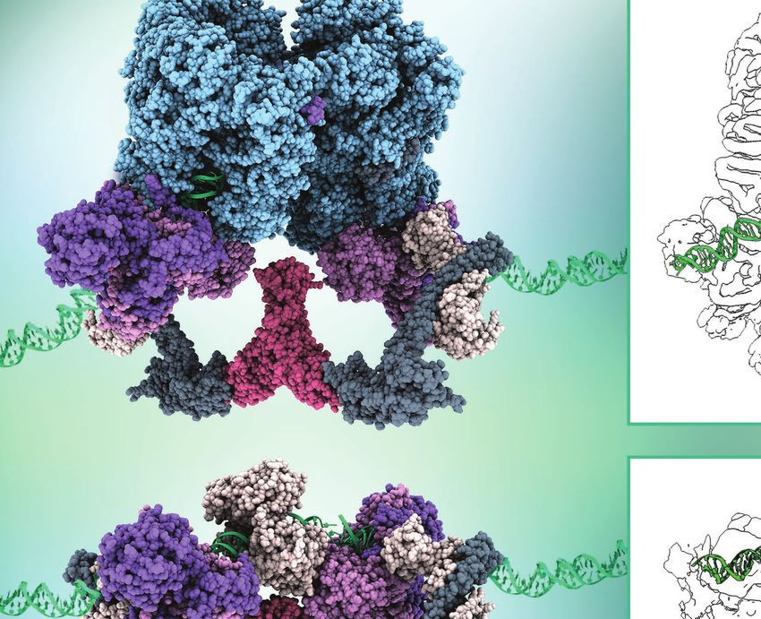
Learn more: rcsb.org | pdb101.rcsb.org
APRIL 2022                                                                 ROQ1 Resistosome
                                                                           Pathogenic bacteria inject plant cells with
Sunday   Monday    Tuesday   Wednesday   Thursday   Friday      Saturday   effector proteins XopQ (red), which alters
                                                                           plant physiology. The immune system of
 27      28        29        30          31         1           2          plants includes the ROQ1 resistosome
                                                                           (green and yellow). The components
                                                                           assemble around foreign proteins and
                                                                           start an orderly process leading to
                                                                           death of the infected cell.
3        4         5         6           7          8           9          First discovered in 1994, it wasn’t until
                                                                           26 years later that a structure of activated
                                                                           ROQ1 would be determined using state-
                                                                           of-the-art 3DEM, providing mechanistic
                                                                           insights into how detection of the XopQ
10       11        12        13          14         15          16         substrate leads to localized cell death
                                                                           and disease resistance.

                                                                           PDB Structures 7JLV, 7JLU, 7JLX
                                                                           3DEM Map EMD-22381
17       18        19        20          21         22          23
                                                                           R. Martin et al. (2020) Structure of the
                                                                           activated ROQ1 resistosome directly
                                                                           recognizing the pathogen effector XopQ.
Easter                                              Earth Day              Science 370: eabd9993.

24       25        26        27          28         29          30

         DNA Day

1        2         3         4           5          6           7

                                                                           Learn more: rcsb.org | pdb101.rcsb.org
Learn more: rcsb.org | pdb101.rcsb.org
MAY 2022                                                                                 Transient Receptor
                                                                                         Potential (TRP)
Sunday         Monday               Tuesday   Wednesday   Thursday   Friday   Saturday   Ion Channel
 24            25                   26        27          28         29       30         TRP ion channels are used by many
                                                                                         different types of organisms to sense pain,
                                                                                         temperature, and some natural toxins.
                                                                                         This tetramer is notoriously difficult to
                                                                                         crystallize owing to its intrinsic flexibility
1              2                    3         4           5          6        7          and low expression level in cells. 3DEM
                                                                                         has been the preferred method for
                                                                                         producing over 70 structures in the past
               Eid al-Fitr begins                                                        8 years, including a 1.8 Å resolution
                                                                                         structure of the wild-type mouse
8              9                    10        11          12         13       14         temperature-sensitive TRPV3
                                                                                         (atomic detail and map overlay
                                                                                         shown in right panel).
Mother’s Day
                                                                                         PDB Structure 7MIJ
15             16                   17        18          19         20       21         3DEM Map EMD-23853
                                                                                         K.D. Nadezhdin et al. (2021) Structural
                                                                                         mechanism of heat-induced opening of a
                                                                                         temperature-sensitive TRP channel.
                                                                                         Nature Structural & Molecular
22             23                   24        25          26         27       28         Biology 28: 564–572.

29             30                   31        1           2          3        4

               Memorial Day
                                                                                         Learn more: rcsb.org | pdb101.rcsb.org
Learn more: rcsb.org | pdb101.rcsb.org
JUNE 2022                                                                    SARS-CoV-2 Spike Protein
                                                                             The genome sequence of SARS-CoV-2
Sunday         Monday   Tuesday   Wednesday   Thursday   Friday   Saturday   was first made publicly available on
                                                                             January 10th 2020, and by March 2020
29             30       31        1           2          3        4          researchers had already produced 3DEM
                                                                             structures of the envelope spike protein.
                                                                             The protein comprises three chains
                                                                             (shown in blue, green and purple). The
                                                                             protein uses a shield of sugars or glycans
5              6        7         8           9          10       11         (some of them highlighted in yellow)
                                                                             to protect itself from the host immune
                                                                             proteins. During the initial steps of the
                                                                             viral infection, the receptor binding domain
                                                                             changes its shape (as shown in the insets)
                                                                             to bind to the angiotensin-converting
12             13       14        15          16         17       18
                                                                             enzyme (ACE2 receptor, sandy brown)
                                                                             on the host cells.
                                                                             Hundreds of 3DEM spike-related structures
                                                                             are available in the PDB, providing insights
19             20       21        22          23         24       25         on the structural mechanisms of SARS-
                                                                             CoV-2 infection, vaccination, and design
                                                                             of therapeutics.
Father’s Day
Juneteenth
                                                                             PDB Structures 6VXX, 6VYB
                                                                             3DEM Maps EMD-21452, EMD-21457
26             27       28        29          30         1        2
                                                                             A.C. Walls et al. (2020) Structure, Function,
                                                                             and Antigenicity of the SARS-CoV-2 Spike
                                                                             Glycoprotein. Cell 181: 281–292.

                                                                             ACE2 Receptor: PDB Structure 6M18
3              4        5         6           7          8        9          R. Yan et al. (2020) Structural basis for the
                                                                             recognition of SARS-CoV-2 by full-length human
                                                                             ACE2. Science 367: 1444–1448.

                                                                             Learn more: rcsb.org | pdb101.rcsb.org
Learn more: rcsb.org | pdb101.rcsb.org
JULY 2022                                                                        NHEJ Complexes
                                                                                 Non-homologous end joining (NHEJ) is the
Sunday   Monday             Tuesday   Wednesday   Thursday   Friday   Saturday   primary pathway by which eukaryotic cells
                                                                                 repair DNA double-strand breaks.
26       27                 28        29          30         1        2          3DEM can provide direct views of the
                                                                                 essential assembly states during NHEJ,
                                                                                 allowing researchers to elucidate the
                                                                                 complete reaction cycle for how broken
                                                                                 DNA ends are stitched together. This
3        4                  5         6           7          8        9          illustration shows a long-range complex
                                                                                 (top) which grabs the broken DNA
         Independence Day                                                        fragments when they are far apart and
         (USA)                                                                   a short-range complex (bottom), which
                                                                                 ultimately joins the DNA ends when
10       11                 12        13          14         15       16         they are close together.

                                                                                 PDB Structures 7LT3, 7LSY
                                                                                 3DEM Maps EMD-23510, EMD-23509

17       18                 19        20          21         22       23         S. Chen et al. (2021) Structural basis of long-
                                                                                 range to short-range synaptic transition in NHEJ.
                                                                                 Nature 593: 294–298.

24       25                 26        27          28         29       30

31       1                  2         3           4          5        6

                                                                                 Learn more: rcsb.org | pdb101.rcsb.org
Learn more: rcsb.org | pdb101.rcsb.org
AUGUST 2022                                                            NompC
                                                                       Specialized mechanoreceptive organelles
Sunday   Monday   Tuesday   Wednesday   Thursday   Friday   Saturday   in fruit flies contain thousands of force-
                                                                       sensitive NompC ion channels that are
31       1        2         3           4          5        6          responsible for the sensations of touch and
                                                                       balance. The channel contains a bundle
                                                                       of ~15-nm-long supercoiled helices that
                                                                       act like springs and open in response
                                                                       to mechanical force on the membrane
7        8        9         10          11         12       13         allowing the ion entry.
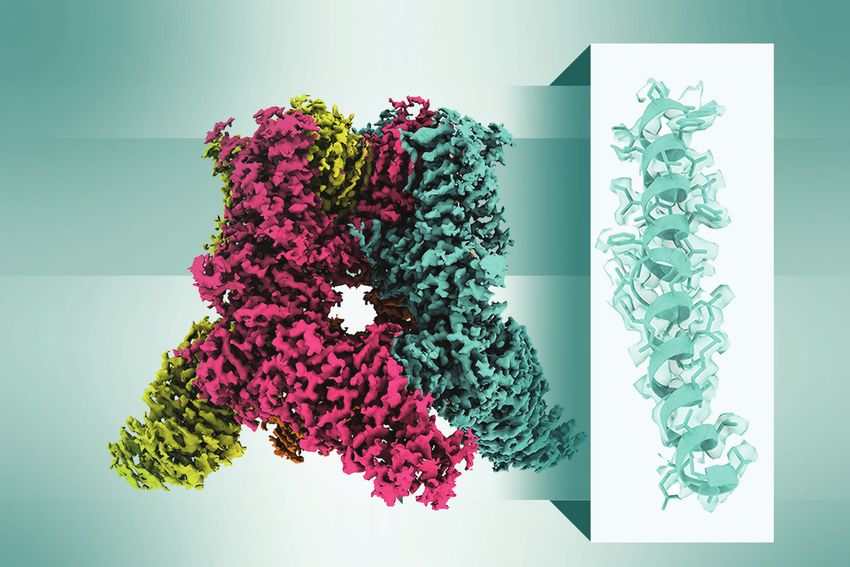
                                                                       The illustration shows the 3DEM density
                                                                       map on the left and 3D atomic model on
                                                                       the right. The sample was reconstituted in
                                                                       a lipid nanodisc. Nanodiscs (shown top left)
14       15       16        17          18         19       20         are often used in EM experiments when
                                                                       determining membrane-bound structures.
                                                                       They are composed of a disc of lipids just
                                                                       big enough to hold one copy of a protein
                                                                       surrounded by a stabilizing belt of proteins.
21       22       23        24          25         26       27
                                                                       PDB Structure 5VKQ
                                                                       3DEM Map EMD-8702
                                                                       P. Jin et al. (2017) Electron cryo-microscopy
                                                                       structure of the mechanotransduction channel
28       29       30        31          1          2        3          NOMPC. Nature 547: 118–122.

4        5        6         7           8          9        10

                                                                       Learn more: rcsb.org | pdb101.rcsb.org
Learn more: rcsb.org | pdb101.rcsb.org
SEPTEMBER 2022                                                                   Phage P68
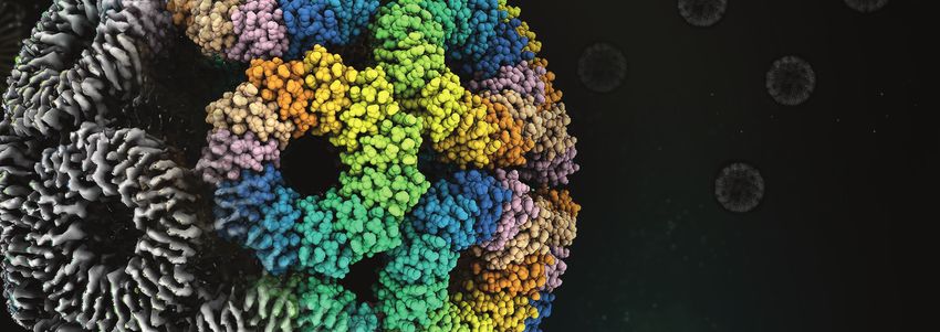
                                                                                 Many bacteriophages (viruses that attack
Sunday          Monday      Tuesday   Wednesday   Thursday   Friday   Saturday   bacteria) have icosahedral capsids (dark
                                                                                 blue) to store and protect their genomes
28              29          30        31          1          2        3          (red) from the harsh extracellular
                                                                                 environment. These viruses use tail fibers
                                                                                 (green) to attach to molecules on the
                                                                                 surface of bacterial cell walls. Thereafter,
                                                                                 the tail spike (yellow) penetrates the thick
4               5           6         7           8          9        10         layer of bacterial peptidoglycan and the
                                                                                 lipid bilayer membrane to inject DNA into
                                                                                 the bacterial cytoplasm. This illustration
                Labor Day
                                                                                 shows Phage P68 in its native form prior
                                                                                 to interaction with the cell wall. The same
                                                                                 study also reported a genome ejection
11              12          13        14          15         16       17
                                                                                 intermediate and empty particle.
                                                                                 The first electron micrographs of raw
                                                                                 unstained phages were published in
                                                                                 Germany in 1940. Less than one hundred
18              19          20        21          22         23       24         years later–thanks to remarkable
                                                                                 developments in 3DEM–researchers are
                                                                                 able to visualize elegant 3D structures
                                                                                 of phages releasing their genomes.

25              26          27        28          29         30       1          PDB Structure 6Q3G
                                                                                 3DEM Map EMD-4459
Rosh Hashanah                                                                    D. Hrebík et al. (2019) Structure and genome
begins                                                                           ejection mechanism of Staphylococcus aureus
                                                                                 phage P68. Science Advances 5: eaaw7414.
2               3           4         5           6          7        8

                                                                                 Learn more: rcsb.org | pdb101.rcsb.org
Learn more: rcsb.org | pdb101.rcsb.org
OCTOBER 2022                                                                                ATP Synthase Dimer
                                                                                            Mitochondrial ATP synthases form
Sunday   Monday         Tuesday             Wednesday   Thursday        Friday   Saturday   dimers and occupy the folds on the inner
                                                                                            membranes of the mitochondria. This
25       26             27                  28          29              30       1          molecular machine links two rotary motors
                                                                                            termed F1 and F0. The F0 motor (cyan) is
                                                                                            nested in the membrane where rotations
                                                                                            are powered by the difference in the proton
                                                                                            gradient across the membrane. A central
2        3              4                   5           6               7        8          stalk connects both motors powering the
                                                                                            F1 motor, which then uses the power of
                                                                                            rotational motion to build ATP from ADP
                        Yom Kippur begins
                                                                                            and inorganic phosphate.
                                                                                            F-type ATP synthases have been studied
9        10             11                  12          13              14       15         extensively over the past 50 years, but only
                                                                                            recently has it become possible to fully
         Indigenous                                                                         understand their molecular mechanisms
         Peoples’ Day                                                                       owing to the determination of complete
                                                                                            complexes by 3DEM.
16       17             18                  19          20              21       22
                                                                                            PDB Structure 6RD4
                                                        PDB announced                       3DEM Map EMD-4805
                                                        in 1971
                                                                                            B.J. Murphy et al. (2019) Rotary substates of
                                                                                            mitochondrial ATP synthase reveal the basis of
23       24             25                  26          27              28       29         flexible F1-Fo coupling. Science 364: eaaw9128.

         Diwali

30       31             1                   2           3               4        5

         Halloween
                                                                                             Learn more: rcsb.org | pdb101.rcsb.org
Learn more: rcsb.org | pdb101.rcsb.org
NOVEMBER 2022                                                                                  Ribosome
                                                                                               Ribosomes are molecular machines built of
Sunday                 Monday   Tuesday   Wednesday   Thursday       Friday         Saturday   proteins (purple) and RNA (red). Ribosomes
                                                                                               are found within all living organisms where
30                     31       1         2           3              4              5          they synthesize new proteins using mRNA
                                                                                               templates. Differences between human and
                                                                                               bacterial ribosomes make them an excellent
                                                                                               target for many antibiotics.
                                                                                               3DEM enables macromolecular structure
6                      7        8         9           10             11             12         determination in vitro and in situ, which
                                                                                               bridges the gap between structural biology
Daylight Saving Time                                                                           and cell biology. This structure shows a
ends (North America)                                                 Veterans Day              snapshot of a ribosome from Mycoplasma
                                                                                               pneumoniae caught inside its native cellular
13                     14       15        16          17             18             19         environment at residue-level resolution.
                                                                                               The structure includes a bound antibiotic
                                                                                               (chloramphenicol).

                                                                                               3DEM Maps EMD-11650, EMD-11998,
20                     21       22        23          24             25             26         EMD-11999
                                                                                               D. Tegunov et al. (2021) Multi-particle
                                                                                               cryo-EM refinement with M visualizes
                                                      Thanksgiving                             ribosome-antibiotic complex at 3.5 Å in cells.
                                                                                               Nature Methods 18: 186–193.
27                     28       29        30          1              2              3

4                      5        6         7           8              9              10

                                                                                               Learn more: rcsb.org | pdb101.rcsb.org
DECEMBER 2022 Nanocage
Tailor-made protein cages are being
Sunday Monday Tuesday Wednesday Thursday Friday Saturday engineered in the laboratory for diverse
applications in materials science,
27 28 29 30 1 2 3 medicine, and synthetic biology.
Several nanocages have been
characterized at near-atomic resolution
by 3DEM, including the gold-laced TRAP
cage that possesses an unusual snub
4 5 6 7 8 9 10 cube geometry long thought
to be impossible.
PDB Structure 6RVV
3DEM Map EMD-4444
11 12 13 14 15 16 17 A.D. Malay et al. (2019) An ultra-stable gold-
coordinated protein cage displaying reversible
assembly. Nature 569: 438–442.
18 19 20 21 22 23 24
Hanukkah begins
25 26 27 28 29 30 31
Christmas Day Kwanzaa New Year’s Eve
1 2 3 4 5 6 7
Learn more: rcsb.org | pdb101.rcsb.orgABOUT THIS CALENDAR
This calendar highlights structures studied by
3D electron microscopy (3DEM) that further our
understanding of biology and serve as a gateway to RCSB.ORG PDB101.RCSB.ORG
advances in technology and medicine.
A Living Digital Data Resource that Enables Molecular Explorations through
Corresponding atomic data are freely available in the
Scientific Breakthroughs Biology and Medicine
Protein Data Bank (PDB; structures) and Electron
Microscopy Data Bank (EMDB; maps) archives. Cells rely on many large molecular machines that carry out PDB-101 is the educational portal of the RCSB PDB
Access these data at RCSB.org by entering the the complex biological and chemical tasks responsible for developed for teachers, students, and the general
PDB or EMDB ID listed each month. sustaining life. 3D structures of these machines are freely public to promote exploration in the 3D world of
available at the Protein Data Bank (PDB), the global storehouse proteins and nucleic acids.
of biomolecular structures central to research and education. Learning about the diverse shapes and functions of
REFERENCES
RCSB.ORG serves millions of users worldwide each year, these biological macromolecules helps us understand
wwPDB: Nature Structural Biology 10: 980 (2003) providing services that all aspects of biomedicine and agriculture, from
doi: 10.1038/nsb1203-980 protein synthesis to human health and disease
• Inform basic and applied research across the sciences
PDB archive: Nucleic Acids Research 47: D520–D528 to biological energy.
• Are central to understanding human, animal, and plant
(2019) doi: 10.1093/nar/gky949 health and disease All resources are freely available, including
EMDB: Nucleic Acids Research 44: D396–D403 (2016) • Are critical for drug discovery/development and curricular materials, paper molecular models,
doi: 10.1093/nar/gkv1126 biotechnology videos/animations, and more.
• Enable education across biology and medicine
RCSB PDB: Visit the Guide to Understanding PDB Data
Nucleic Acids Research 28: 235–242 (2000) (accessible from the Learn menu) to learn
RCSB PDB is a member of the Worldwide PDB (wwPDB.ORG). more about 3DEM and other techniques for
doi: 10.1093/nar/28.1.235
structure determination.
Nucleic Acids Research 49: D437–D451 (2021) The RCSB PDB is managed by the members of the Research
doi: 10.1093/nar/gkaa1038 Collaboratory for Structural Bioinformatics:
Molecular images were created using UCSF ChimeraX
(E. F. Pettersen et al. (2021) UCSF ChimeraX: Structure
visualization for researchers, educators, and developers.
Protein Sci. 30: 70–82.) RCSB PDB is funded by the National Science Foundation (DBI-1832184), the US Department of Energy (DE-SC0019749),
and the National Cancer Institute, National Institute of Allergy and Infectious Diseases, and National Institute of General
Medical Sciences of the National Institutes of Health under grant R01GM133198. Learn more: rcsb.org | pdb101.rcsb.orgYou can also read


