A rare case of gallstone ileus-the unanswered question
←
→
Page content transcription
If your browser does not render page correctly, please read the page content below
Journal of Surgical Case Reports, 2021;4, 1–3

                                                                              doi: 10.1093/jscr/rjab164
                                                                              Case Report

CASE REPORT

A rare case of gallstone ileus—the unanswered
question
Chi Fai Tsang*
Department of General Surgery, Prince of Wales Hospital and Community Health Services, Sydney, New South
Wales 2002, Australia
*Correspondence address: Department of General Surgery, Prince of Wales Hospital and Community Health Services, P.O. Box 21502, World Square,
Sydney, New South Wales 2002, Australia. Tel: +61404737803; E-mail: chifai.tsang@health.nsw.gov.au

Abstract
Gallstone ileus is caused by an impaction of one or more gallstones within the gastrointestinal tract leading to mechanical
intestinal obstruction. It is a rare complication of cholelithiasis and found in 2–3% of all cases associated with recurrent
episodes of cholecystitis. This case study demonstrates an atypical presentation of gallstone ileus. A 57-year-old woman was
presented with abdominal pain and vomiting without previous history of gallstone disease. The features of gallstone ileus are
evident on computed tomography. She underwent an emergency laparotomy and enterotomy for the removal of impacting
gallstones, followed by an interval cholecystectomy and cholecystoduodenal fistula closure. This case report aims to explore
the proper surgical management of gallstone ileus. Unfortunately, the question of whether interval biliary surgery should be
performed remains unanswered, and surgeons will continue to make the decision based on their clinical judgement.

INTRODUCTION                                                                  and the duodenum, cholecystoduodenal fistula is the most com-
                                                                              mon (32.5–96.5%). Less commonly, the stomach (0–13.3%), small
Gallstone ileus accounts for 1–4% of all presentations to hospital
                                                                              bowel (0–2.5%) and the colon (0–10.9%) may also be involved [5].
with mechanical small bowel obstruction. It occurs predomi-
                                                                              This case report aims to explore the most acceptable operative
nantly in female. In elderly patients (>65 years) it consists of
                                                                              management in the literature for gallstone ileus.
25% of all cases of small bowel obstruction [1, 2]. Gallstone ileus
is caused by an impaction of one or more gallstones within the
gastrointestinal tract leading to mechanical intestinal obstruc-
                                                                              CASE STUDY
tion. It is a rare complication of cholelithiasis and found in 2–3%
of all cases associated with recurrent episodes of cholecystitis              A 57-year-old woman presented to emergency department (ED)
[2]. However, it carries five times the risk of morbidity (20–57%)            with 3 days history of left lower quadrant abdominal pain. The
compared with other causes of small bowel obstruction. The                    pain was constant, cramping, and non-radiating.
mortality is ∼7–18%. In addition, biliary malignancy may be the                  There were associated vomiting and abdominal distension.
underlying cause in up to 15% of such cases [3]. Often following              Her last bowel movement was 12 hours prior to the presentation,
an episode of cholecystitis, the pressure effect, inflammation                and she passed minimal flatus since. She has a background
and ischemia from the offending gallstone causes the erosion                  of chronic kidney disease. She was slightly tachycardic in ED.
through the gallbladder wall and subsequent fistula formation                 Abdominal examination demonstrated a mildly distended and
between the gallbladder and the adhered portion of the gastroin-              soft abdomen with left lower quadrant tenderness without peri-
testinal tract [4]. Due to the proximity between the gallbladder              tonism. Blood work showed an acute on chronic renal failure

Received: February 8, 2021. Revised: April 4, 2021. Accepted: April 7, 2021
Published by Oxford University Press and JSCR Publishing Ltd. © The Author(s) 2021.
This is an Open Access article distributed under the terms of the Creative Commons Attribution License (http://creativecommons.org/licenses/by/4.0/),
which permits unrestricted reuse, distribution, and reproduction in any medium, provided the original work is properly cited.
                                                                                                                                                1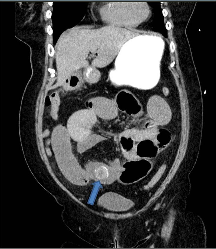
2 C.F. Tsang
Figure 1: Axial CT image demonstrates two 25-mm gallstones (blue arrows) in a
loop of small bowel in the right lower quadrant.
(creatinine 198 μmol/l and estimated glomerular filtration rate
24 ml/min/1.73 m2 ). Liver function tests were mildly deranged Figure 2: Coronal CT image demonstrates gallstones (blue arrow) (only one
gallstone shown on this image) in a loop of small bowel in the right lower
(bilirubin 27 μmol/l, alkaline phosphatase 105 U/l, GGT 85 U/l,
quadrant.
alanine aminotransferase 38 U/l, aspartate aminotransferase
37 U/l) and white cell count was slightly elevated (10.7 × 10⁹ /l;
reference range 3.7–9.5 × 10⁹ /l). Computed tomography (CT) scan
(Figs 1 and 2) demonstrated multiple loops of moderated dilated
small bowel. There were two 25-mm gallstones in a loop of small
bowel in the right lower quadrant. The gallbladder is collapsed
with intraluminal gas. There was pneumobilia in the biliary
tree. The features on CT are in keeping with a gallstone ileus.
Initial management included fluid resuscitation, insertion of a
nasogastric tube, analgesia and nil by mouth. A Foley catheter
was inserted to monitor urine output. A laparotomy proceeded
within 24 hours. Two palpable gallstones were found within the
distal ileum. A longitudinal enterotomy was made immediately
proximal to the point of calculus impaction and two calculi were
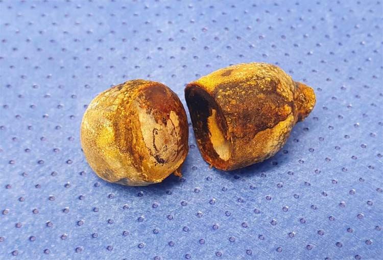
delivered from the bowel lumen (Fig. 3). There was a suspected
Figure 3: Two gallstones were delivered via enterolithotomy.
calculus contained within the gallbladder. However, the decision
was made not to proceed for further reduction via the fistula due
to significant risk of duodenal injury. Her recovery was unevent-
ful, and renal function returned to baseline. She was discharged into three subgroups—enterolithotomy alone, one-stage proce-
home 7 days after her operation. During the outpatient clinic dure of enterolithotomy, cholecystectomy and fistula closure
follow-up, an interval cholecystectomy and cholecystoduodenal and two-stage procedure of enterolithotomy with an interval
fistula closure had been arranged in 4–6 months after her initial cholecystectomy and fistula closure [5]. In a review of 1001 cases
operation. [6], the one-stage procedure had a higher mortality rate com-
pared with simple enterolithotomy (16.9 versus 11.7%). In the
simple enterolithotomy group, 15% of patients had remaining
DISCUSSION biliary symptoms, of which only 10% required further surgeries
Gallstone ileus is often the result of recurrent cholecystitis. In for symptomatic relief. The recurrence for gallstone ileus was
addition to the inflammation in the gallbladder, the pressure
A rare case of gallstone ileus   3

REFERENCES                                                                diagnostic and treatment approach. World J Gastrointest
1. Ravikumar R, Williams JG. The operative management of                  Surg 2016;8:65–76.
   gallstone ileus. Ann R Coll Surg Engl 2010;92:279–81.             6.   Reisner RM, Cohen JR. Gallstone ileus: a review of 1001
2. Al-Mudares S, Kurer M, Koshy RM, El-Menyar A. An unusual               reported cases. Am Surg 1994;60:441–6.
   presentation of gallstone ileus: a red-herring or missed diag-    7.   Scuderi V, Adamo V, Naddeo M, Di Natale W, Boglione
   nosis. Am J Case Rep 2016;17:301–4.                                    L, Cavalli S. Gallstone ileus: monocentric experience look-
3. Mikhaylov-Schrank Y, Jordan M, Chowdhary S, Chowdhary S,               ing for the adequate approach. Updates Surg 2018;70:
   Pullattrana C. Laparoscopic management of gallstone ileus.             503–11.
   Am Surg 2018;84:89–90.                                            8.   Doko M, Zovak M, Kopljar M, Glavan E, Ljubicic N,
4. Zaliekas J, Munson JL. Complications of gallstones: the                Hochstädter H. Comparison of surgical treatments of gall-
   Mirizzi syndrome, gallstone ileus, gallstone pancreatitis,             stone ileus: preliminary report. World J Surg 2003;27:400–4.
   complications of "lost" gallstones. Surg Clin North Am            9.   Clavien PA, Richon J, Burgan S, Rohner A. Gallstone ileus. Br
   2008;88:1345–68.                                                       J Surg 1990;77:737–42.
5. Nuño-Guzmán CM, Marín-Contreras ME, Figueroa-Sánchez             10.   Warshaw AL, Bartlett MK. Choice of operation for gallstone
   M, Corona JL. Gallstone ileus, clinical presentation,                  intestinal obstruction. Ann Surg 1966;164:1051–5.
You can also read