A rare cause of elbow stiffness after internal fixation of proximal ulna fracture: a case report of heterotopic ossification
←
→
Page content transcription
If your browser does not render page correctly, please read the page content below
Case Report
                                                                                                                                        Page 1 of 7

A rare cause of elbow stiffness after internal fixation of proximal
ulna fracture: a case report of heterotopic ossification
Chang-Song Mun1, Tae-Song So1, Chol-Ho Jang2
1
    Department of Trauma and Orthopaedic Surgery, 2Department of Radiology, Pyongyang Medical College Hospital, Kim Il Sung University,
Pyongyang, Democratic People’s Republic of Korea
Correspondence to: Chang-Song Mun. Department of Trauma and Orthopaedic Surgery, Pyongyang Medical College Hospital, Kim Il Sung
University, Pyongyang, Democratic People’s Republic of Korea. Email: pmed5@ryongnamsan.edu.kp.

                  Abstract: Heterotopic ossification (HO), as a complication of internal fixation of proximal ulna, has seldom
                  reported. In this case we experienced the HO in the triceps after operating the woman on the proximal ulna
                  fracture. After locking plating on the proximal ulna the patient didn’t recover her functional range of motion
                  (ROM) in time, although anatomical reduction of the articular surface was correct and early mobilization was
                  done. Elbow stiffness, especially extension loss was notable. Six months later, HO was found in the triceps
                  over the olecranon, which significantly resisted the extension of elbow. Thus, excision of ossified tissue was
                  performed simultaneously with the implant removal, and then rehabilitation therapy and pharmacologic
                  therapy was performed. Final outcomes of excision of HO was good, showing the satisfactory recovery
                  of functional ROM. Through this experience we suggest that after open reduction and plate fixation of
                  comminuted fracture of proximal ulna, HO can be developed around the elbow, disturbing the recovery
                  of elbow function. In order to prevent postoperative HO, intraoperatively, surgical dissection should be
                  performed as carefully as possible, and postoperative, management should involve all effective measures.

                  Keywords: Heterotopic ossification (HO); ectopic calcification; elbow stiffness; proximal ulna; internal fixation;
                  case report

                  Received: 16 October 2019; Accepted: 02 January 2020; Published: 25 March 2020.
                  doi: 10.21037/dmr.2020.01.01
                  View this article at: http://dx.doi.org/10.21037/dmr.2020.01.01

Introduction                                                                  been used principally for the management of comminuted
                                                                              olecranon fractures in which TBW is not appropriate (4).
Olecranon fractures comprise approximately 10% of all
                                                                              Plates are generally applied to the dorsal surface of the
fractures around the elbow (1). They vary in their complexity
                                                                              ulna because this is the tension side of the olecranon which
from relatively straightforward transverse fractures to
                                                                              makes the construct most biomechanically sound, and
comminuted and unstable configurations. As in other articular                 because screws can be passed into the coronoid (9,10).
fractures, the aims of treatment, as defined by the AO group,                    Anatomically contoured locking plates are one of the
are to restore the articular surface, achieve absolute stability of           newest developments in olecranon plate technology and are
the fracture, and commence early active motion, and, finally,                 being marketed as offering superior fixation as a result of
the restoration of function without pain (2,3).                               the fixed angle construct (5).
   This is only possible with ORIF (open reduction and                           Whilst good results have been shown with the use of these
internal fixation) such as TBW (tension band wiring),                         plates, there is currently insufficient evidence to suggest they
plate and intramedullary fixation techniques as well as                       are superior to other forms of plate fixation (5,11).
fragment excision with triceps advancement (4-8). The                            This report describes heterotopic ossification (HO) as a
TBW described by Weber and Vasey (3) has been widely                          complication related to the internal fixation of a proximal
used in the ORIF of olecranon, however, plate fixation has                    ulna comminuted fracture.

© Digestive Medicine Research. All rights reserved.                                 Dig Med Res 2020;3:9 | http://dx.doi.org/10.21037/dmr.2020.01.01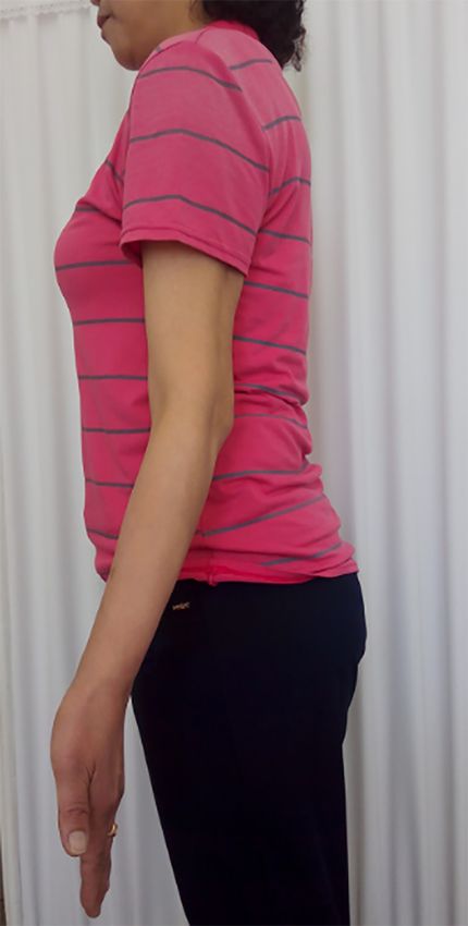
Page 2 of 7 Digestive Medicine Research, 2020
A B Case presentation
A 35-year-old, right-hand–dominant woman fell from a
height sustaining an intra-articular, comminuted, fracture
of the left proximal ulna (Figure 1). As shown in Figure 1,
direct forces generated comminution (fragmentation) of
the central portion of the proximal part of ulna including
olecranon articular surface and, also avulsions of the
coronoid process including the extension of the ulna shaft.
The triceps brachii inserted into the posterior third of the
olecranon and the proximal ulna separated the olecranon,
the brachialis inserted into the coronoid process of the
ulna produces tensile forces across the elbow joint during
contraction, so as to separate the coronoid, thus resulting in
complex fracture of proximal ulna and severe dysfunction of
the elbow.
There was no abnormal findings in her medical, family
Figure 1 Preoperative X-ray view of the patient sustaining an
and psycho-social history including relevant genetic
intra-articular, comminuted, fracture of the left proximal ulna.
information.
Olecranon, coronoid process and the shaft of ulna are separated. (A)
According to the AO principle of articular fracture
Lateral view. (B) Anterioposterior view.
management, ORIF using anatomical locking plate
was performed, one day after the injury, without any
complication.
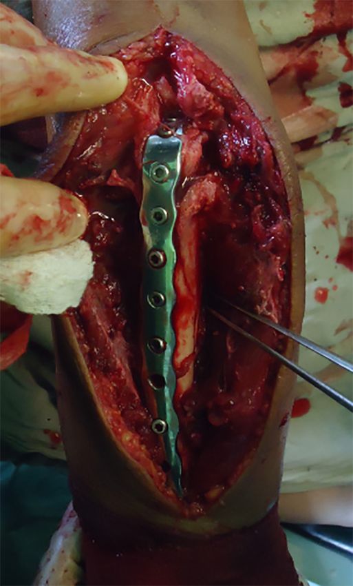
Surgical technique
A direct reduction was performed using hooks, pointed
reduction forceps, and K-wires. With exposure of the
major fragments under direct vision, reduction was
achieved and maintained with temporary pin fixation. The
fracture reduction was visualized on each of the sigmoid
notch and dorsally. Visualization of the coronoid could be
made through the fracture site of the separated olecranon
fragments before reduction.
After reduction and temporary fixation, a posterior
locking plate 3.5 which was contoured to the anatomical
shape of proximal ulna was applied for this comminuted
fractures.
Coronoid process was fixed with 3 screws and subsequent
bicortical screws were placed in a locking plate (Figure 2).
Postoperative management
No external device like a dorsal splint was applied. The
patient was started on an early rehabilitation program. The
patient was allowed to use her elbow as tolerated. Active
assisted exercises are started the day after, including gravity
Figure 2 Intraoperative view (competition of internal fixation).
assisted elbow flexion with the patient lying supine.
© Digestive Medicine Research. All rights reserved. Dig Med Res 2020;3:9 | http://dx.doi.org/10.21037/dmr.2020.01.01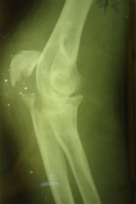
Digestive Medicine Research, 2020 Page 3 of 7
that was resistant to formal therapy.
At 16 weeks after surgery, his elbow flexion arc was
not returned to functional range yet, ROM was 45° to
100°. Physical examination showed a loss of about 45° of
extension, and about 50° of flexion compared with the
uninjured side. Forearm rotation was maintained.
Radiographs taken at 6 months showed a union of
proximal ulna and also profound heterotopic ossification
posteriorly within the triceps muscle just over the olecranon
and posterior to olecranon fossa within the triceps muscle
(Figure 3).
Revision surgery & final results
Six months after the initial procedure we performed the
second surgery, which included removal of plate and screws
and excision of heterotopic ossification at the same time.
Intraoperatively we could find the 2 cm long bony mass
surrounding the first and second screw hole of plate on the
Figure 3 Occurence of heterotopic ossification in the triceps
olecranon. Intraoperatively, we confirmed a full passive
muscle.
range of movement under anaesthesia (Figure 4).
Postoperative radiographs confirmed that the entire mass
has been excised. 75 mg indomethacin was prescribed daily
A B
for 6 weeks after the second surgery. The patient started
a regimen of active assisted elbow movements from the
second postoperative day.
She was pain-free and obtained functional range of
motion 3 weeks after the second procedure.
Two months after revision surgery, the patient was
asymptomatic and had regained a range of elbow motion
from 20° to 130°. The functional assessment revealed
possibilities for global nutrition (hand-mouth), hygiene
(hand-face) and grooming (hand-neck). On the basis of
plain radiographic findings, we didn’t find any sign of
recurrent ossification after an 18-month follow-up period.
The evaluation of 24-month follow-up period showed relief
of pain and maintenance of functional range of motion
(Figure 5).
Figure 4 X-ray view of the elbow after removal of HO and At the final follow-up, there was no adverse and
implants. (A) Lateral radiograph view of the extended elbow. (B) unanticipated events.
Lateral radiograph view of the flexed elbow. Injury on the 15th Dec. 2015.
ORIF and then early rehabilitation program.
Postop.8th week—union, but not enough elbow
At approximately 8 weeks the evidence of union was ROM.
revealed on plain radiograph, but she didn`t recover her Postop.8th week—ROM 45° to 100°.
functional range of elbow motion, although professional Postop.6th month—union of proximal ulna and also
physical therapy was applied. profound heterotopic ossification posteriorly within
She subsequently developed progressive elbow stiffness the triceps muscle just over the olecranon.
© Digestive Medicine Research. All rights reserved. Dig Med Res 2020;3:9 | http://dx.doi.org/10.21037/dmr.2020.01.01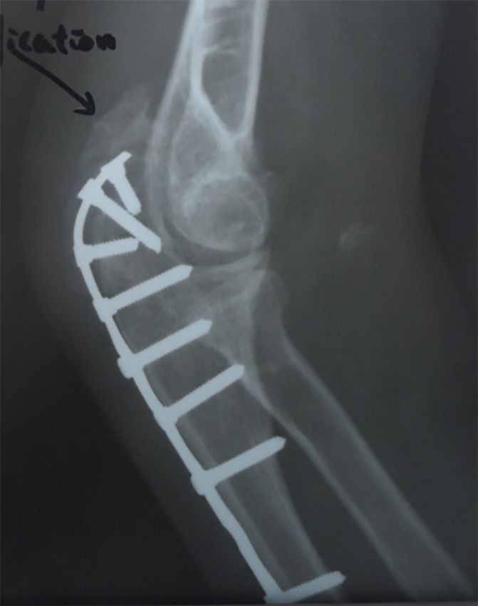
Page 4 of 7 Digestive Medicine Research, 2020
A B The majority of patients recover a functional range of
motion, frequently with small losses of extension, usually
with no associated disability.
Previous studies of proximal ulna comminuted fracture,
however, to our knowledge, have reported no cases of HO
as a complication, despite the potential development of
ectopic bone in the elbow.
Heterotopic ossification (HO), which is first described
by Patin in 1962, is the formation of mature lamellar bone
in nonosseous tissue. It is also termed heterotopic bone
or ectopic osteogenesis (ossification and calcification).
The most common cause of HO is trauma such as
musculoskeletal injury, surgical trauma, or warfare injuries.
When it forms outside the joint capsule and periosteum, it
causes pain, swelling and is usually associated with limited
range of motion. that is why, this is an important problem
throughout orthopaedic surgery (14-24).
Heterotopic ossification may follow intracranial damage
and is made more likely by delayed fixation and by passive
stretching of the elbow (25).
Figure 5 Recovered functional arc of the elbow. (A) Extension It was reported that HO developed in approximately
loss is approximately 20 degree. (B) Flexion is approximately 3% of patients with a local injury of the elbow (26-29), and
130 degree. Elbow HO occurs in 20% of patients with Traumatic Brain
Injury and forearm fractures (27,30-32).
Otherwise, Robert W. Wysocki (33) reported a case
Second procedure which included removal of of triceps muscle ectopic ossification and resultant elbow
implants and excision of heterotopic ossification. stiffness after application of recombinant human bone
Indomethacin administration. morphogenetic protein to a distal humerus nonunion site.
Postop.8th month (2 months after revision surgery)— This case was directly related to the use of OP-1.
elbow ROM 20° to 130°. Most investigators agreed that the most frequent cause of
Postop.18th month—not any sign of recurrent heterotopic bone about the elbow is direct trauma, however,
ossification. HO after ORIF of proximal ulna has not been reported yet,
Postop.24th month—relief of pain and maintenance to our knowledge.
of functional ROM, no adverse and unanticipated So now we are reporting a case of HO affecting the
events. elbow motion after ORIF of proximal ulna comminuted
fracture.
Discussion According to Hastings and Graham (27), this case seems
class IIA.
The functional outcome following olecranon fracture In the present case we have experienced a complication of
fixation is generally good or excellent whatever method of massive HO within the triceps directly over the olecranon
fixation is used. The main complication following internal where anatomical locking plate was lied, leading to marked
fixation of olecranon fractures is related to irritation caused loss of elbow motion. We believe that anatomical locking
by hardware. plate fixation is much superior over other techniques, but
Loss of motion is commonly encountered with patients this unpredicted complication which was directly related to
typically losing 10°–15° of extension. The loss of elbow the operation developed.
motion is worse in cases associated with fractures of the The internal fixation in the present case has been rigid
radial head, capitellum, coronoid or Monteggia fracture- enough to ensure stability of fracture site, but we guess that
dislocations (12,13). the microtrauma caused by friction between the plate and
© Digestive Medicine Research. All rights reserved. Dig Med Res 2020;3:9 | http://dx.doi.org/10.21037/dmr.2020.01.01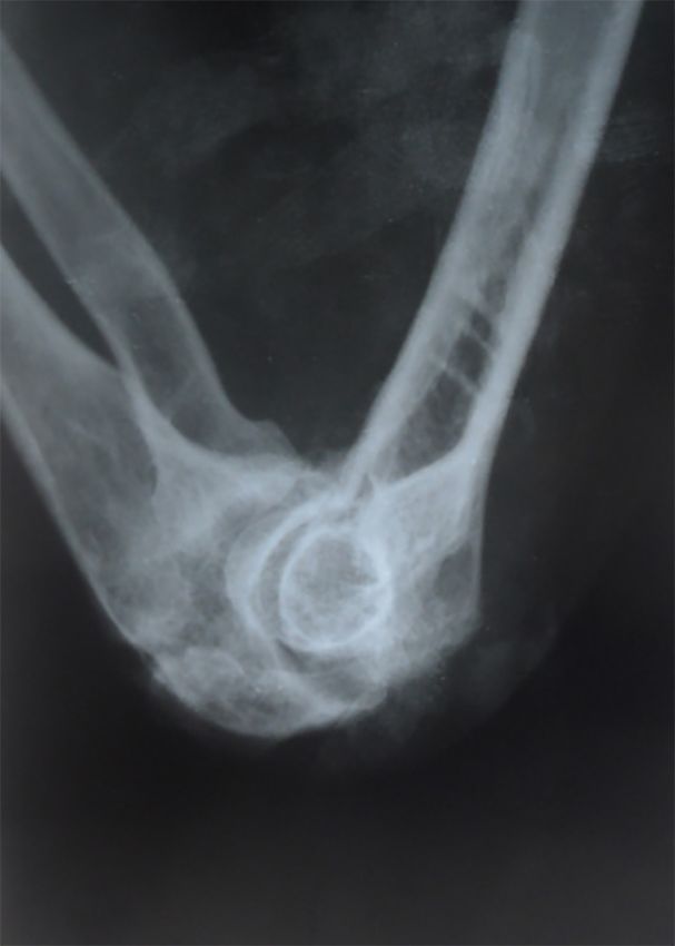
Digestive Medicine Research, 2020                                                                                         Page 5 of 7

triceps may have been the main factor. After operation we           They suggest that patients usually regain a full active
allowed the patient to use her elbow as tolerated, and then      range of motion within a few days of excision of the bony
formal physical therapy which was thought to aggravate the       mass (21,24). In this case, the result of surgical excision was
injury caused by incision and muscular dissection.               similar to the previous data.
    This is supported by the pattern and extent of the ectopic      The patient was pain-free and obtained functional range
bone that formed in this case.                                   of motion 3 weeks after the second procedure.
    Furthermore, we’ve not taken any measure to prevent             Although this is a very rare case, we recommend that
the HO, which could be also a cause of this unique case, to      postoperative therapy should be performed with caution in
our knowledge.                                                   the elbow where formation of excess bone restrict motion.
    Careful handling of tissue during any surgical procedure,    Unless the recovery of functional ROM is achieved as
especially around the hip and elbow, is also of importance       predicted, HO should be doubted, refraining from passively
to minimize any trauma and subsequent inflammation.              stretching the injured elbow.
But the role of proper surgical technique in the formation          In order to prevent postoperative HO, intraoperatively,
of heterotopic bone is difficult to quantitate, so various       surgical dissection should be performed as carefully as
preventive modalities of HO have been discussed in the           possible, and postoperative, management should involve all
literature (34).                                                 effective measures such as abovementioned ones.
    Different modalities used include diphosphonates,               The patient felt satisfactory about her final outcome of
etidronate, and NSAID’s such as indomethacin and                 the treatment and consent to this case report.
naproxen. radiotherapy, Noggin, a BMP inhibitor, pulsed
electromagnetic fields (PEMF), and free radical scavengers
                                                                 Acknowledgments
and N-acetylcysteine (35-39).
    But we’ve not taken any the abovementioned measures          None.
because this complication has been very rarely reported. We
thought postoperative therapy was not proper in this case,
                                                                 Footnote
either. The principal of the HO treatment is appropriate
immobilization for immature bone and physical therapy for        Conflicts of Interest: The authors have no conflicts of interest
a mature mass (38).                                              to declare.
    Just only focusing on the early motion of the injured
elbow was more likely to promote the development of HO.          Ethical Statement: The authors are accountable for all
    In the present case, we wonder if immobilization for         aspects of the work in ensuring that questions related
3–4 weeks rather than early motion would be beneficial.          to the accuracy or integrity of any part of the work are
Although early motion and manipulation has been essential        appropriately investigated and resolved. Written informed
to prevent stiffness and has been reported to improve range      consent was obtained from the patient for publication of
of elbow motion, in this case they may have predisposed to       this manuscript and any accompanying images.
hematoma formation, scarring, and heterotopic ossification.
    From this experience, we do not recommend isolated
                                                                 References
physical therapy in patients with elbow injury, rather
we recommend combined physical therapy with other                1.   Rommens PM, Kuchle R, Schneider RU, et al. Olecranon
preventive measures. Many authors agree that if ectopic               fractures in adults: factors influencing outcome. Injury
bone around the elbow is causing or contributing to a loss            2004;35:1149-57.
of functional elbow motion, an operative procedure is            2.   Mueller ME, Allgower M, Schneider R. Manual of internal
warranted to remove the offending bone and release the                fixation: techniques recommended by the AO-ASIF group,
joint capsule whether the motion limitation is partial or             3rd ed., Berlin, Germany: Springer–Verlag; 1991:1-158.
complete (ankylosis) (16,27).                                    3.   Weber BG, Vasey H. Osteosynthese bei Olecranonfraktur.
    Results of surgical excision of heterotopic ossification          Unfallmedizinische Berufskrankheiten 1963;2:90-6.
about the elbow have shown significant improvement in            4.   Anderson ML, Larson AL, Merton SM, et al. Congruent
range of motion, independence, and quality of life in most            elbow plate fixation of olecranon fractures. J Orthop
cases (40-44).                                                        Trauma 2007;21:386-93.

© Digestive Medicine Research. All rights reserved.                   Dig Med Res 2020;3:9 | http://dx.doi.org/10.21037/dmr.2020.01.01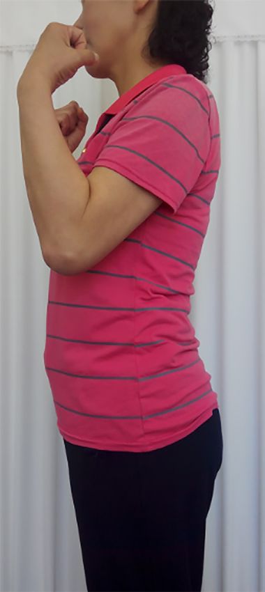
Page 6 of 7                                                                                          Digestive Medicine Research, 2020

5.    Hume MC, Wiss DA. Olecranon fractures. A clinical and                total hip arthroplasty. Risk factors and consequences. Clin
      radiographic comparison of tension band wiring and plate             Orthop Relat Res 1991;(263):49-58.
      fixation. Clin Orthop Relat Res 1992;285:229-35.               23.   Ritter MA, Vaughan RB. Ectopic ossification after total hip
6.    Lavigne G, Baratz M. Fractures of the olecranon. J Am                arthroplasty. Predisposing factors, frequency, and effect on
      Soc Surg Hand 2004;4:94-102.                                         results. J Bone Joint Surg 1977;59:345-51.
7.    Nowinski RJ, Nork SE, Segina DN, et al. Comminuted             24.   Thorseth K. A case of traumatic myositis ossificans in the
      fracture-dislocations of the elbow treated with an AO wrist          iliopsoas muscle. Acta Orthop Scand 1968;39:73-5.
      fusion plate. Clin Orthop 2000;378:238-44.                     25.   Summerfield SL, DiGiovanni C, Weiss AP. Heterotopic
8.    Wolfgang G, Burke F, Bush D, et al. Surgical treatment               ossification of the elbow. Shoulder Elbow Surg
      of displaced olecranon fractures by tension band wiring              1997;6:321-31.
      technique. Clin Orthop Relat Res 1987;224:192-204.             26.   Garland DE, Blum C, Waters R. Periarticular Heterotopic
9.    Bailey CS, MacDermid J, Patterson SD, et al. Outcome                 Ossification in Head Injured Adults. J Bone Joint Surg
      of plate fixation of olecranon fractures. J Orthop Trauma            1980;62:1143-6.
      2001;15:542-8.                                                 27.   Hastings H, Graham T. The Classification and Treatment
10.   Simpson NS, Goodman LA, Jupiter JB. Contoured LC–                    of Heterotopic Ossification about the Elbow and Forearm.
      DC plating of the proximal ulna. Injury 1996;27:411-7.               Hand Clin 1994;10:417-37.
11.   Hak DJ, Golladay GJ. Olecranon fractures: treatment            28.   Peterson SL, Mani MM, Crawford CM, et al. Postburn
      options. J Am Acad Orthop Surg 2000;8:266-75.                        Heterotopic Ossification: Insights for Management
12.   Ring D, Jupiter JB. Fracture dislocation of the elbow. J             Decision Making. J Trauma 1989;29:365-9.
      Bone Joint Surg Am 1998;80:566-80.                             29.   Thompson HC, Garcia A. Myositis Ossificans: Aftermath
13.   Newman SD, Mauffrey C, Krikler S. Olecranon fractures.               of Elbow Injuries. Clin Orthop 1967;50:129-34.
      Injury 2009;40:575-81.                                         30.   Cohen MS. Heterotopic ossification of the elbow. In:
14.   Jupiter JB, O’Driscoll SW, Cohen MS. The assessment                  Jupiter JB, ed. The stiff elbow. Rosemont: American
      and management of the stiff elbow. Instr Course Lect                 Academy of Orthopaedic Surgeons, 2006:31-40.
      2003;52:93-111.                                                31.   Garland DE, O'Hollaren R. Fractures and Dislocations
15.   Furukawa K. Pharmacological aspect of ectopic                        about the Elbow in the Head-injured Adult. Clin Orthop
      ossification in spinal ligament tissues. Pharmacol Ther              1982;168:38-41.
      2008;118:352-8.                                                32.   Garland DE, Dowling V. Forearm Fractures in the Head-
16.   Tonbul M, Ozen S, Tonbul AT. Bilateral simultaneous                  Injured Adult. Clin Orthop 1983;176:190-6.
      heterotopic ossification of the reflected head of rectus       33.   Wysocki RW, Cohen MS. Ectopic ossification of the
      femoris muscle: a case report and review of the literature.          triceps muscle after application of bone morphogenetic
      Case Rep Orthop 2014;2014:497075.                                    protein-7 to the distal humerus for recalcitrant nonunion:
17.   Zimmermann SM, Würgler-Hauri CC, Wanner GA, et al.                   a case report. J Hand Surg Am 2007;32:647-50.
      Echinomycin in the prevention of heterotopic ossification      34.   Baird EO, Kang QK. Prophylaxis of heterotopic
      - an experimental antibiotic agent shows promising results           ossification - an updated review. J Orthop Surg Res
      in a murine model. Injury 2013;44:570-5.                             2009;4:12.
18.   Chouhan DK, Dhillon M, Bachhal V, et al. Atraumatic            35.   Bek D, Beksaç B, Della Valle AG, et al. Aspirin decreases
      heterotopic ossification of iliopsoas muscle: a case report.         the prevalence and severity of heterotopic ossification after
      Orthop Surg 2012;4:197-201.                                          1-stage bilateral total hip arthroplasty for osteoarthrosis. J
19.   Onder K, Muhammed B, Saime U, et al. Post-traumatic                  Arthroplasty 2009;24:226-32.
      heterotopic ossification of the crus. A case study. Ortop      36.   Vavken P, Castellani L, Sculco TP. Prophylaxis of
      Traumatol Rehabil 2011;13:299-301.                                   heterotopic ossification of the hip: systematic review and
20.   Hsu JE, Keenan MA. Current review of heterotopic                     meta-analysis. Clin Orthop Relat Res 2009;467:3283-9.
      ossification. J Orthop 2010;20:126-30.                         37.   Pakos EE, Tsekeris PG, Paschos NK, et al. The role of
21.   McCulloch PC, Bush-Joseph CA. Massive heterotopic                    radiation dose in a combined therapeutic protocol for
      ossification complicating iliopsoas tendon lengthening: a            the prevention of heterotopic ossification after total hip
      case report. Am J Sports Med 2006;34:2022-5.                         replacement. J BUON 2010;15:74-8.
22.   Ahrengart L. Periarticular heterotopic ossification after      38.   Banovac K. The effect of etidronate on late development

© Digestive Medicine Research. All rights reserved.                        Dig Med Res 2020;3:9 | http://dx.doi.org/10.21037/dmr.2020.01.01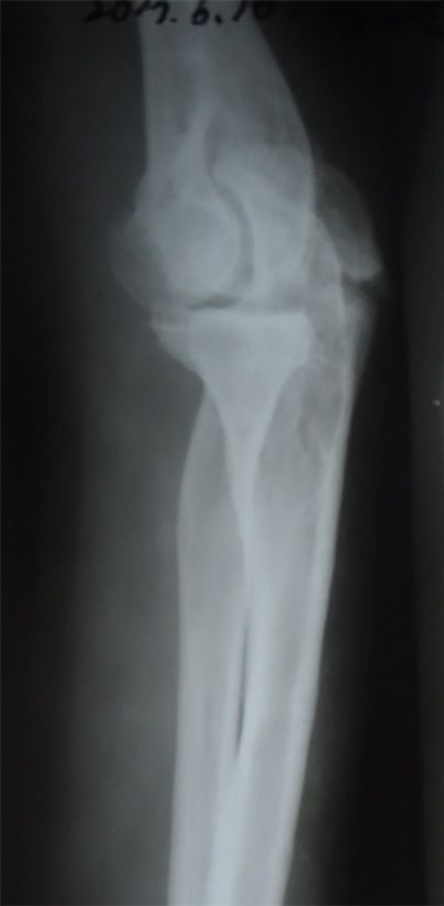
Digestive Medicine Research, 2020                                                                                            Page 7 of 7

    of heterotopic ossification after spinal cord injury. J Spinal    42. Garland DE. Surgical Approaches for Resection of
    Cord Med 2000;23:40-4.                                                Heterotopic Ossification in Traumatic Brain-injured
39. McAuliffe J. Early Excision of Heterotopic Ossification               Adults. Clin Orthop 1991;263:59-70.
    about the Elbow followed by Radiation Therapy. J Bone             43. Garland DE, Hanscom DA, Keenan MA, et al. Resection
    Joint Surg 1997;79:749-55.                                            of Heterotopic Ossification in the Adult with Head
40. Djurickovic S, Meek RN, Snelling CF, et al. Range of                  Trauma. J Bone Joint Surg Am 1985;67:1261-9.
    Motion and Complications after Postburn Heterotopic               44. Kolessar DJ, Katz SD, Keenan ME. Functional Outcome
    Bone Excision about the Elbow. J Trauma 1996;41:825-30.               Following Surgical Resection of Heterotopic Ossification
41. Garland DE. A Clinical Perspective on Common Forms                    in Patients with Brain Injury. J Head Trauma Rehabil
    of Acquired Heterotopic Ossification. Clin Orthop                     1996;44:4.
    1991;263:13-29.

 doi: 10.21037/dmr.2020.01.01
 Cite this article as: Mun CS, So TS, Jang CH. A rare cause of
 elbow stiffness after internal fixation of proximal ulna fracture:
 a case report of heterotopic ossification. Dig Med Res 2020;3:9.

© Digestive Medicine Research. All rights reserved.                      Dig Med Res 2020;3:9 | http://dx.doi.org/10.21037/dmr.2020.01.01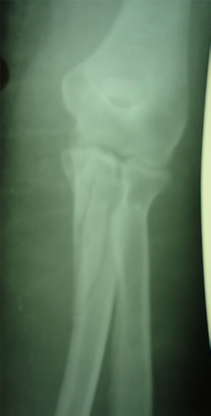
You can also read