Pediatric Supracondylar Humerus Fractures - OCTOBER 2020 Caroline Tougas, MD Children's Mercy Hospital, Kansas City Clinical Assistant Professor ...
←
→
Page content transcription
If your browser does not render page correctly, please read the page content below
Pediatric Supracondylar
  Humerus Fractures

                  Caroline Tougas, MD
         Children's Mercy Hospital, Kansas City
Clinical Assistant Professor - UMKC School of Medicine

                   OCTOBER 2020

                                                         Core Curriculum V5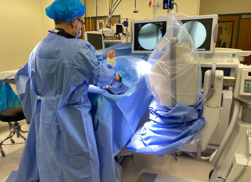
Disclaimer
• All clinical and radiographic images provided are used with
  permission of Caroline Tougas, MD unless otherwise specified.

                                                              Core Curriculum V5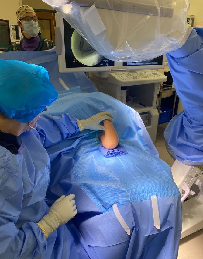
OBJECTIVES
By the end of this presentation, learners will be better able to:
• Recognize the signs and symptoms of more severe pediatric supracondylar humerus
  fractures (SCHF)
• Assess the degree of displacement of pediatric SCHF on radiographs
• Determine the type of fracture according to the modified Gartland classification
• Prescribe appropriate treatment for SCHF based on fracture characteristics
• Describe the technique of closed reduction and percutaneous pinning of pediatric
  SCHF
• Recognize SCHF that may require more complex care and manage them appropriately

                                                                    Core Curriculum V5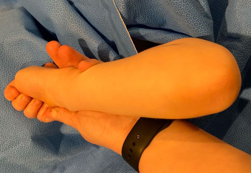
PEDIATRIC SUPRACONDYLAR HUMERUS
FRACTURES (SCHF)
                                                           What goes up...
• Most common elbow fracture in children
• Most commonly occurs in 5-7yo children
• Most common mechanism of injury is from a
  low energy fall
  • FOOSH for extension types (common)
     • Monkeybars, trampolines, cartwheels, etc
  • Fall on flexed elbow for flexion types (uncommon)

                                                        Core Curriculum V5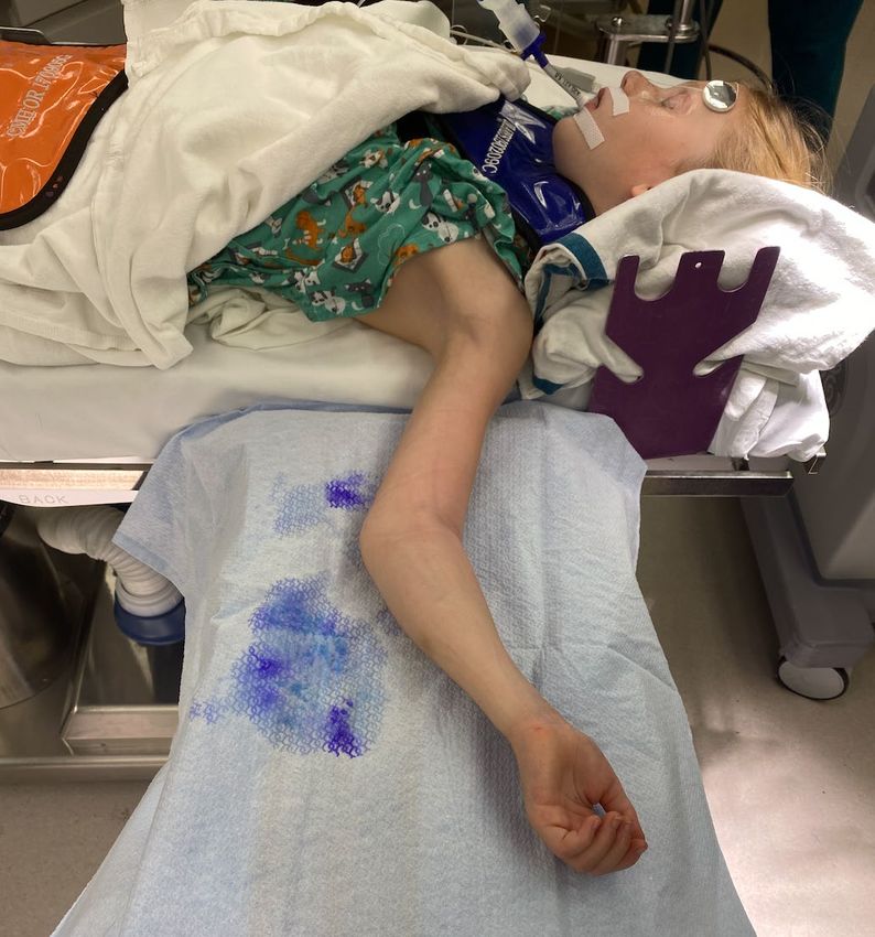
80% of
                                                                  longitudinal

PEDIATRIC SCHF                                                    growth of the
                                                                  arm

• Most common surgical pediatric fracture
   • Frequently require surgical treatment to avoid
     complications due to:
      • Limited contribution of growth of distal humerus =
        limited remodeling potential

   • Displaced SCHF are unstable and require
     reduction and stabilization to heal in                         only 20%
     appropriate alignment

                                                             Core Curriculum V5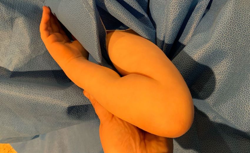
PHYSICAL EXAM
• Pain
• Refusal/inability to move the elbow
• Deformity proportional to displacement
• Swelling & bruising
• Skin integrity
   • Tenting/compromise
   • Open fractures

                                           Core Curriculum V5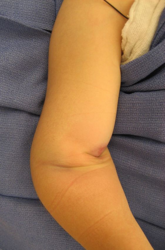
PHYSICAL EXAM
• Brachialis sign:
   • Antecubital ecchymosis
   • Skin puckering
   • Subcutaneous bone fragment (soft-tissue
    interposition)
   • Indicator of:
       • Significant injury and swelling
       • Potential failure of closed reduction
     *Will require milking maneuver (discussed
     later)
                                                 Courtesy of Mark Sinclair, MD
                                                                   Core Curriculum V5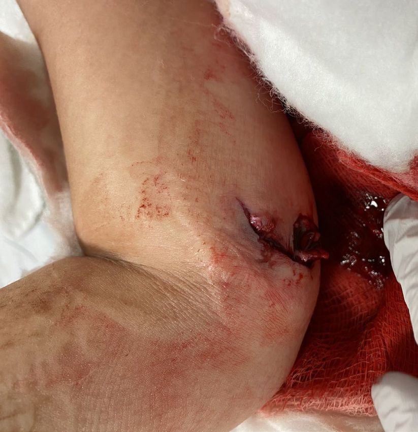
NEUROVASCULAR EXAM
                                                Neurovascular structures around the elbow

• Relatively high rate of neurovascular
  injuries due to intimate relationship of
  nerves and artery to displaced fracture
  fragments
• Neurologic exam can be challening in
  injured child but important to document
  pre-manipulation exam
• Pulseless hand may still be perfused
  because of excellent collateral circulation
  in pediatric elbow
                                                           Rockwood and Green, Fig 33-7
                                                                         Core Curriculum V5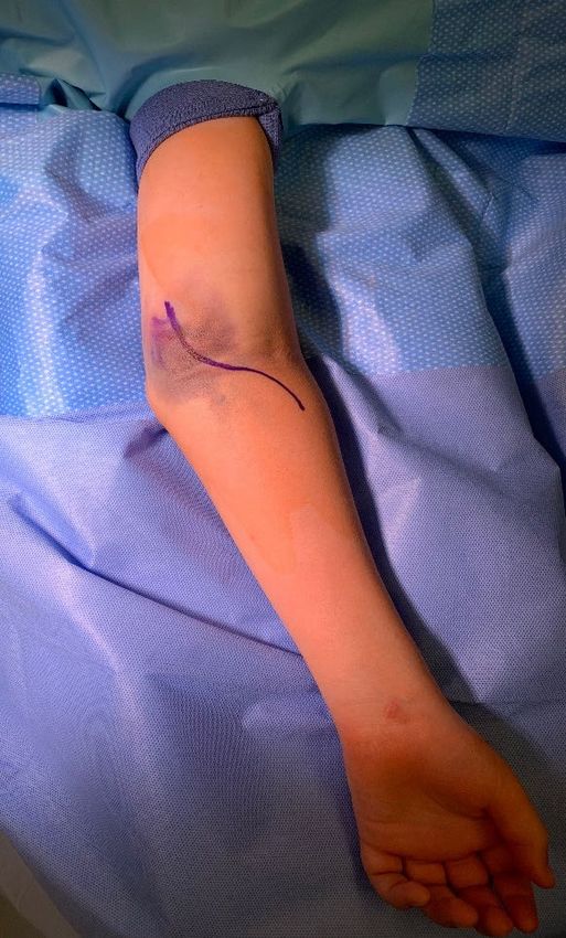
VASCULAR INJURY
• Occurs in 0.5-5%
• Vascular status
   • Assess pulse (palpation or doppler)
   • Assess perfusion
      • Capillary refill (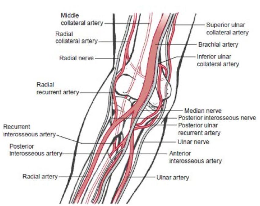
VASCULAR STATUS
• 3 categories:
    Pulse present, perfused hand
    Pulse absent, perfused hand
    Pulse absent, nonperfused hand

                               Absent pulse
                                                              Artery draped
                                                              over humerus

              Fingers pink & warm
              Brisk capillary refill
                                              Courtesy of Micah Sinclair, MD

                                                                               Core Curriculum V5
NEUROLOGIC EXAM
• What to assess:
• Median nerve: sensation pulp of index finger
• Anterior interosseus nerve: flexion IP thumb and DIP index
• Radial nerve: sensation dorsum of thumb
• Posterior interosseus nerve: extension IP thumb
• Don't be fooled by intrinsics (extension finger IPs)
• Ulnar nerves: finger abduction/adduction
BEDSIDE TEST (many options):
Thumbs up (PIN) - Cross Fingers (Ulnar N) - AOK (AIN)
Core Curriculum V5NEUROLOGIC INJURY
• Occurs almost exclusively in Type 3 or Flexion Types
• RISK FACTOR:
• Median N/AIN: posterolateral
displacement
• Radial N:
posteromedial displacement
• Ulnar N:
flexion types
Babal JC, et al. JPO 2010 Core Curriculum V5OSTEOLOGY
• Distal humerus composed of medial
and lateral columns connected by the Medial Column Lateral Column
articular segment
Typical
Fracture Line
• Displaced fractures inherently unstable
• Medial/lateral columns displace easily Articular
Segment
Tornetta P, Ricci WM, eds. Rockwood and Green's Fractures in Adults,
9e. Philadelphia, PA. Wolters Kluwer Health, Inc; 2019. Figure 33-7
Core Curriculum V5OSTEOLOGY • Medial and lateral columns
connected by a thin wafer of bone
through olecranon fossa
• Point of weakness, prone to fracture
• Muscles lose mechanical advantage
when elbow extended past neutral
(hyperextension common in children)
2-3 mm wide Olecranon Fossa • Olecranon acts as a fulcrum
• Capsule transmits an extension force to
distal humerus just proximal to the
Articular Surface physis
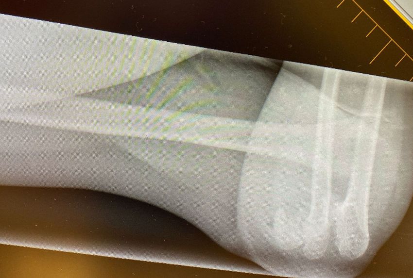
Core Curriculum V5IMAGING
• XR usually sufficient
• AP + LAT of elbow
• Ipsilateral forearm/wrist Fat pad sign
• Look for posterior fat pad
sign in non displaced
fractures (arrow)
• Advanced imaging rarely Fat pad sign
indicated (intra-articular
variant)
Type 3 Type 1 - fat
pad sign
Core Curriculum V5AHL
IMAGING
Posterior
Distal humerus alignment (true lateral): fat pad
LCHA
• Anterior humeral line (AHL): should
intersect capitellar ossific nucleus
• Anterior tilt of capitellum (30-40º)
• Lateral capitellohumeral angle
(LCHA)IMAGING
Distal humerus alignment (AP):
• Baumann's Angle: formed by a line
perpendicular to the axis of the
humerus, and a line that goes through
the physis of the capitellum
• Wide range of normal for this value
(9-26 deg)
• Best judge of normal is to obtain
contralateral comparison views
Core Curriculum V5CLASSIFICATION 95-98%
FOOSH
• Two Major Fracture Types:
• Extension:
• Gartland Classification (1959)
• Wilkins Modification (1991)
2-5%
• Flexion: Considered seperately Direct blow to
flexed elbow
Core Curriculum V5GARTLAND CLASSIFICATION
• Fracture Type: Characteristic
Type 1
• Type 1: Nondisplaced
• Type 2:
• Angulation
Type 2
• Posterior hinge intact
• Type 3:
• Complete displacement
• Loss of posterior hinge
Type 3
Core Curriculum V5GARTLAND CLASSIFICATION
• Type 1: Nondisplaced
• Fat pad sign +
• No angulation
• +/- Impaction
• Treat with immobilization
• Long-arm cast (LAC)
• 3-4weeks
Core Curriculum V5GARTLAND CLASSIFICATION
• Type 2:
Anterior Humeral Line
• Sagittal angulation
• Posterior hinge intact
• If anterior humeral line (AHL) does
not intersect at least anterior 1/3rd
of capitellum can require CR +/- PP
LAC ok Needs CR
Core Curriculum V5MODIFIED GARTLAND CLASSIFICATION
• Type 2A: Sagittal angulation only
TYPE 2A: TYPE 2B:
No coronal Sagittal angulation • Amenable to CR + LAC
deformity Lateral comminution • Requires close follow-up
Valgus
• Type 2B: + rotation, coronal
angulation (varus, valgus),
translation +/- comminution or
impaction present
• Higher rate of failure with CR without
percutaneous pinning
• Recomend CRPP
Core Curriculum V5MODIFIED GARTLAND CLASSIFICATION
• Type 3:
• Complete posterior displacement
• Loss of posterior hinge
• Maintains periosteal sleeve
• Type 4:
• Instability in extension and flexion
• Disruption of periosteal sleeve
• Type 3 vs. 4 based on fluoroscopic
examination with patient under anesthesia
--> intraoperative distinction Core Curriculum V5FLEXION TYPES
• Generally more unstable
• Higher complication rates
• Association with ulnar nerve palsy
• TREATMENT:
• Any displacement --> CRPP
• Higher rate of ORPP than extension
types
Core Curriculum V5IPSILATERAL FRACTURES
• Radius and/or Ulna (shaft or distal)
• “Floating Elbow”
• Occurs in 5% of Type 3s
• Can be missed by distracting SCHF
• Rate of complications proportional to severity of
injury Floating Elbow
• Compartment syndrome rate 2%
• Consider urgent fixation for higher energy injuries
• Consider distal fixation if closed reduction required
• Difficult to hold reduction in LAC with swelling
Baghdadi et al. JPO 2020 Lucas DE, et al. JOT 2013 Core Curriculum V5MANAGEMENT • AAOS adopted appropriate use criteria (AUC) for the management of: • Pediatric supracondylar humerus fractures (2014) • Pediatric supracondylar humerus fractures with vascular injury (2015) • Can be referenced in the treatment of a pediatric supracondylar humerus fracture. Appropriate Use Criteria: Management of Pediatric Supracondylar Humerus Fractures. Journal of the American Academy of Orthopaedic Surgeons, 2015. 23(10): p. e52-e55 Core Curriculum V5
MANAGEMENT

         swelling

                    Core Curriculum V5
NON OPERATIVE CONSIDERATIONS
• Avoid casting > 90 deg in swollen elbows
• Consider splitting cast 1 week
• Close follow-up
• Especially for Type 2s
• Especially if CR performed
• Up to 48% rate of loss of reduction
• Risk factors for displacement:
• Greater initial displacement 3 weeks
• Type 2B
• Large arm (circumference)
Lucas DE, et al. JOT 2013 Fitzgibbons, et al. JPO 2011 Camus T, et al. JPO 2011 Core Curriculum V5TIMING OF OPERATIVE TREATMENT
• Dependent on:
• Fracture pattern and displacement
• Distal vascular status and limb perfusion
• Neurologic function distal to the fracture
• Soft tissue swelling
• Associated fractures
• Access to OR
• Type 2s can safely be treated as outpatients in delayed manner
• Type 3s should be admitted for monitoring if surgery is delayed
Core Curriculum V5TIMING OF OPERATIVE TREATMENT
• Closed Type 3 SCHF with normal neurovascular exam can be treated
safely in a delayed fashion
• No difference in rates of:
• Conversion to open reduction
• Compartment syndrome
*over 21 hours in some studies
• Iatrogenic nerve injury
• Vascular complications
• Fractures with distal neurologic deficits are more controversial
• May indicate more significant injury with increased risk of complications with
delayed surgery
Ramachandran, et al. JBJS Br 2008 Bales et al. JPO 2010 Core Curriculum V5TIMING OF OPERATIVE TREATMENT
• Emergent (immediately limb- or life- threatening)
• NONPERFUSED limb
• Urgent
• Open fractures
• Skin puckering/compromise
• Ipsilateral forearm/wrist fractures
• Significant displacement and/or swelling
• Neurologic injury?
• Pulseless but perfused hand?
Core Curriculum V5CLOSED REDUCTION AND PERCUTANEOUS
PINNING SCHF
• https://otaonline.org/video-library/45036/procedures-and-
techniques/multimedia/17165284/closed-reduction-percutaneous-
pinning-of-a
• TECHNIQUE VIDEO with case example
Core Curriculum V5OR SETUP
• Armboard vs C-arm as table
• Ability to swing through for lateral in very unstable reductions
• +/- Invert C-Arm
• Increases radiation doses
• Place lead over patient
• Secure head
• Tape forehead
• Tube tree
Core Curriculum V5CLOSED REDUCTION
• Longitudinal traction to reestablish length
+/- milking maneuver
• Rotation correction
• Coronal plane correction
• Translation
• Varus/valgus
• Sagittal plane correction
• Anterior translation and hyperflexion of distal segment with
pressure on olecranon
• Forearm position
• Hyperpronation vs Supination
Core Curriculum V5CLOSED REDUCTION
• Brachialis sign --> Milking maneuver
pre post
Courtesy of Mark Sinclair, MD
Archibeck. J Pediatr Orthop. 1997 Core Curriculum V5CLOSED REDUCTION
• Rule of Thumb:
• Thumb points in direction of initial
displacement of distal segment
• Posteromedial
• Pronation tightens medial soft-
tissue sleeve
• Posterolateral
• Supination tightens lateral soft-
tissue sleeve
Core Curriculum V5DIFFICULT CLOSED REDUCTION
ex: Type 4 and Flexion Types
• Swing through laterals to avoid rotating
through elbow
*advantage of using arm board
• Bump underneath the proximal fragment
• Lessen elbow flexion and/or apply posteriorly
directed force to distal segment through
forearm
• May use joystick pins in the distal fragment to
help control and manipulate it
Core Curriculum V5ACCEPTABLE ALIGNMENT
• Anterior humeral line intersects capitellum
• No significant gapping (suggestive of soft-tissue
interposition)
• No clear parameters otherwise:
• Avoid varus (increased Baumann's angle)
• Mild rotational deformity acceptable
• Slight valgus or translation better tolerated
• Upper limit of acceptable undefined
Core Curriculum V5OPEN REDUCTION
• Variable rates in literature: 1-10%
• Indications:
• Unable to achieve acceptable alignment
• Association with posterolateral displacement
• Flexion types
• Open fracture
• Vascular exploration required
Core Curriculum V5OPEN REDUCTION
• Choice of approach: follow metaphyseal spike
• Anterior: posterior displacement or vascular injury
and/or median nerve injury
• Medial: Posterolateral displacement or flexion type
injuries
• Lateral: Posteromedial displacement
• Posterior: Generally avoided; poorer outcomes Medial
(stiffness, AVN, cosmesis) approach
• Avoid compromised tissues
• Avoid further disruption of soft-tissues
Core Curriculum V5MANAGEMENT OF VASCULAR INJURIES
Core Curriculum V5VASCULAR INJURY
• 1266 consecutive operatively treated supracondylar humerus
fractures over 5 years (Texas Scottish Rite)
• 54 (4%) lacked a palpable radial pulse on admission
• All Type 3s
• 5 (0.4%) were ischemic and underwent direct vascular repair
• 29/54 regained their radial pulse after CRPP of the fracture
• 20 were still pulseless after CRPP, but had perfused hands
• 1/20 became ischemic and required vascular repair
Weller A et al. J Bone Joint Surg Am 2013 Core Curriculum V5VASCULAR EXPLORATION
• Indications:
• Persistent nonperfused hand after
adequate CRPP
• Loss of pulse after fracture reduction
• Perfused pulseless associated with
median nerve injury management
controversial
• To explore or not to explore?
• Anterior approach preferred
• Consider UE vascular surgeon
Courtesy of John Anderson, MD
consultation early
Core Curriculum V5NEUROVASCULAR REPAIR
Brachial Artery Repair
Median Nerve
Laceration
Courtesy of Micah Sinclair, MD
Core Curriculum V5PIN CONFIGURATION OPTIONS
• Laterally based Laterally
• **MOST COMMON technique Based
• 2 vs 3 are lateral pins
• Cross-pinning
• Medial and Lateral
• Ulnar nerve at risk Ulnar N
at risk
• All-Lateral
• Radial nerve at risk
• Less commonly used
• Antegrade ESIN technique also
described
• High SCH fx
Shenoy et al. Cureus 2020 Core Curriculum V5PIN CONFIGURATION OPTIONS
Laterally
• Laterally based Based
• **MOST COMMON technique
• 2 vs 3 laterally-based pins
NB: Antegrade ESIN technique also described
but less common
• High SCH fx
Shenoy et al. Cureus 2020 Core Curriculum V5PIN CONFIGURATION OPTIONS...
OPTIONS
Cross-Pinning Cross-Pinning
• Cross-pinning (medial AND Antegrade
(All Lateral)
ESIN
• Medial and Lateral lateral)
• Ulnar nerve at risk
• All-Lateral
• Radial nerve at risk
Ulnar N
at risk
Shenoy et al. Cureus 2020 Core Curriculum V5PIN CONFIGURATION
• Cross-pinning most stable biomechanically
• No clear CLINICAL advantage to cross pinning over
lateral pinning for most Type 3 fractures with
• Greater risk of iatrogenic ulnar nerve injury (4.3X)
• Indications for medial pin:
• Medial comminution
• Proximal medial to distal lateral oblique fracture
pattern (reverse oblique)
• Intra-articular variants
Woratanarat P, et al. JOT 2012 Core Curriculum V5PIN CONFIGURATION
Medial Pin Technique:
• Fix with 2 lateral pins
• Extend elbow 45deg to relax ulnar nerve
• Beware of ulnar nerve subluxation
• 16% of children (Zaltz 1996)
• Thumb pressure or small incision to
protect ulnar nerve as pins inserted
Silva M, Knutsen AR, Kalma JJ, et al. Biomechanical Testing of Pin Configurations in If iatrogenic nerve palsy postop,
Supracondylar Humeral Fractures: The Effect of Medial Column Comminution. Journal
of Orthopaedic Trauma: May 2013 - Volume 27 - Issue 5 - p 275-280
controversy re: leave or remove
pin
Core Curriculum V51.6mm k wires
PERCUTANEOUS PINNING
• IDEALLY:
• 1.6-2mm k-wires
• Engage lateral and medial columns
• Divergent
*Greater pin spread = Greater stability
Type 2A: Type 3:
2 pins 3 pins
Type 2B: 2-3 pins
Core Curriculum V5PIN CONSTRUCT
• Wide spread at fracture site
• Control lateral column with pin
along metaphyseal flare
• Control medial column with
laterally based pin
• Engage distal humerus just above
fracture site
• A 3rd pin can be added between
these two for additional
stability
Core Curriculum V5FLUOROSCOPY
extension stress view
• After stable reduction and pinning:
• Review AP alignment with elbow extended
• Obtain true lateral view to assess alignment
• Oblique views to assess reduction of medial and lateral
columns
• Consider stress views under fluoroscopy to
assess stability of contruct/reduction
*Especially if considering limited follow-up
• On AP: rotational stress, varus/valgus stress
• On LAT: flexion/extension arc
Bauer JM, et al. JPO 2019
Core Curriculum V5POSTOPERATIVE CARE
• Type 2: Outpatient
• Type 3: Monitoring for 12-24h
• NV exams
• Compartment checks
• Split cast or splint
• Especially if acute or early discharge
• Pain control:
• Ibuprofen + Acetaminophen often sufficient
• Narcotics may not be necessary
Nelson SE, et al. JBJS 2019 Core Curriculum V5POSTOPERATIVE ANALGESIA
• n = 81 Type 2 & 3 SCHF --> CRPP
• Pain levels decreased to clinically unimportant
levels by POD 3
• Rx of 7 opioid doses postop should be sufficient
• Pain scores >6 after d/c are outliers and should
be screened for compartment syndrome or
ischemia
Nelson SE, et al. JBJS 2019 Core Curriculum V5FOLLOW UP
• Pin removal generally at 3-4 weeks
• Frequency of follow-up variable per surgeon and/or fracture type
• PT/ROM exercises generally not required
• Post-pin removal radiographs may not provide clinical utility in the
absence of other clinical findings.
Core Curriculum V5COMPLICATIONS
• Pin site infections
• Loss of fixation, pin migration
• Malunion
• Cubitus varus - thought to be only
esthetic, however may contribute to
loss of motion and posterolateral
rotatory instability
• Nonunion: very rare
Courtesy of Mark Sinclair, MD
• Stiffness: uncommon long term
O'Driscoll SW, et al. JBJS Am 2001 Ho, CA. JPO 2017
Core Curriculum V5COMPLICATIONS Perineural fibrosis
• Nerve injury
• Traumatic
• Mostly neuropraxias with full recovery
• Nerve transection is rare
• Prolonged deficit (>6 months) may be due to
perineural fibrosis (neurolysis helpful)
• Iatrogenic from pin placement or entrapment in
fracture during reduction
• Vascular injury
• Compartment syndrome (rare)
• Increased risk with “floating elbow”
• Can lead to Volkmann ischemic contracture Courtesy of Micah Sinclair, MD
Core Curriculum V5SUMMARY - SCHF
• Very common pediatric elbow injury
• Careful pre-operative neurovascular exam is essential
• Don't miss ipsilateral fractures (the “floating elbow”)
• Closed reduction and casting possible for Type 2A fractures
• Close follow-up for some nonoperatively treated fractures
• Surgical timing only emergent if vascular compromise
• Surgical treatment generally some variation of CRPP
• Variation in the approach to managing pediatric SCHF
Core Curriculum V5REFERENCES
• Appropriate Use Criteria: Management of Pediatric Supracondylar Humerus Fractures. Journal of the American Academy of Orthopaedic
Surgeons, 2015. 23(10): p. e52-e55
• Alton TB, Werner SE, Gee AO. Classifications in brief : the Gartland classification of supracondylar humerus fractures. Clin Orthop Relat Res.
2015;473:738–741.
• Archibeck MJ, Scott SM, Peters CL. Brachialis muscle entrapment in displaced supracondylar humerus fractures: a technique of closed reduction
and report of initial results. J Pediatr Orthop. 1997 Apr.;17(3):298–302.
• Baghdadi S;CORTICES. Pediatric floating elbow injuries are not as problematic as they were once thought to be: A systematic review. J Petri
Orthop 2020;40(8):380-389.
• Bales JG, Spencer HT, Wong MA, et al. The effects of surgical delay on the outcome of pediatric supracondylar humeral fractures. J Pediatr
Orthop. 2010;30:785–791)
• Bauer JM, Stutz CM, Schoenecker JG, Lovejoy SA, Mencio GA, Martus JE. Internal rotation stress testing improves radiographic outcomes of
Type 3 supracondylar humerus fractures. J Pediatr Orthop 2019;39(1):8-13.
• Camus T, MacLellan B, Cook PC, Leahey JL, Hyndman JC, El-Hawary R. Extension type II pediatric supracondylar humerus fractures: a
radiographic outcomes study of closed reduction and cast immobilization. J Pediatr Orthop 2011;31(4):366-371.
• Edmonds EW, Roocroft JH, Mubarak SJ. Treatment of displaced pediatric supracondylar humerus fracture patterns requiring medial fixation: a
reliable and safer cross-pinning technique. J Pediatr Orthop. 2012;32: 346–351
• Fitzgibbons PG, Bruce B, Got C, Reinert S, Solga P, Katarincic J, et al. Predictors of failure of nonoperative treatment for type-2 supracondylar
humerus fractures. J Pediatr Orthop 2011;31(4):372–376.
• Frick SL, Mehlman CT. The community orthopaedic surgeon taking trauma call: pediatric supracondylar humeral fracture pearls and pitfalls. J
Orthop Trauma 2017;31:S11–S15
• Gartland. Management of supracondylar fractures of the humerus in children. Surg Gynecol Obstet. 1959;109:145-54.
Core Curriculum V5REFERENCES • Ho, CA. Cubitus Varus - It's more than just a crooked arm! J Pediatr Orthop 2017;37(Suppl 2):S37-S41 • Karalius VP, Stanfield J, Ashley P, et al: The utility of routine postoperative radiographs after pinning of pediatric supracondylar humerus fractures. J Pediatr Orthop 2017;37:e309-e312 • Lee SS, Mahar AT, Miesen D, Newton PO. Displaced pediatric supracondylar humerus fractures: biomechanical analysis of percutaneous pinning techniques. J Pediatr Orthop. 2002 Jun.;22(4):440–443. • Lucas DE, Willis LM, Klingele KE. Factors Predictive of Early Radiographic Failure After Closed Reduction of Gartland Type II Supracondylar Humeral Fractures. Journal of Orthopaedic Trauma: August 2013 - Volume 27 - Issue 8 - p 457-461 • Mohammad S, Rymaszewski LA, Runciman J. The Baumann angle in supracondylar fractures of the distal humerus in children. J Pediatr Orthop. 1999;19(1):65–69. • Muchow RD et al. Neurological and vascular injury associated with supracondylar humerus fractures and ipsilateral forearm fractures in children. J Pediatr Orthop 2015;35:121-125 • O'Driscoll SW, Spinner RJ, McKee MD, et al. Tardy posterolateral rotatory instability of the elbow due to cubitus varus, J Bone Joint Surg Am. 2001;83:1358-1369 • Ramachandran M, Skaggs DL, Crawford HA, et al. Delaying treatment of supracondylar fractures in children: has the pendulum swung too far? J Bone Joint Surg Br. 2008;90:1228–1233 • Shenoy PM, Islam A, Puri R. Current management of paediatric supracondylar fractures of the humerus. Cureus 2020;12(5):e8137 Core Curriculum V5
REFERENCES
• Silva M, Knutsen AR, Kalma JJ, et al. Biomechanical Testing of Pin Configurations in Supracondylar Humeral Fractures: The Effect of
  Medial Column Comminution. Journal of Orthopaedic Trauma: May 2013 - Volume 27 - Issue 5 - p 275-280
• Tremains MR, Georgiadis GM, Dennis MJ. Radiation exposure with use of the inverted-c-arm technique in upper-extremity
  surgery. J Bone Joint Surg Am. 2001 May;83-A(5):674–678.
• Tuomilehto N, Kivisaari R, Sommarhem A, et al: Outcome after pin fixation of supracondylar humerus fractures in children:
  Postoperative radiographic examinations are unnecessary. Acta Orthop 2017;88:109-115.
• Weller A et al. Management of the Pediatric Pulseless Supracondylar Humeral Fracture: Is Vascular Exploration
  Necessary? J Bone Joint Surg Am 2013;95:1906-12.
• Wilkins KE. Changes in the management of monteggia fractures. J Pediatr Orthop. 2002;22(4):548–554.
• Wingfield JJ, Ho CA, Abzug JM, et al. Open reduction techniques for supracondylar humerus fractures in chil dren. Instr Course
  Lect. 2016;65: 361–370
• Woratanarat P, Angsanuntsukh C, et al. Meta-Analysis of Pinning in Supracondylar Fracture of the Humerus in Children. J Orthop
  Trauma 2012;26:48–53
• Zusman NL, Barney NA, Halsey MF, Yang S. Utility of Follow-up Radiographs After Pin Removal in Supracondylar Humerus
  Fractures: A Retrospective Cohort Study. JAAOS 2020; 28(2):e71-e76

                                                                                                              Core Curriculum V5
You can also read