Anterograde fixation of inverted oblique medial malleolus fractures: case report
←
→
Page content transcription
If your browser does not render page correctly, please read the page content below
DOI: https://doi.org/10.30795/jfootankle.2021.v15.1230
Case Report
Anterograde fixation of inverted oblique medial
malleolus fractures: case report
Sávio Manhães Chami1 , Thiago Lopes Lima1 , Alexandre Bustamante Pallottino1 , Breno Jorge Scorza1 , José Sérgio Franco 1,2
,
Rogério Carneiro Bitar3
1. Casa de Saúde São José, Rio de Janeiro, RJ, Brazil.
2. Universidade Federal do Rio de Janeiro, Rio de Janeiro, RJ, Brazil.
3.Hospital das Clínicas, Faculdade de Medicina de Ribeirão Preto, Ribeirão Preto, SP, Brazil.
Abstract
Fractures of the medial malleolus are common, with avulsion being the main trauma mechanism. In simple transverse fractures, re-
trograde fixation with interfragmentary screws is the most common means of achieving anatomical reduction and absolute stability.
However, greater attention must be paid in cases of inverted oblique fractures, which make traditional fixation difficult. We report a
case in which anatomical reduction and stabilization were achieved using a reduction clamp and two headless compression screws
placed anteriorly, resulting in a mechanically stable, safe and effective repair.
Level of Evidence V, Therapeutic Studies; Expert Opinion.
Keywords: Ankle Injuries/surgery; Bone screws; Fracture fixation, internal/methods; Range of motion, articular; Treatment outcome.
Introduction In the medial malleolus, variation in the direction of the
fracture line is responsible for different bone injury presen-
Different mechanisms of trauma to the ankle result in diffe-
tations, as well as the involvement of the deltoid ligament,
rent fracture patterns and associated ligament injuries(1-2). The especially its deep portion(6).
precise identification of these injuries, as well as defining the
Several techniques have been described for the internal fi-
direction of the fracture line, is essential for the best surgical xation of medial malleolus fractures, the most common being
planning and treatment. osteosynthesis with two partial thread screws for interfrag-
In joint fractures, interfragmentary compression is more mentary compression and the tension band technique. The
effective when the forces act perpendicular to the fracture latter is more indicated in cases of fragmentation, poor bone
line, otherwise shear forces and fracture deviation can occur(1). quality or small avulsed fragments(7).
In these small deviations, incongruence in the tibiotalar joint In supracollicular avulsion fractures of the medial malleolus,
and joint instability can result in residual pain and joint de the tension band technique has greater biomechanical re-
generation(3). sistance to pullout and can be used in all cases(7). However,
these conventional fixation methods present several com-
The fracture classification system allows us to identify pos- plications, mainly related to irritation to soft tissue and the
sible mechanisms of injury and determine which surgical deltoid ligament, which in some cases requires removal of
technique is necessary(2). In an assessment of Herscovici’s the osteosynthesis material(8).
classification(5) for fractures of the medial malleolus, Aitken The use of headless double compression screws for me-
et al.(4) observed greater disagreement in interpretation dial malleolus fractures has proven efficient and seems to be
between types B and C due to the obliquity of the fracture line, related to a shorter consolidation time and less soft tissue
and there was no subtype for inverted oblique type B fractu- irritation(9-10). In addition, the anterograde approach allows bi-
res, which, in our understanding, require a different approach cortical fixation, which provides greater resistance and less
from conventional type B treatment (Figure 1). aggression to the deltoid ligament(8-10).
Study performed at the Casa de Saúde São José, Rio de Janeiro, RJ, Brazil. How to cite this article: Chami SM, Lima TL,
Pallottino AB, Scorza BJ, Franco JS, Bitar RC.
Correspondence: Rogerio Carneiro Bitar. 181 Cezario Goncalves St., Ribeirao
Anterograde fixation of inverted oblique medial
Preto, SP, Brazil. Zip Code: 14021-656. E-mail: rogeriobitar@gmail.com. Conflicts
of interest: none. Source of funding: none. Date received: March 14, 2021 malleolus fractures: case report.
Date accepted: March 25, 2021. Online: April 30, 2021. J Foot Ankle. 2021;15(1):66-9.
66 J Foot Ankle. 2021;15(1):66-9 Copyright © 2021 - Journal of the Foot&Ankle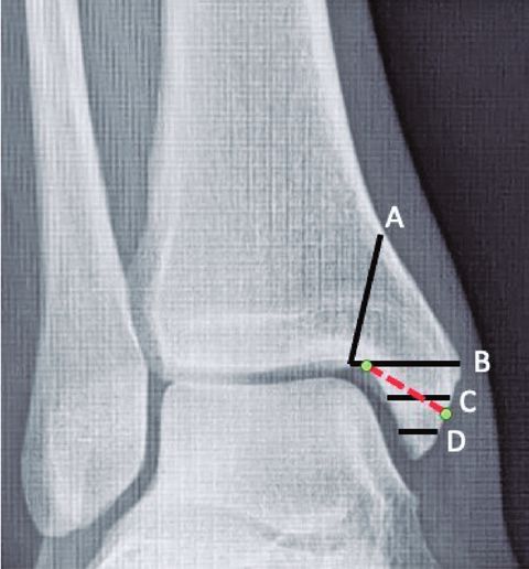
Chami et al. Anterograde fixation of inverted oblique medial malleolus fractures: case report

  The purpose of this case report is to draw attention to              Surgical technique
medial malleolus fractures with an inverted oblique pattern
                                                                        The patient was placed in the supine position on a radiolu-
in the anteroposterior view and the use of an anterograde
                                                                       cent table under spinal anesthesia and peripheral nerve block.
approach with headless double compression screws to                    The pneumatic cuff was positioned in the proximal third of
achieve osteosynthesis.                                                the right thigh and we followed the usual steps for asepsis
                                                                       and placement of the surgical fields.
Case report                                                              The reduction and fixation of the fibular fracture was per-
                                                                       formed according to AO foundation guidelines regarding
 This study was approved by the Institutional Review Board
                                                                       anatomical fracture reduction, fixation with interfragmentary
under the protocol number: CSJ000-688-21 and the patient
                                                                       screws, and the use of a neutralization plate.
provided written informed consent.
                                                                         After osteosynthesis of the fibular fracture, a large opening
  This 21-year-old female patient suffered torsional trauma in         was observed in the joint clamp, which hindered reduction
the right ankle and was referred to the emergency depart-              of the medial malleolus due to the integrity of the deltoid
ment. Physical examination revealed deformity of the right             ligament. Thus, it was necessary to reduce and stabilize the
ankle, severe pain, functional limitation, excoriation and             syndesmosis with two screws.
edema (+++/4+). Radiographs and computed tomography
showed a trimalleolar fracture-dislocation (Figures 2 and 3).
As comorbidities, the patient was obese and glucose intole-
rant, and was using an oral hypoglycemic agent.
  The patient was immediately taken to the operating room
and underwent surgery for fracture dislocation reduction
and definitive osteosynthesis, since the condition of the
soft tissue was good. We will describe the fixation of the
medial malleolus fracture, classified as a Herscovici type B
with an inverted oblique profile, which was directly redu-
ced with a reduction clamp and fixed with two 3.0mm No.
16 long-thread headless compression screws, which were
inserted anterograde, as detailed below.

                                                                        Figure 2. AP and ankle profile radiographs showing the tri-
                                                                        malleolar dislocation fracture. The red lines show the inverted
                                                                        oblique fracture pattern in the medial malleolus.

                                                                         A                                       B
                                                                        Figure 3. 3D tomographic reconstruction. (A) Trimalleolar frac-
 Figure 1. Medial malleolus fracture lines classified by Hersco-        ture in the anteroposterior view. (B) Posteroanterior view. In
 vici (types A, B, C and D); the red dotted line illustrates the        the medial malleolus, the entire anterior colliculus is fractured,
 patient’s inverted oblique fracture (subtype not classified).          including a small fragment of the posterior colliculus.

                                                                                                               J Foot Ankle. 2021;15(1):66-9          67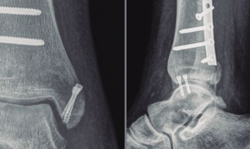
Chami et al. Anterograde fixation of inverted oblique medial malleolus fractures: case report
A medial incision of approximately 3-4cm was made using Discussion
the anteromedial cortex of the distal tibia as a reference and
To classify the medial malleolus fracture, we observed the
curving distally and posteriorly, preserving the saphenous
size of the fragment, relating it to the height of the horizontal
vein and nerve. An anterior colliculus fragment and a small
line of the fracture (Herscovici types A, B and C) and the ver-
posterior colliculus fragment were identified, with the intact
tical direction (shear - Herscovici type D)(5).
deltoid ligament attached to the fracture fragment. The frac-
ture was reduced using a Backhaus clamp placed perpendi- Pancovich and Shivran identified 6 main medial malleolus
cular to the fracture line (Figure 4). injury patterns(6), which facilitates selection of the osteosyn-
thesis type (all retrograde) for each fracture pattern.
Two guidewires were then introduced from the proximal
to the distal end, perpendicular to the fracture line and with We have observed that fractures involving the entire ante-
a good angle of attack, since this technique allows greater rior colliculus and a small fragment of the posterior colliculus
freedom of inclination than a distal-to-proximal placement. may be associated with an inverted oblique pattern.
After measurement, two cannulated 3.0mm No. 16 long-thread The Herscovici classification differentiates four main frac-
headless compression screws were inserted anterograde, re- ture patterns and helps determine the treatment. However,
sulting in perfect interfragmentary compression between the as found by Aitken in 2016, there is a high prevalence of di-
fracture fragments (Figure 5). Through fluoroscopy we con- sagreement between types B and C due to the obliquity of
firmed the correct placement of the guidewires and screws, the fracture, which we believe is essential for selecting the
which prevented them from becoming intra-articular. surgical technique(4,5).
After osteosynthesis of the medial malleolus, intraoperative Fractures in which the medial cortex fracture pattern is
stress radiographs showed no instability and that the ankle compatible with Herscovici type B and the joint face frac-
anatomy was restored. ture pattern is compatible with Herscovici type C can be
At the end of the procedure, the pneumatic cuff was ope- characterized as an inverted oblique pattern, a subtype not
ned and the blood perfusion was checked. The skin was su- considered in this classification system.
tured after revision of hemostasis. The patient was initially In this fracture “subtype”, it is difficult to obtain perpen-
immobilized with a plaster cast and then a short ankle ortho- dicularity during compression with any conventional re-
sis at discharge. trograde technique. Anterograde fixation with cannulated
headless screws is a more mechanically stable alternative
that involves less consolidation time, less pain due to the
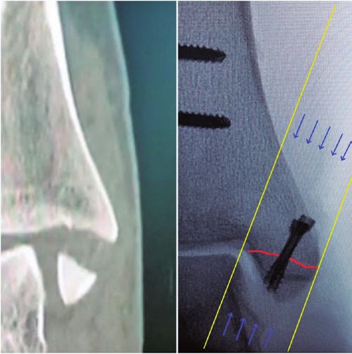
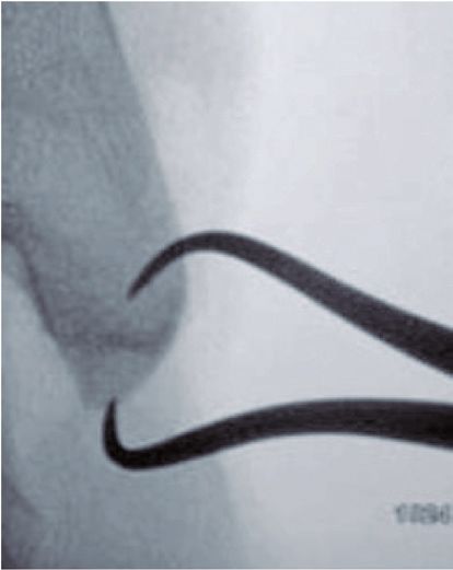
Figure 4. Intraoperative fluoroscopy image showing
A B
anatomical reduction of the medial malleolus fracture
with Backhaus forceps, whose upper end was intro- Figure 5. (A) Coronal CT section showing the inverted oblique
duced through the medial cortex after drilling with a fracture line; (B) AP radiograph of intraoperative control showing
1.5mm bit. screws inserted in the perpendicular “corridor” to the fracture line.
68 J Foot Ankle. 2021;15(1):66-9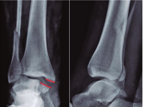
Chami et al. Anterograde fixation of inverted oblique medial malleolus fractures: case report

implants (reducing the need for removal), an easier surgical
approach, and less damage to the medial ligament complex
(Figure 6).

Conclusion
  Inverted oblique type B Herscovici fractures hinder conven-
tional retrograde osteosynthesis and are a challenge due to
difficulties in intraoperative reduction and fixation. The sur-
geon must prepare for these fractures with good planning
and a collection of images that enable identification of this
unusual pattern. Reduction and provisional stabilization that
applies the correct compression force in the correct direc-                      A                                        B
tion is fundamental for success. The anterograde approach
allows the compression screw to be inserted perpendicular                       Figure 6. (A) AP radiograph showing the consolidated medial
to the fracture line, which is adequate for interfragmentary                    malleolus 4 weeks after surgery. (B) Ankle profile 30 days after
compression and absolute stability.                                             surgery.

Authors’ contributions: Each author contributed individually and significantly to the development of this article: SMC *(https://orcid.org/0000 0001 6416
5865) performed the surgeries; TLL *(https://orcid.org/0000-0002-5242-4548) Participated in the review process; ABP *(https://orcid.org/0000-0001-
9785-1642) performed the surgeries; BJS *(https://orcid.org/0000-0001-5817-2743) participated in the review process; JSF *(https://orcid.org/0000-
0002-4964-0979) conceived and planned the activities that led to the study; RCB *(https://orcid.org/0000-0003-3199-4055) participated in the review
process. All authors read and approved the final manuscript. * ORCID (Open Researcher and Contributor ID)        .

References
1.   Buckley R, Moran C, Apivatthakakul T. Princípios AO do tratamento         6.   Pankovich AM, Shivaram MS. Anatomical basis of variability in
     das fraturas. 3ed. Porto Alegre: Artmed; 2020.                                  injuries of the medial malleolus and the deltoid ligament. II. Clinical
                                                                                     studies. Acta Orthop Scand. 1979;50(2):225-36.
2.   Lauge-Hansen N. Fractures of the ankle. II. Combined experimental-         7.   Johnson BA, Fallat LM. Comparison of tension band wire and
     surgical and experimental- roentgenologic investigations. Arch                  cancellous bone screw fixation for medial malleolar fractures. J
     Surg. 1950;60:957-85.                                                           Foot Ankle Surg. 1997;36(4):284-9.
                                                                                8.   Bulut T, Gursoy M. Isolated medial malleolus fractures: conventional
3.   Ramsey PL, Hamilton W. Changes in tibiotalar area of contact
                                                                                     techniques versus headless compression screw fixation. J Foot
     caused by lateral talar shift. J Bone Joint Surg Am. 1976;58(3):356-7.
                                                                                     Ankle Surg. 2018;57(3):552-6.
4.   Aitken SA, Johnston I, Jennings AC, Chua ITH, Buckley RE. An               9.   Tekin AÇ, Çabuk H, Dedeoğlu SS, Saygılı MS, Adaş M, Büyükkurt
     evaluation of the Herscovici classification for fractures of the                CD, et al. Anterograde headless cannulated screw fixation in
     medial malleolus. Foot Ankle Surg. 2017;23(4):317-20.                           the treatment of medial malleolar fractures: evaluation of a new
                                                                                     technique and its outcomes. Med Princ Pract. 2016;25(5):429-34.
5.   Herscovici D Jr, Scaduto JM, Infante A. Conservative treatment of          10. Parada SA, Krieg JC, Benirschke SK, Nork SE. Bicortical fixation
     isolated fractures of the medial malleolus. J Bone Joint Surg Br.              of medial malleolar fractures. Am J Orthop (Belle Mead NJ). 2013;
     2007;89(1):89-93.                                                              42(2):90-2.

                                                                                                                        J Foot Ankle. 2021;15(1):66-9          69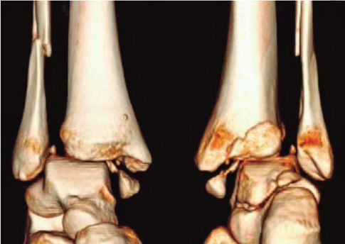
You can also read