Acute aortic dissection with highly compressed true lumen: unanticipated pitfall of point- of- care ultrasonography
←
→
Page content transcription
If your browser does not render page correctly, please read the page content below
Case report
BMJ Case Rep: first published as 10.1136/bcr-2020-239328 on 2 March 2021. Downloaded from http://casereports.bmj.com/ on March 16, 2021 by guest. Protected by copyright.
Acute aortic dissection with highly compressed true
lumen: unanticipated pitfall of point-of-care
ultrasonography
Hisashi Dote ,1 Masaaki Koide,2 Shunsuke Kobayashi,1 Takahiro Atsumi1
1
Department of Emergency SUMMARY
and Critical Care Medicine, A 46-year-old man presented with sudden onset of
Seirei Hamamatsu Hospital, chest pain. He was in cardiogenic shock at arrival.
Hamamatsu, Japan
2 Based on the results of ECG and echocardiogram, he
Department of Cardiovascular
was diagnosed with ST-segment elevation myocardial
Surgery, Seirei Hamamatsu
infarction. Point-of-care ultrasonography (POCUS) did
Hospital, Hamamatsu, Japan Figure 2 Point-of-care ultrasonogram. (A) Left
not reveal acute aortic dissection (AAD). During an
parasternal long-axis view of the heart. (B) The
Correspondence to emergency coronary angiography, aortic dissection was
descending aorta is posterior to the left ventricle. (C) The
Dr Hisashi Dote; detected and computed tomographic angiography (CTA)
suprasternal view. Aortic dissection cannot be identified
hisashi.dote@gmail.com revealed Stanford type A AAD with a highly compressed
on any image. Ao, aorta; LA, left atrium; LBV, left
true lumen. Because of this form of aortic dissection, the
brachiocephalic vein; LV, left ventricle.
Accepted 16 February 2021 enlarged false lumen could be potentially misidentified
as a normal aorta in POCUS. Although POCUS is useful
when AAD is suspected, we should not overestimate its rate, 98 beats/min; respiratory rate, 26 breaths/
findings and lower the threshold for CTA. min; oxygen saturation, 99% on 10 L of oxygen per
minute. He was in a state of restlessness and had
large amounts of cold sweat. On auscultation, no
BACKGROUND abnormal heart or breathing sounds were observed.
Acute aortic dissection (AAD) is a fatal disease Further, there was no difference in blood pressure
that presents in the emergency department (ED). or radial arterial palpitation in either of the upper
However, the symptoms and severity of AAD at extremities.
the time of presentation vary and are often difficult
to diagnose. As AAD sometimes mimics myocar- INVESTIGATIONS
dial infarction, emergency physicians (EPs) are The laboratory data showed a normal troponin
faced with a difficult decision. Although point-of- I level of under 10 pg/mL (normal range: under
care ultrasonography (POCUS) has proven to help 26.2 pg/mL) and elevated lactate level 32 mg/dL
distinguish between these two diseases,1 we expe- (normal range: 4.5–18.0 mg/dL). ECG showed ST
rienced a case in which the form of the dissection elevation in the II, III, aVF, V1, V2 and V4R leads
made it difficult to diagnose using POCUS. (figure 1). Thoracic echocardiography showed
decreased contraction of the inferior left ventric-
CASE PRESENTATION ular wall. Chest radiography revealed mediastinal
A 46- year-
old Japanese man suddenly developed widening (97 mm). Aortic dissection- oriented
severe chest pain. He had no significant medical POCUS with left parasternal, apical, suprasternal,
or family history, and smoked one pack of ciga- subcostal and abdominal views did not reveal an
rettes per day. The symptoms appeared while he intimal flap, a thoracic aorta dilation, a massive
was working in a restaurant. He arrived at the ED pericardial effusion or an aortic valve regurgitation
30 min after onset.
The initial vital signs of the subject were as
follows: blood pressure, 183/120 mm Hg; heart
© BMJ Publishing Group
Limited 2021. Re-use
permitted under CC BY-NC. No
commercial re-use. See rights
and permissions. Published
by BMJ.
Figure 3 Coronary angiography shows the
To cite: Dote H, Koide M,
Kobayashi S, et al. BMJ Case complete occlusion of right coronary artery (pre).
Rep 2021;14:e239328. After percutaneous coronary intervention, TIMI-3 flow
doi:10.1136/bcr-2020- Figure 1 Initial ECG showing ischaemic changes (ST was obtained (post). TIMI, thrombolysis in myocardial
239328 elevation in the II, III, aVF, V1 and V2 leads). infarction.
Dote H, et al. BMJ Case Rep 2021;14:e239328. doi:10.1136/bcr-2020-239328 1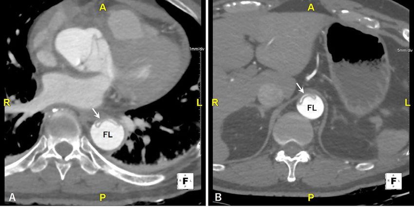
Case report
BMJ Case Rep: first published as 10.1136/bcr-2020-239328 on 2 March 2021. Downloaded from http://casereports.bmj.com/ on March 16, 2021 by guest. Protected by copyright.
Figure 4 Computed tomographic angiography showing aortic
Figure 6 Computed tomographic angiography showing aortic
dissection in the aortic arch in coronal view (A) and sagittal view (B).
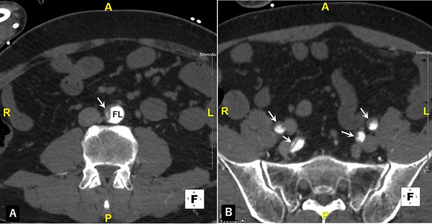
dissection at the abdominal aorta below the renal artery branch points
Intimal flap is indicated by black arrow. FL, false lumen; TL, true lumen.
(A), and the external and internal iliac arteries (B). White arrows show
highly compressed true lumen. FL, false lumen.
(AR; figure 2). The D-dimer levels were not determined during
the ED stay.
rate of Stanford type A AAD is aproximately 22%.1 This large
DIFFERENTIAL DIAGNOSIS multicenter detabase also revealed that the 25% of AAD are
He was diagnosed with ST-segment elevation myocardial infarc- initially diagnosed by thoracic echocardiography.1
tion (STEMI). After the administration of aspirin and prasugrel, Occasionally, the dissection of the ascending aorta occludes
an emergency coronary angiography (CAG) was performed. the coronary artery and mimics an STEMI. Discriminating
During CAG, a dissection was detected in the ascending aorta. between an AAD and a primary STEMI is very important for
The right coronary artery was obstructed and drug- eluting decisions regarding treatment plans. Aortic dissection-oriented
coronary stents were placed (figure 3). After CAG and percu- POCUS has been reported as a useful tool to help discriminate
taneous coronary intervention, computed tomographic angiog- these conditions.2 Another report suggests that the dissection of
raphy (CTA) was performed. It revealed a Stanford type A AAD the aortic arch could be diagnosed using the suprasternal view.3
with a highly compressed true lumen (figures 4 and 5), large Many parts of the thoracic aorta can be evaluated using POCUS.
intestinal ischemia and left renal infarction. The DeBakey type I Direct POCUS findings suggesting AAD are the presence of an
aortic dissection extended to the bilateral internal iliac arteries intimal flap, intramural aortic haematoma (circular or crescentic
(figure 6). thickening of the aortic wall >5 mm) and penetrating aortic
ulcer (crater-like outpouching with jagged edges in the aortic
TREATMENT wall). Indirect findings include dilation of the aorta (>40 mm),
He underwent emergency thoracic aortic graft replacement and pericardial effusion and cardiac tamponade and AR more than
open stent grafting. moderate levels.4 Detection of any POCUS findings suggesting
AAD has a sensitivity of 88% (95% CI: 76% to 95%).5
OUTCOME AND FOLLOW-UP CTA findings revealed that our patient had a highly compressed
After an extensive bowel resection, he was discharged from the true lumen and enlarged false lumen. Because of this form of
intensive care unit. aortic dissection, there was potential to misidentify the enlarged
false lumen as a normal aorta on POCUS. Although the dissec-
tion of the aortic arch was relatively clear of the true lumen from
DISCUSSION
CTA, given that evaluation of the aortic arch from the supra-
AAD is a lethal condition in the ED. The International Registry
sternal notch view using ultrasonography is not so easy for EPs,6
of Acute Aortic Dissection reported that the overall mortality
we could not visualise adequate findings.
Guidelines for the diagnosis and management of patients with
thoracic aortic disease recommend estimating pretest risk during
initial management.7 Our patient presented with an Aortic
Dissection Detection Risk Score >1 and should have been rated
as high risk.7 Mediastinal widening on chest radiography was
also observed. A mediastinal width >87 mm demonstrated high
sensitivity in the diagnosis of a probable Stanford type A AAD.8
If we had focused on these findings and had performed CTA
before CAG, the treatment plan would have changed to emer-
gency surgery. If AAD is suggested in an ED, POCUS is useful;
however, detecting aortic dissection is occasionally difficult
depending on the form. In our case, the true lumen from the
abdominal aorta to the iliac artery was also highly compressed,
Figure 5 Computed tomographic angiography showing aortic thus we think that diagnosis by POCUS was difficult if we tested
dissection at the descending aorta with a highly compressed true lumen these area. Furthermore, the diagnostic accuracy of POCUS
(white arrow) and false lumen (FL) resembling a normal aorta. Findings depends in no small part on the skill of the operator. Therefore,
have been detected at the same level as the left parasternal long-axis we should not overestimate the POCUS findings and lower the
view (A) and subcostal view (B) by point-of-care ultrasonography. threshold for CTA.
2 Dote H, et al. BMJ Case Rep 2021;14:e239328. doi:10.1136/bcr-2020-239328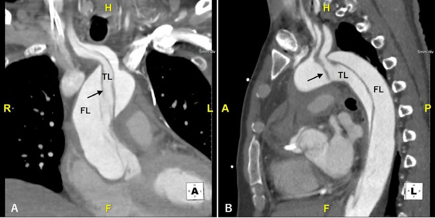
Case report
BMJ Case Rep: first published as 10.1136/bcr-2020-239328 on 2 March 2021. Downloaded from http://casereports.bmj.com/ on March 16, 2021 by guest. Protected by copyright.
ORCID iD
Learning points Hisashi Dote http://orcid.org/0000-0002-9323-656X
►► Point-of-care ultrasonography (POCUS) is a useful tool to REFERENCES
discriminate acute aortic dissection (AAD) and other mimics. 1 Evangelista A, Isselbacher EM, Bossone E, et al. Insights from the International registry
►► One of the pitfalls of POCUS is that AAD with a highly of acute aortic dissection: a 20-year experience of collaborative clinical research.
compressed true lumen and enlarged false lumen could be Circulation 2018;137:1846–60.
2 Chenkin J. Diagnosis of aortic dissection presenting as ST-elevation myocardial
difficult to identify. infarction using point-of-care ultrasound. J Emerg Med 2017;53:880–4.
►► We should not overestimate the POCUS findings and lower 3 Fernando SM, Kisilewicz M, Millington SJ. View from the top: point-of-care ultrasound
the threshold for computed tomographic angiography. diagnosis of type A aortic dissection using the suprasternal view. J Emerg Med
2018;54:e13–14.
4 Morello F, Santoro M, Fargion AT, et al. Diagnosis and management of acute aortic
Contributors HD drafted the article, and all authors contributed substantially to its syndromes in the emergency department. Intern Emerg Med 2021;16:171–81.
revision. TA supervised the conduct of the reports. 5 Nazerian P, Vanni S, Castelli M, et al. Diagnostic performance of emergency
transthoracic focus cardiac ultrasound in suspected acute type A aortic dissection.
Funding The authors have not declared a specific grant for this research from any Intern Emerg Med 2014;9:665–70.
funding agency in the public, commercial or not-for-profit sectors. 6 Kinnaman KA, Kimberly HH, Pivetta E, et al. Evaluation of the aortic arch from
Competing interests None declared. the suprasternal Notch view using focused cardiac ultrasound. J Emerg Med
2016;50:643–50.
Patient consent for publication Obtained. 7 Hiratzka LF, Bakris GL, Beckman JA. 2010 ACCF/AHA/AATS/ACR/ASA/SCA/SCAI/SIR/
Provenance and peer review Not commissioned; externally peer reviewed. STS/SVM guidelines for the diagnosis and management of patients with thoracic
aortic disease: Executive summary: a report of the American College of cardiology
Open access This is an open access article distributed in accordance with the Foundation/American heart association Task force on practice guidelines, American
Creative Commons Attribution Non Commercial (CC BY-NC 4.0) license, which association for thoracic surgery, American College of radiology, American stroke
permits others to distribute, remix, adapt, build upon this work non-commercially, association. Circulation 2010;121:266–369.
and license their derivative works on different terms, provided the original work 8 Funakoshi H, Mizobe M, Homma Y, et al. The diagnostic accuracy of the mediastinal
is properly cited and the use is non-commercial. See: http://creativecommons.org/ width on supine anteroposterior chest radiographs with nontraumatic Stanford type A
licenses/by-nc/4 .0/. acute aortic dissection. J Gen Fam Med 2018;19:45–9.
Copyright 2021 BMJ Publishing Group. All rights reserved. For permission to reuse any of this content visit
https://www.bmj.com/company/products-services/rights-and-licensing/permissions/
BMJ Case Report Fellows may re-use this article for personal use and teaching without any further permission.
Become a Fellow of BMJ Case Reports today and you can:
►► Submit as many cases as you like
►► Enjoy fast sympathetic peer review and rapid publication of accepted articles
►► Access all the published articles
►► Re-use any of the published material for personal use and teaching without further permission
Customer Service
If you have any further queries about your subscription, please contact our customer services team on +44 (0) 207111 1105 or via email at support@bmj.com.
Visit casereports.bmj.com for more articles like this and to become a Fellow
Dote H, et al. BMJ Case Rep 2021;14:e239328. doi:10.1136/bcr-2020-239328 3

You can also read