Role of mycology in accurate diagnosis of various fungal aetiologies in rhino/orbital diseases: 'needle in a haystack'
←
→
Page content transcription
If your browser does not render page correctly, please read the page content below
Images in…
BMJ Case Rep: first published as 10.1136/bcr-2021-242684 on 11 May 2021. Downloaded from http://casereports.bmj.com/ on September 19, 2021 by guest. Protected by copyright.
Role of mycology in accurate diagnosis of various
fungal aetiologies in rhino/orbital diseases: ‘needle in
a haystack’
Surya Ravichandran ,1 Saranya Thangavel ,1 Rakesh Singh ,2
Sivaraman Ganesan 1
1
ENT, Jawaharlal Institute of DESCRIPTION
Postgraduate Medical Education A 37- year-old man with no known comorbidi-
and Research, Puducherry, India ties presented to our department with complaints
2
Microbiology, Jawaharlal
of left eye proptosis and diminution of vision for
Institute of Post Graduate
1 month and left facial swelling for the past 2 years.
Medical Education and
Research, Puducherry, He underwent left- sided nasal surgery thrice—
Pondicherry, India functional endoscopic sinus surgery (FESS) in
2018, followed by revision FESS in 2019, followed
Correspondence to by Caldwell Luc procedure in 2020. In 2018, the
Dr Saranya Thangavel; biopsy report was caseating granulomatous lesion,
softsaran.nrp@gmail.com and so he was started on antitubercular drugs for
6 months, but there was no response to treatment.
Accepted 29 April 2021 In 2020, it was reported as a fungal granuloma.
He was not started on antifungal drugs despite the
persistence of symptoms. On external examination,
left diffuse facial swelling with depression at nasal
ala and left eye proptosis with reduced infraorbital
sensations (figure 1A,B). Diagnostic nasal endos-
copy showed left vestibular stenosis with status
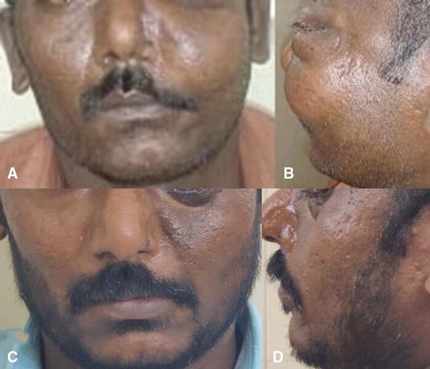
postoperative FESS with no evident growth or Figure 1 Showing external facial appearance. (A,
mass inside the sinuses. Visual acuity was 5/60 on B) pretreatment with left facial swelling with left eye
right and hand movements close to face on the left proptosis and chemosis. (C, D): post-treatment with
with abaxial proptosis, non-reacting pupils, corneal completely reduced facial swelling and proptosis.
ulcer with restriction of extraocular movements
in all sides. Contrast-enhanced CT (CECT) of the
nose and paranasal sinuses showed left lobulated
hyper dense non enhancing lesion with extensive
destruction involving the orbit, maxillary, bilat-
eral sphenoid and ethmoid sinuses and extending
into intracranial and left infratemporal fossa with
encasement of bilateral cavernous part of Internal
Carotid Artery (ICA) (figure 2A,B).
Because of its extensive involvement in the skin
and subcutaneous tissues, phaeohypomycosis was
considered the diagnosis. Fine- needle aspiration
cytology from the left facial swelling was reported
as a possibility of aspergillosis with epithelioid
granuloma with septate, fungal hyphae. Periodic
acid-Schiff and Gomori methenamine silver were
positive for the fungus. Fungal culture and staining
were suggestive of Aspergillus flavus (figures 3 and
4). When the culture report was awaited, the patient
© BMJ Publishing Group was started on injection liposomal amphotericin B
Limited 2021. No commercial
re-use. See rights and with the cumulative dosage of 1800 mg. The patient
permissions. Published by BMJ. was started on an injection voriconazole 200 mg
stat dose, followed by tablet 200 mg. The patient
To cite: Ravichandran S,
responded very well, and the left facial swelling and Figure 2 CECT nose and PNS showing. (A, B):
Thangavel S, Singh R,
et al. BMJ Case Rep proptosis had reduced (figure 1C,D) with repeat Pretreatment left hyper dense non-enhancing lesion with
2021;14:e242684. CECT showing resolution of disease (figure 2C,D). maxillary swelling in axial view(A) and coronal view (B).
doi:10.1136/bcr-2021- A maxillary sinus aspirate material and a conjunc- (C, D) Post-treatment resolution of disease in axial (C)
242684 tival swab sample were collected and sent to the and coronal views (D). CECT, contrast-enhanced CT.
Ravichandran S, et al. BMJ Case Rep 2021;14:e242684. doi:10.1136/bcr-2021-242684 1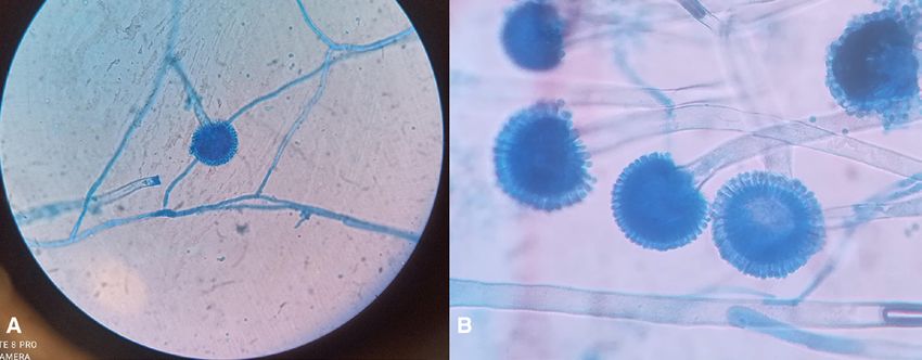
Images in…
BMJ Case Rep: first published as 10.1136/bcr-2021-242684 on 11 May 2021. Downloaded from http://casereports.bmj.com/ on September 19, 2021 by guest. Protected by copyright.
Patient’s perspective
I was taking treatment for my condition for the past 2 years. I
feel delighted after my swelling got reduced with treatment here.
Now I am on regular follow-up and feeling happy.
Learning points
►► Phaeohypomycosis means the condition of fungi with dark
hyphae and is very rare in immunocompetent individuals.
►► The high index of suspicion of aspergillosis should be kept in
mind in immunocompetent individuals and confirmed with a
fungal culture.
►► Aspergillus grows from haematogenous lesions in tissues in a
radial fashion, like wheel spokes, with the hyphae appearing
nearly parallel to one another.
Figure 3 SDA growing Aspergillus flavus with the characteristic ►► The parallel arrangement of aspergillus is absent if the tissue
yellow-green to an olive colony with a white border and a velvety or is not intact or if the specimen is more liquid.
cottony texture. SDA, sabouraud dextrose agar.
microscopy at 10–40 × magnification. In this case, thin hyaline
department of microbiology. Ten per cent potassium hydroxide septate hyphae with acute-angled branching were noted sugges-
(KOH) mount was prepared from sinus aspirate material tive of Aspergillosis.
which was negative. The conjunctival swab was inoculated into
sabouraud dextrose agar (SDA) culture plate. The sinus aspirate
Fungal culture
sample was inoculated into multiple tubes of SDA. Inoculated
Aside from the fact that fungal culture helps in identifying the
culture media were incubated at 25℃. A yellow-green, velvety
species of Aspergillus infection, it also helps in identifying the
and powdery growth with a white margin was observed within
resistance to antifungal therapy; for example, Aspergillus terreus
5 days of incubation. No pigmentation was seen on the reverse
and Aspergillus nidulans are found to have an inherent resistance
side of the fungal growth. Lactophenol cotton blue (LPCB)
to amphotericin-b.1–3 All medically important fungi samples
tease mount was prepared and observed under the microscope.
must be sent in a sealed, leak-proof, sterile container marked
Hyaline septate hyphae were observed with spiny conidiophore,
with a biohazard sticker to comply with occupational safety and
which was ending in the vesicle. Phialides and metulae entirely
health administration safety standards.4 It should be transported
covered the vesicle. Chains of conidia were seen arising from
within 2 hours; otherwise, it should be stored at 4℃.
metulae, which was morphologically suggestive of A. flavus
The sample after KOH staining is inoculated in SDA medium
species complex.
either in plates or tubes. SDA is preferred as it is more efficient
A. flavus has a conidiophore bearing vesicle with a typical bise-
and has been found a high detection rate for a wide range of
riate structure with metulae and phialide. The conidiophore also
fungi.5 After incubating at 25℃ for 3–4 days, an olive green
has this rough or gently spiked texture, particularly at the apex
coloured growth with velvety or cottony texture with clear
(where it meets the vesicle), a diagnostic feature of A. flavus.
margins will be noted in the medium.
Phialides radiate from the vesicle in all directions.
A sample taken from the nasal cavity—nasal crust—is sent for
Fungal staining
fungal staining and fungal culture.
On examining, this cultured growth under light microscopy after
LPCB staining, thin septate hyphae with conidiophores bearing
KOH examination vesicle at its apex will be noted. The wall of the conidiophores is
KOH (10%) lyses the fungal cell wall’s protein content and
rough or spiny, especially below the vesicle. Uniserate or biserate
reveals fungal elements. After lysis, it is examined under light
phialides are formed, pointing out in all directions covering the
entire vesicle.
Acknowledgements I would like to thank Professor Dr. Sunil Kumar Saxena
for supporting in submitting this article and Dr. Mukundan for providing valuable
information regarding mycological data.
Contributors SR: conception and design, acquisition of data or analysis and
interpretation of data. ST: drafting the article or revising it critically for important
intellectual content. SG and RS: final approval of the version published. ST:
agreement to be accountable for the article and to ensure that all questions
regarding the accuracy or integrity of the article are investigated and resolved.
Funding The authors have not declared a specific grant for this research from any
funding agency in the public, commercial or not-for-profit sectors.
Figure 4 LPCB staining showing thin long conidiophore and vesicle
Competing interests None declared.
at the apex bearing phialides radiating from the entirety of the vesicle,
Patient consent for publication Obtained.
characteristic of Aspergillus flavus (A) ×40 and (B) ×100. LPCB,
lactophenol cotton blue. Provenance and peer review Not commissioned; externally peer reviewed.
2 Ravichandran S, et al. BMJ Case Rep 2021;14:e242684. doi:10.1136/bcr-2021-242684
Images in…
BMJ Case Rep: first published as 10.1136/bcr-2021-242684 on 11 May 2021. Downloaded from http://casereports.bmj.com/ on September 19, 2021 by guest. Protected by copyright.
ORCID iDs 2 Kontoyiannis DP, Lewis RE, May GS, et al. Aspergillus nidulans is frequently resistant to
Surya Ravichandran http://orcid.org/0000-0002-4579-9835 amphotericin B. Mycoses 2002;45:406–7.
Saranya Thangavel http://orcid.org/0000-0001-6954-1364 3 Van Der Linden JWM, Warris A, Verweij PE. Aspergillus species intrinsically resistant to
Rakesh Singh http://orcid.org/0000-0002-2213-3993 antifungal agents. Med Mycol 2011;49:S82–9.
Sivaraman Ganesan http://orcid.org/0000-0002-7065-0258 4 Miller JM, Astles R, Baszler T, et al. Guidelines for safe work practices in human and
animal medical diagnostic laboratories. MMWR Surveill Summ 2012;6:13.
5 Gebala B, Sandle T. Comparison of different fungal agar for the environmental
REFERENCES monitoring of pharmaceutical-grade cleanrooms. PDA J Pharm Sci Technol
1 Walsh TJ, Petraitis V, Petraitiene R, et al. Experimental pulmonary aspergillosis due 2013;67:621–33.
to Aspergillus terreus: pathogenesis and treatment of an emerging fungal pathogen
resistant to amphotericin B. J Infect Dis 2003;188:305–19.
Copyright 2021 BMJ Publishing Group. All rights reserved. For permission to reuse any of this content visit
https://www.bmj.com/company/products-services/rights-and-licensing/permissions/
BMJ Case Report Fellows may re-use this article for personal use and teaching without any further permission.
Become a Fellow of BMJ Case Reports today and you can:
►► Submit as many cases as you like
►► Enjoy fast sympathetic peer review and rapid publication of accepted articles
►► Access all the published articles
►► Re-use any of the published material for personal use and teaching without further permission
Customer Service
If you have any further queries about your subscription, please contact our customer services team on +44 (0) 207111 1105 or via email at support@bmj.com.
Visit casereports.bmj.com for more articles like this and to become a Fellow
Ravichandran S, et al. BMJ Case Rep 2021;14:e242684. doi:10.1136/bcr-2021-242684 3You can also read