A retrospective study of clinical and laboratory features and treatment on cats highly suspected of feline infectious peritonitis in Wuhan, China ...
←
→
Page content transcription
If your browser does not render page correctly, please read the page content below
www.nature.com/scientificreports
OPEN A retrospective study of clinical
and laboratory features
and treatment on cats highly
suspected of feline infectious
peritonitis in Wuhan, China
Yiya Yin, Ting Li, Chaohao Wang, Xiaoya Liu, Hehao Ouyang, Wanfeng Ji, Jiahao Liu,
Xueyu Liao, Junyi Li & Changmin Hu*
Feline infectious peritonitis (FIP) is a systemic, potentially fatal viral disease. The objectives of this
study were to review clinical and laboratory features and treatment of cats highly suspected of FIP
in Wuhan, China. The clinical records of 127 cats highly suspected of FIP were reviewed for history,
clinical signs, physical findings, and diagnostic test results. Sex, neutering status, breed, age, and
month of onset of disease were compared with the characteristics of the clinic population. Age and
neutering status were significantly correlated with FIP-suspicion. Sex, breed and onset month were
not associated with FIP. There were many more FIP-suspected cases in cats in young cats or male
intact cats. Effusion was observed in 85.8% of the FIP-suspected cats. Increased serum amyloid A
(SAA) and lymphopenia were common laboratory abnormalities in the FIP cases. Furthermore, 91.7%
of the cats highly suspected of FIP had an albumin/globulin (A/G) ratio < 0.6, while 85.3% had an A/G
ratio < 0.5. The mortality rate for FIP-suspected cats was 67%, and six submitted cases were confirmed
by FIP-specific immunohistochemistry. Of the 30 cats treated with GS-441524 and/or GC376, 29
were clinically cured. The study highlights the diverse range of clinical manifestations by clinicians
in diagnosing this potentially fatal disease. A/G ratio and SAA were of higher diagnostic value.
GS-441524 and GC376 were efficient for the treatment of FIP-suspected cats.
Feline infectious peritonitis (FIP) is a worldwide disease of domestic and wild felids. The pathogen of FIP is
feline infectious peritonitis virus (FIPV), which is mutated from feline coronavirus (FCoV)1,2. Although much
research into FIP has been performed, FIP remains one of the most prevalent and fatal infectious diseases of cats.
Except for non-specific clinical signs, such as lassitude, inappetence, fluctuating fever and weight loss, the
clinical presentation of FIP is complex and variable. Different clinical features may be a result of variations in the
virus and in the nature of the individual host’s immune response. FIP can be divided into three types—effusive,
non-effusive and mixed—according to the different clinical features that emerge, but these types can change over
time in any individual cat. Effusive FIP is mainly manifested as ascites and/or pleural effusion, while non-effusive
FIP is mainly manifested as granuloma formation involving the central nervous system, eyes and abdominal
organs, and it does not produce body cavity effusion3. When effusive and non-effusive signs occur together, it is
mixed FIP4. To our knowledge, there are few studies about the clinical and laboratory features of FIP in China.
Ante-mortem diagnosis of FIP is a controversial and complicated issue. It is more difficult to diagnose FIP
definitively in vivo in cats without effusion as clinical signs and laboratory features are relatively vague. Immu-
nofluorescence staining of FCoV antigen in macrophages is highly specific, but it is difficult to detected due
to the low numbers of macrophages in the effusion. At present, the gold standard for the diagnostic of FIP is
immunohistochemistry (IHC) to identify FCoV antigens in diseased tissues. However, this invasive method has
low operability and requires professional detection equipment and personnel. Also, a negative IHC result does
not exclude the diagnosis of FIP, adding to the challenges of diagnosing this disease5. FIP has an extremely high
mortality rate and was once considered a terminal disease. In recent years, 3C-like protease inhibitor (GC376)
and nucleoside analogue (GS-441524) have been proven to have a certain therapeutic effect on FIP6–9. The
College of Veterinary Medicine, Huazhong Agricultural University, Rm 321 Vet. Teaching Hospital BLDG, No.1
Shizishan St., Hongshan District, Wuhan City 430070, Hubei Province, China. *email: hcm@mail.hzau.edu.cn
Scientific Reports | (2021) 11:5208 | https://doi.org/10.1038/s41598-021-84754-0 1
Vol.:(0123456789)
www.nature.com/scientificreports/
purpose of this study is to determine the epidemiological features and related risk factors of FIP, summarise
common clinical signs and laboratory features, and evaluate the therapeutic effects of GS-441524 and GC376.
Materials and methods
Selection of cases. This study retrospectively evaluated the medical records in the database of the 12 veter-
inary hospitals in Wuhan, China, from 1 January 2019 to 1 January 2020. Due to the limitations of the diagnostic
facilities in veterinary hospitals and the cat owners were reluctant to consent to invasive diagnostic procedures
such as biopsies, most of the cases were not confirmed by IHC. Therefore the following inclusion criteria for the
diagnosis of suspected FIP cases were established based on the previously published literature in this fi eld5–15
Those cases meeting at least four of the following characteristics were defined as highly suspected of FIP: 1)
Typical clinical signs of effusive FIP (pleural effusion and/or ascites) or typical clinical signs of non-effusive FIP
(ocular and neurological signs); 2) A decreased albumin/globulin (A/G) ratio of the cut-off (0.6); 3) Rivalta’s
test showing positive results in effusion; 4) Positive reverse transcription–polymerase chain reaction (RT-PCR)
detection of FCoV RNA in effusion; 5) Tissue samples collected by autopsy showing typical histological features
of FIP, namely systemic vasculitis and pyogranulomatous lesions.
Out of 20 984 cats registered in the 12 veterinary hospitals, 127 cases with a high suspicion of FIP were
included according to the criteria above. Through basic information collection, history investigation, clinical
signs, haematological and biochemical analyses, diagnostic imaging, RT-PCR, Rivalta’s test, exploratory laparot-
omy, and histopathology, the cases were comprehensively diagnosed and analysed, and detailed information and
examination results were recorded (see Supplementary Table S1 for the FIP case record sheet). All information
was collected with the consent and cooperation of the veterinarians and cat owners. The protocol was approved
by the Animal Management and Ethics Committee of the Laboratory Animal Centre, Huazhong Agriculture
University (approval no.: HZAUCA-2018–005).
Statistical analysis. Statistical analysis was performed using commercial software (SPSS Version 21.0
[IBM]). Descriptive statistics were employed for all evaluated variables. Categorical data were analysed using
a Pearson chi-square (χ2) test. In 2 × 2 contingency tables with any expected cell values < 5, Fisher’s exact two-
tailed results were used. Sex, neutering status, breed, age and month of onset of highly suspected of FIP were
compared with the feline clinic population (20 984 cats) presenting to the 12 veterinary hospitals from January
2019 to January 2020 to determine whether these risk factors were associated with FIP.
The history, clinical signs and laboratory examination results of FIP-suspected cases were retrospectively
analysed to summarise the common clinical features and laboratory examination abnormalities of the disease.
Samples were randomly selected from the cats undergoing exploratory laparotomy and sent to LABOKLIN
GMBH & CO. KG (Bad Kissingen, Germany) for IHC to verify whether the inclusion criteria for FIP cases were
correct and reasonable. In addition, the therapeutic effect of GS-441524 and GC376 was evaluated.
Ethical approval. All applicable international, national and institutional guidelines for the care and use of
animals were followed. All procedures performed in studies involving animals were in accordance with the ethi-
cal standards of the institution at which the studies were conducted.
Results
Risk factors. There was no significant correlation between FIP and sex (p = 0.083). However, FIP was signifi-
cantly correlated with neutering status (p < 0.001), and male intact cats were more susceptible to disease. As for
breed, whether classified as crossbred or purebred (p = 0.069) or specific to each breed (p = 0.246), no correlation
with the FIP was found. There was a significant correlation between age and FIP (p < 0.001), and young cats were
more susceptible. The age of the FIP-suspected cases ranged from 1 month to 8 years and 2 months, with an
average age of 13.1 months and a median age of 8 months. Within these cases, 40.2% were under 6 months old,
67% were under 1 year old and 90.6% were under 2 years old; moreover, the prevalence of older cats was signifi-
cantly lower than that of young cats (Table 1). The correlation of onset month was studied for the first time, and
the number of FIP-suspected cats and clinic population in each month was counted. The results showed that the
highest prevalence was 1% in May. The prevalence in January (0.95%), February (0.87%), September (0.66%) and
November (0.77%) were higher than the total prevalence, and the lowest prevalence was 0.35% in July. However,
in the statistical analysis, there was no correlation between FIP and month (p = 0.135; Table 1).
Stressful events before the diagnosis were documented for 60/127 cats. Since some cats had multiple stressors,
a total of 77 stressors were recorded. Of these events, changing environment was the most common factor lead-
ing to stress (40.3%), followed by new pets in the household, coexisting with other diseases, being frightened,
changing food (Fig. 1). In addition, information on the housing density of 83 cats was collected, of which 43
(51.8%) were single-cat households and 40 (48.2%) were multi-cat households.
Clinical signs. Of the 127 cats highly suspected of FIP, 12 (9.4%) presented with non-effusive FIP and 109
(85.8%) presented with effusive FIP, of which 92 presented with ascites (84.4%), 11 with pleural effusion (10.1%),
and 6 with both ascites and pleural effusion (5.5%). In addition, 6 cats (4.7%) presented with mixed FIP. Some
presented with non-effusive FIP first, followed by ascites and the development of effusive FIP. Some cats showed
effusive FIP first and develop neurological signs a few days before dying.
The clinical signs of cats highly suspected of FIP are shown in Table 2. The most common non-specific signs
were weight loss (93.8%), lassitude (86.2%) and inappetence (86%). Icterus and fever were also present in more
than half of the cases, 59.4% and 52.9%, respectively. Of the 85 cats highly suspected of FIP for which the temper-
ature was documented on physical examination, 52.9% had a temperature above 39.5℃, 36.5% had a temperature
Scientific Reports | (2021) 11:5208 | https://doi.org/10.1038/s41598-021-84754-0 2
Vol:.(1234567890)www.nature.com/scientificreports/
FIP-suspected population Clinic population
Risk factors (n = 127) (n = 20,984) χ2 p value OR 95% CI
Sex n = 127 (%) n = 17,494 (%) 3.009 0.083
Male 78 (61.4) 9397 (53.7) 1.372 0.958–1.963
Female 49 (38.6) 8097 (46.3) 0.729 0.509–1.043
Neutering status n = 127 (%) n = 16,041 (%) 45.684 < 0.001***
Intact male 69 (54.3) 4887 (30.5) Ref
Neutered male 9 (7.1) 3791 (23.6) 0.168 0.084–0.337
Intact female 39 (30.7) 4472 (27.9) 0.618 0.416–0.917
Spayed female 10 (7.9) 2891 (18.0) 0.245 0.126–0.476
Breed n = 127 (%) n = 16,469 (%) 3.307 0.069
Crossbred 36 (28.3) 5953 (36.1) 0.699 0.474–1.029
Purebred 91 (71.7) 10,516 (63.9) 1.431 0.972–2.108
Breed (specific) n = 127 (%) n = 16,469 (%) 16.752 0.053
Crossbred 36 (28.3) 5953 (36.1) Ref
British Shorthair 60 (47.2) 6558 (39.8) 1.513 0.999–2.290
American Shorthair 8 (6.3) 1275 (7.7) 1.038 0.481–2.237
Ragdoll 9 (7.1) 877 (5.3) 1.697 0.815–3.535
Siamese 6 (4.7) 430 (2.6) 2.307 0.967–5.506
Exotic Shorthair 3 (2.4) 1019 (6.2) 0.487 0.150–1.584
Chinchilla 2 (1.6) 258 (1.6) 1.282 0.307–5.353
Longhair Scottish Fold 1 (0.8) 44 (0.3) 3.758 0.504–28.021
American Curl 1 (0.8) 28 (0.2) 5.906 0.782–44.581
Bengal 1 (0.8) 27 (0.2) 6.124 0.810–46.291
Age groups n = 127 (%) n = 19,772 (%) 220.276 < 0.001***
≤6 M 51 (40.2) 1530 (7.7) Ref
6 M < A ≤ 1Y 34 (26.8) 3254 (16.5) 0.313 0.202–0.486
1Y < A ≤ 2Y 30 (23.6) 4660 (23.6) 0.193 0.123–0.304
2Y < A ≤ 4Y 8 (6.3) 4027 (20.4) 0.060 0.028–0.126
4Y < A ≤ 7Y 3 (2.4) 3507 (17.7) 0.026 0.008–0.082
A > 7Y 1 (0.8) 2794 (14.1) 0.011 0.001–0.078
Onset months n = 127 (%) n = 20,984 (%) 16.166 0.135
January 16 (12.6) 1685 (8.0) Ref
February 13 (10.2) 1500 (7.1) 0.913 0.438–1.904
March 7 (5.5) 1768 (8.4) 0.417 0.171–1.016
April 7 (5.5) 1656 (7.9) 0.445 0.183–1.085
May 17 (13.4) 1689 (8.1) 1.054 0.531–2.094
June 10 (7.9) 2109 (10.1) 0.499 0.226–1.103
July 7 (5.5) 2022 (9.6) 0.365 0.150–0.888
August 9 (7.1) 1773 (8.4) 0.535 0.236–1.213
September 12 (9.4) 1812 (8.6) 0.697 0.329–1.479
October 8 (6.3) 1734 (8.3) 0.486 0.207–1.138
November 13 (10.2) 1698 (8.1) 0.806 0.387–1.681
December 8 (6.3) 1529 (7.3) 0.551 0.235–1.291
Table 1. Risk factors of cats highly suspected of FIP compared with the clinic population. Data were given as n
(%); χ2 = Chi-square; OR = Odds Ratio; CI = Confidence Interval; Ref = Reference Category; A = Age; Y = Years;
***: p < 0.001, indicating a statistically significant difference.
above 40℃ and 10.6% had a temperature above 40.5℃ (see Supplementary Fig. S1). In the results, 41.3% of the
cats had a palpable mass on abdominal palpation, which could be either mesenteric lymph node enlargement or
accretion of the intestinal wall. Diarrhoea was uncommon, occurring in only 17.6%, while dyspnoea occurred in
35.2%, and was most common in cats with pleural effusion. In addition, ocular and neurological signs generally
occurred only in non-effusive or mixed forms of suspected FIP cases.
Haematology and serum biochemistry. As shown in Table 3, haematocrit (HCT) decreased in 40.2%
of the cats, but only 15.9% of the cats had a decrease in red blood cell (RBC) count. Half of the cats developed
lymphocytopenia, 33.9% had an increase in white blood cells (WBC), and 34.3% had an increased neutrophil
(NEU) count.
Scientific Reports | (2021) 11:5208 | https://doi.org/10.1038/s41598-021-84754-0 3
Vol.:(0123456789)www.nature.com/scientificreports/
Figure 1. Frequency of documented stress situation of cats highly suspected of FIP.
Clinical signs Animals examined (n) Number of cats with clinical signs (n) Percentage of cats with clinical signs (%)
Weight loss 64 60 93.8
Lassitude 94 81 86.2
Inappetence 100 86 86
Icterus 64 38 59.4
Fever 85 45 52.9
Abdominal mass 63 26 41.3
Dyspnoea 71 25 35.2
Diarrhoea 85 15 17.6
Ocular signs 103 9 8.7
Neurological signs 103 9 8.7
FIP types
Non-effusive 127 12 9.4
Ascites 127 92
Effusive Pleural effusion 127 11 85.8
Both 127 6
Mixed 127 6 4.7
Table 2. Clinical signs of cats highly suspected of FIP. Ocular signs included uveitis, corneal oedema,
hyphema, anisocoria, and retinal detachment. Neurological signs included ataxia, paralysis of the hind limbs,
nystagmus, twitching, and salivation. Data were given as n.
In serum biochemistry, the total protein (TP) levels were mostly normal (75.2%); a few (14.7%) cats had an
increased level and a very few had a decreased level. More than half of the cats highly suspected of FIP showed
hypoalbuminemia (58.7%). Although the albumin (ALB) levels of the remaining cats were normal, they were also
near to the lower limit of the reference range. Hyperglobulinemia was also common in cats highly suspected of
FIP (57.8%), and the remainder of the cats had normal globulin (GLOB) levels. The A/G ratio ranged from 0.137
to 0.767, with an mean of 0.399 and a median of 0.385. Among the results, 91.7% were lower than 0.6, 85.3%
were lower than 0.5 and 54.1% were lower than 0.4 (see Supplementary Fig. S2). An increase in total bilirubin
Scientific Reports | (2021) 11:5208 | https://doi.org/10.1038/s41598-021-84754-0 4
Vol:.(1234567890)www.nature.com/scientificreports/
Reference Animals
Measurement Unit interval examined Range Mean Median Increased Decreased Normal
Red blood cells × 1012/L 5–10 107 3.22–12.5 6.78 6.81 7 (6.5) 17 (15.9) 83 (77.6)
Haematocrit % 24–45 82 13.8–46.6 26.68 26 2 (2.4) 33 (40.2) 47 (57.3)
White blood 9
× 10 /L 5–18.9 109 0–71.9 17.23 15.6 37 (33.9) 8 (7.3) 64 (58.7)
cells
Lymphocytes × 109/L 1.5–7.8 106 0–27.8 3.42 1.45 11 (10.4) 53 (50.0) 42 (39.6)
Neutrophils × 109/L 2.5–12.5 35 0–30.83 9.7 7.93 12 (34.3) 8 (22.9) 15 (42.9)
Total protein g/L 57–89 109 47–120 75.77 76 16 (14.7) 11 (10.1) 82 (75.2)
Albumin g/L 22–40 109 10–33 20.96 21 0 64 (58.7) 45 (41.3)
Globulin g/L 28–51 109 30–96 55.03 53 63 (57.8) 0 46 (42.2)
Albumin/
/ / 109 0.137–0.767 0.399 0.385 / / /
globulin ratio
Total bilirubin µmol/L 0–15 76 0.8–147.1 25.2 15 37 (48.7) 0 39 (51.3)
Alanine ami-
U/L 12–130 101 10–488 49.31 35 5 (5.0) 13 (12.9) 83 (82.2)
notransferase
Alkaline phos-
U/L 14–111 74 0–155 35.53 27.5 3 (4.1) 19 (25.7) 52 (70.3)
phatase
Creatinine µmol/L 71–212 94 7.3–302 79.04 71 1 (1.1) 45 (47.9) 48 (51.1)
Urea mmol/L 5.7–12.9 89 2.2–44.27 6.05 5.1 2 (2.2) 54 (60.7) 33 (37.1)
Amylase U/L 500–1500 39 26–3178 1356.01 1187 13 (33.3) 2 (5.1) 24 (61.5)
Lipase U/L 100–1400 11 81–1163 460.91 470 0 2 (18.2) 9 (81.8)
Feline serum
mg/L 0–8 44 16.9–253 121.82 118.35 44 (100.0) 0 0
amyloid A
Table 3. Haematology and serum biochemistry of cats highly suspected of FIP. Data were given as n (%).
(TBIL) was observed in nearly half of the cats highly suspected of FIP (48.7%), and alanine aminotransferase
(ALT) and alkaline phosphatase (ALP) were mostly normal. Creatinine (CRE) decreased in 47.9% of the cats,
while urea decreased in 60.7% of the cats and rarely increased. Amylase (AMYL) and lipase (LIPA) were mostly
negative and only positive in fewer cats. In addition, feline serum amyloid A (SAA) increased in all cats (Table 3).
Imaging and molecular biology. Of the 127 cats, 90 were examined by ultrasound, which showed dif-
ferent degrees of ascites, and a few cats demonstrated loss of corticomedullary distinction within the kidneys.
Radiography was performed in 21 cats, which showed pleural effusion and/or ascites (Fig. 2). Among 109 cats
with effusive disease, 98 had extracted effusion. Then we detected FCoV RNA of 98 effusion by RT-PCR, 83 cats
(84.7%) showing FCoV-positive. FCoV mutation was detected in 34 FCoV-positive effusions, with 12 success-
fully sequenced and partial S gene sequences obtained, all of which had M1058L or S1060A mutations. Further-
more, Rivalta’s tests were performed on 85 cases with a positivity rate of 100%; FCoV antibody tests were carried
out in 12 cases, and all were positive. Microscopic examination of smears of the effusions of 25 cats showed many
pink protein granules and an increase in inflammatory cells.
Histopathology and immunohistochemistry. 13 cats underwent exploratory laparotomy, and histo-
pathologic diagnosis was performed in 9 cats (see Supplementary Table S2 for details on 13 cats). After explora-
tory laparotomy, the samples were subjected to histopathological examination and we found the typical patho-
logical changes of FIP, namely systemic vasculitis and pyogranulomatous lesions (Fig. 2). Tissue samples of six
cats were randomly selected and sent to LABOKLIN GMBH & CO. KG for IHC. This testing revealed the tissue
macrophages were positive-staining for the FCoV antigen, confirming the diagnosis of FIP (see Supplementary
Fig. S3–S8).
Treatment and outcomes. The outcomes of 88 cats were recorded on follow up. Among them, 59 cats
eventually died, with a mortality rate of 67%. Thirty-two cats were euthanised directly. Twenty-six cats did not
take any treatment or only received treatment for symptoms, and they eventually died naturally. Only one non-
effusive FIP cat was treated with GS-441524 for 4 weeks. After remission of the disease, the owner chose to stop
the drug, and the cat eventually relapsed and was euthanised. Moreover, 29 cats (25 effusive, 3 non-effusive and
1 mixed) were cured with GS-441524 (2-4 mg/kg, once a day for at least 4 weeks) or GC-376 (6–8 mg/kg, once a
day for at least 4 weeks), with a cure rate of 33%. The most obvious signs were rapid reduction of ascites and/or
pleural effusion (within 1 week), recovery of mental and appetite and weight gain. The longest survivor was an
effusive FIP cat, which was diagnosed on 1 January, 2019 and has survived for more than a year.
Scientific Reports | (2021) 11:5208 | https://doi.org/10.1038/s41598-021-84754-0 5
Vol.:(0123456789)www.nature.com/scientificreports/
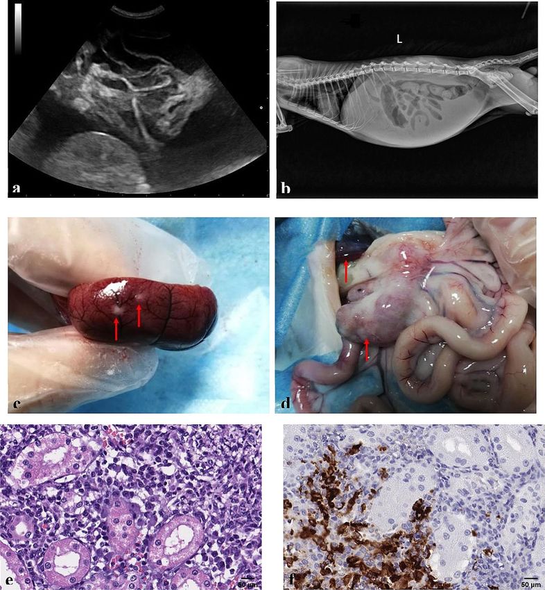
Figure 2. Clinical examination results of a 4-month-old intact female British shorthair cat with effusive FIP.
(a) The ultrasound showed a large amount of ascites, in which the intestine was floating; (b) radiography
revealed a large amount of effusion in the abdominal cavity; (c) the surface of kidney was scattered with
miliary white nodules; (d) ascites and enlarged mesenteric lymph nodes were observed. (e) Moderate focal
pyogranulomatous interstitial nephritis (haematoxylin and eosin stain, × 400). The interstitium of the cortex
showed a focal marked infiltration with macrophages, lymphocytes, neutrophils and some plasma cells; (f) renal
macrophages were stained strongly positive for FCoV antigen with immunohistochemistry, × 400.
Discussion
In the past, FIP was considered a fatal disease by researchers. Meanwhile, it is difficult to apply the IHC gold
standard in the diagnosis of FIP in China. With the development of research, there were gradually found break-
throughs in treatment on FIP, such as studies on GS-441524 and GC3766–9,16. But the side effects of these two
drugs are not clear, and cannot be used for clinical treatment immediately. Therefore, the establishment of a more
accurate diagnostic system is crucial for FIP in China. This paper reviewed 127 FIP cases in Wuhan, China, and
studied the diagnostic methods of several hospitals. It emphasizes the clinical manifestations of clinicians in the
diagnosis of the disease, and perfects the FIP diagnosis system.
Data on 127 cases highly suspected of FIP were collected in this study, where 13 cats underwent exploratory
laparotomy, 9 samples from the cats highly suspected of FIP underwent histopathology, and 6 samples underwent
IHC, all of which were finally confirmed as FIP. Therefore, the inclusion criteria for the cases in this study were
reasonable. There were also many investigations and studies on FIP based on the analysis of highly suspected
cases, which could not be diagnosed by gold standard methods (IHC), but still provided a great reference value
for the diagnosis and treatment of F IP17–22. In addition, the implementation of invasive diagnostic methods
was difficult in clinical practice. Even cat owners who chose euthanasia could not accept autopsy after death.
Therefore, when IHC is not available, how to diagnose a FIP-suspected case more accurately is a problem that
veterinarians pay close attention to at present.
Scientific Reports | (2021) 11:5208 | https://doi.org/10.1038/s41598-021-84754-0 6
Vol:.(1234567890)www.nature.com/scientificreports/
In this study, the prevalence of cats highly suspected of FIP was 0.61%, and Riemer’s research in Germany
showed that the prevalence of FIP was 1.42%14. Earlier studies also showed that the prevalence of FIP was 0.02%
in single-cat or double-cat households, while the prevalence was 5%–10% in catteries23,24. There was no cor-
relation between sex and FIP. However, some studies indicated that male cats were more prone to F IP25. The
prevalence of intact males was significantly higher than that of other groups, and the prevalence of neutered cats
was less, consistent with Rohrbach’s findings26. This may be because sex hormones, especially androgens, have
a negative effect on the immune system, increasing the risk of virus proliferation and m utation27. As reported
previously, this study observed that FIP was significantly associated with age, such that young cats were more
susceptible, possibly because the immature immune system and various stressors often resulted in high viral
loads in young cats. It is possible that the increased rate of unneutered cats may also have been due in part to the
young age of infected cats, meaning that the disease occurred before the age of neutering. Purebred cats were
previously considered to be more prone to FIP. However, a growing number of studies, including this study,
have shown that the proportion of purebred cats was not excessive when compared with the clinic population14.
This study also explored the regularity of the month of FIP onset, which was not considered in other FIP-related
epidemiological surveys, and finally found no significant difference in the prevalence of FIP in each month of
the year. This study recorded stressors in 60 cats, the most common of which was changing environment. There
was no doubt that stress could inhibit immune function and make viruses more susceptible to mutation and
proliferation10. Housing density was considered one of the major risk factors for FIP as well, and overcrowding
could lead to virus mutation and disease development14. However, more of the cats highly suspected of FIP in this
study came from single-cat households. This may be because these cats were exposed to FCoV whilst they lived
in a multicat household during their infancy, and then moved into single-cat household prior to developing FIP.
Most cases of FIP presented as effusive (up to 85.8%), in which ascites accounted for the majority. FIP could
affect systemic organs, most commonly the abdominal organs, including the intestine, mesenteric lymph nodes,
liver, kidney, and spleen. Neurological and ocular signs were typical signs of non-effusive FIP. A retrospective
study of 286 cats with neurological disease showed that FIP was one of the main causes of neurological disease
in cats28. In this study, 52.9% of the infected cats had a temperature over 39.5℃, and 36.5% had a temperature
over 40℃. Other research has shown that the proportion of cats with fever was larger, where 82% of the cats had
a temperature of over 39.5℃ and 39% had a temperature of over 40℃; fever was also found to be more common
in effusive F IP14. There were no cases of pericardial effusion, scrotal swelling or skin papules, which were not
common but could not be ignored in routine examination. The clinical signs of FIP always change over time
(such as the gradual production of effusion and changes in the fundus), so repeated clinical examination was
highly important to avoid delay in diagnosis and treatment.
Changes in haematology and serum biochemistry could only increase or decrease the suspicion of FIP, but
could not be used as a definitive diagnostic method. In this study, there was no obvious indication of hema-
tological changes in the diagnosis of the FIP cases, with HCT decreased in 40.2% of cats, and LYM decreased
in 50% of cats. However, many studies have shown that lymphocytopenia is the most common haematologi-
cal abnormality of FIP induced by natural infection or experiment29,30, caused by virus-induced apoptosis of
T-cells31. In this study, we guess the protein level testing plays an important role in diagnosing FIP cases. ALB
decreased in 58.7% of the cats and GLOB increased in 57.8% of the cats, so when both occurred at the same
time, TP often remained within the normal range. This was why only 14.7% of the cats showed an increase in
TP. Both hyperglobulinemia and hypoalbuminemia could increase the suspicion of FIP, but the most important
serum biochemical abnormality was the A/G ratio. The A/G ratio reduction occurred in almost all cases of FIP.
Different studies had different results on the effective critical value of the A/G r atio32. According to the A/G
ratios in this study (91.7% < 0.6, 85.3% < 0.5, and 54.1% < 0.4), the critical value could be considered as 0.5. When
the A/G ratio > 0.8, the possibility of FIP was very small; when 0.5 < A/G ratio < 0.8, FIP remained a possibility;
when A/G ratio < 0.5, FIP was highly suspected. In addition, FIP was accompanied by inflammatory response,
resulting in increased protein concentration in the acute phase protein33. Therefore, we guess that the increase
in SAA may assist in the diagnosis of FIP.
Compared with serum detection, the detection and analysis of body cavity effusion in cats has better pre-
dictive value32. The positivity rate of Rivalta’s test was 100% in this study. A study indicated that the sensitivity
of Rivalta’s test was 91.3%, the specificity was 65.5%, the positive predictive value was 58.4% and the negative
predictive value was 93.4%34. Rivalta’s test is a cheap and rapid detection method that does not need expensive
instruments and is simple to operate. In view of the good sensitivity and negative predictive value, Rivalta’s
test should be included in the diagnosis of every cat with e ffusion15. The positive rate of FCoV RNA detected
by RT-PCR was 84.7%. However, all coronaviruses, including FCoV, often mutate and recombine, so RT-PCR
designed for specific sequences could not amplify all F CoVs35, and the positive results only represented that the
cat carried FCoV, not the exact FIPV.
In this study a total of 24 cats were treated with GS-441524, of which 23 were cured. Only 1 cat relapsed after
4 weeks of treatment and was eventually euthanised. Treatment with GS-441524 for more than 8 weeks was highly
effective. Treatment with GC376 alone or in combination with GS-441524 was also effective. In fact, most of the
final deaths were due to no treatment, symptomatic treatment alone or euthanasia. In addition, Pruijssers and
Denison indicated that a combination of potent, broad-spectrum anti-coronavirus drugs can enhance efficacy
and reduce the emergence of drug resistance36.
In conclusion, FIP was not correlated with sex, breed or month, but it was significantly correlated with age
and neutering status. FIP occurred significantly more often in young cats or male intact cats. Effusive FIP was the
most common clinical feature. A decreased A/G ratio, increased SAA and lymphopenia were common laboratory
abnormalities. GS-441524 and GC376 were efficient for the treatment of FIP-suspected cats.
Scientific Reports | (2021) 11:5208 | https://doi.org/10.1038/s41598-021-84754-0 7
Vol.:(0123456789)www.nature.com/scientificreports/
Data availability
All data generated or analysed during this study are included in this published article (and its Supplementary
Information files).
Received: 19 June 2020; Accepted: 18 February 2021
References
1. Bank-Wolf, B. R. et al. Mutations of 3c and spike protein genes correlate with the occurrence of feline infectious peritonitis. Vet.
Microbiol. 173, 177–188 (2014).
2. Lewis, C. S. et al. Genotyping coronaviruses associated with feline infectious peritonitis. J. Gen. Virol. 96, 1358 (2015).
3. Norsworthy, G. D., Grace, S.F., Crystal, M.A. & Tilley, L.P. The feline patient (4th ed), 181–183 (John Wiley & Sons, New York, 2011).
4. Pedersen, N. C. A review of feline infectious peritonitis virus infection: 1963–2008. J. Feline Med. Surg. 11, 225–258 (2009).
5. Sykes, J. E. Canine and Feline Infectious Diseases, 195–208 (W. B. Saunders, 2014).
6. Kim, Y. et al. Reversal of the progression of fatal coronavirus infection in cats by a broad-spectrum coronavirus protease inhibitor.
PLoS Pathog. 12, 1–18 (2016).
7. Murphy, B. G. et al. The nucleoside analog GS-441524 strongly inhibits feline infectious peritonitis (FIP) virus in tissue culture
and experimental cat infection studies. Vet. Microbiol. 219, 226–233 (2018).
8. Pedersen, N. C. et al. Efficacy of a 3C-like protease inhibitor in treating various forms of acquired feline infectious peritonitis. J.
Feline Med. Surg. 20, 378–392 (2018).
9. Pedersen, N. C. et al. Efficacy and safety of the nucleoside analog GS-441524 for treatment of cats with naturally occurring feline
infectious peritonitis. J. Feline Med. Surg. 21, 271–281 (2019).
10. Addie, D. D. et al. Feline infectious peritonitis. ABCD guidelines on prevention and management. J. Feline Med. Surg. 11, 594–604
(2009).
11. Greene, C. E. Infectious diseases of the dog and cat, 92–108 (W. B. Saunders, 2012).
12. Kipar, A. & Meli, M. L. Feline infectious peritonitis: still an enigma?. Vet. Pathol. 51, 505–526 (2014).
13. Pedersen, N. C. An update on feline infectious peritonitis: diagnostics and therapeutics. Vet. J. 201, 133–141 (2014).
14. Riemer, F., Kuehner, K. A., Ritz, S., Sauter-Louis, C. & Hartmann, K. Clinical and laboratory features of cats with feline infectious
peritonitis–a retrospective study of 231 confirmed cases (2000–2010). J. Feline Med. Surg. 18, 348–356 (2016).
15. Felten, S. & Hartmann, K. Diagnosis of feline infectious peritonitis: a review of the current literature. Viruses. 11, 1068 (2019).
16. Dickinson, P. J. et al. Antiviral treatment using the adenosine nucleoside analogue GS-441524 in cats with clinically diagnosed
neurological feline infectious peritonitis. J. Vet. Intern. Med. 34, 1587–1593 (2020).
17. Soma, T. & Ishii, H. Detection of feline coronavirus antibody, feline immunodeficiency virus antibody, and feline leukemia virus
antigen in ascites from cats with effusive feline infectious peritonitis. J. Vet. Med. Sci. 66, 89–90 (2004).
18. Simons, F. A. et al. A mRNA PCR for the diagnosis of feline infectious peritonitis. J. Virol. Methods. 124, 111–116 (2005).
19. Can-Şahna, K., Ataseven, V. S., Pınar, D. & Oğuzoğlu, T. Ç. The detection of feline coronaviruses in blood samples from cats by
mRNA RT-PCR. J. Feline Med. Surg. 9, 369–372 (2007).
20. Sharif, S. et al. Evaluation of feline coronavirus viraemia in clinically healthy and ill cats with feline infectious peritonitis. J. Anim.
Vet. Adv. 10, 18–22 (2011).
21. Li, C. et al. Circulation and genetic diversity of Feline coronavirus type I and II from clinically healthy and FIP-suspected cats in
China. Transbound. Emerg. Dis. 66, 763–775 (2019).
22. Lorusso, E. et al. Discrepancies between feline coronavirus antibody and nucleic acid detection in effusions of cats with suspected
feline infectious peritonitis. Res. Vet. Sci. 125, 421–424 (2019).
23. Pedersen, N. C. Serologic studies of naturally occurring feline infectious peritonitis. Am. J. Vet. Res. 37, 1449–1453 (1976).
24. Addie, D. D. & Jarrett, O. A study of naturally occurring feline coronavirus infections in kittens. Vet. Rec. 130, 133–137 (1992).
25. Worthing, K. A. et al. Risk factors for feline infectious peritonitis in Australian cats. J. Feline Med. Surg. 14, 405–412 (2012).
26. Rohrbach, B. W. et al. Epidemiology of feline infectious peritonitis among cats examined at veterinary medical teaching hospitals.
J. Am. Vet. Med. Assoc. 218, 1111–1115 (2001).
27. Grossman, C. J. Interactions between the gonadal steroids and the immune system. Science 227, 257–261 (1985).
28. Bradshaw, J. M., Pearson, G. R. & Gruffydd-Jones, T. J. A Retrospective Study of 286 Cases of Neurological Disorders of the Cat.
J. Comp. Pathol. 131, 112–120.
29. Dean, G. A., Olivry, T., Stanton, C. & Pedersen, N. C. In vivo cytokine response to experimental feline infectious peritonitis virus
infection. Vet. Microbiol. 97, 1–12 (2003).
30. de Groot-Mijnes, J. D. F., van Dun, J. M., van der Most, R. G. & de Groot, R. J. Natural history of a recurrent feline coronavirus
infection and the role of cellular immunity in survival and disease. J. Virol. 79, 1036–1044 (2005).
31. Vermeulen, B. L. et al. Suppression of NK cells and regulatory T lymphocytes in cats naturally infected with feline infectious
peritonitis virus. Vet. Microbiol. 164, 46–59 (2013).
32. Hartmann, K. et al. Comparison of different tests to diagnose feline infectious peritonitis. J. Vet. Intern. Med. 17, 781–790 (2003).
33. Hazuchova, K., Held, S. & Neiger, R. Usefulness of acute phase proteins in differentiating between feline infectious peritonitis and
other diseases in cats with body cavity effusions. J. Feline Med. Surg. 19, 809–816 (2017).
34. Fischer, Y., Sauter-Louis, C. & Hartmann, K. Diagnostic accuracy of the Rivalta test for feline infectious peritonitis. Vet. Clin.
Pathol. 41, 558–567 (2012).
35. Tasker, S. Diagnosis of feline infectious peritonitis: Update on evidence supporting available tests. J. Feline Med. Surg. 20, 228–243
(2018).
36. Pruijssers, A. J. & Denison, M. R. Nucleoside analogues for the treatment of coronavirus infections. Curr. Opin. Virol. 35, 57–62
(2019).
Acknowledgements
The authors would like to thank the veterinarians and cat owners, without whose help this study could not
have been completed. The study was funded by the National Key Research and Development Plan (No.
2020YFC08845600) and the teaching hospital of Huazhong Agricultural University. The study was carried out
in compliance with the ARRIVE (Animal Research: Reporting of In Vivo Experiments) guidelines (https://arriv
eguidelines.org).
Author contributions
Study conception and design: C.H. and Y.Yin. Data collection: Y.Y., H.O., W.J., J.L., X.L. and J.L. Data analysis:
Y.Y., T.L., C.W. and X.L. Manuscript writing: Y.Y. and C.H. All authors reviewed the manuscript.
Scientific Reports | (2021) 11:5208 | https://doi.org/10.1038/s41598-021-84754-0 8
Vol:.(1234567890)www.nature.com/scientificreports/
Competing interests
The authors declare no competing interests.
Additional information
Supplementary Information The online version contains supplementary material available at https://doi.
org/10.1038/s41598-021-84754-0.
Correspondence and requests for materials should be addressed to C.H.
Reprints and permissions information is available at www.nature.com/reprints.
Publisher’s note Springer Nature remains neutral with regard to jurisdictional claims in published maps and
institutional affiliations.
Open Access This article is licensed under a Creative Commons Attribution 4.0 International
License, which permits use, sharing, adaptation, distribution and reproduction in any medium or
format, as long as you give appropriate credit to the original author(s) and the source, provide a link to the
Creative Commons licence, and indicate if changes were made. The images or other third party material in this
article are included in the article’s Creative Commons licence, unless indicated otherwise in a credit line to the
material. If material is not included in the article’s Creative Commons licence and your intended use is not
permitted by statutory regulation or exceeds the permitted use, you will need to obtain permission directly from
the copyright holder. To view a copy of this licence, visit http://creativecommons.org/licenses/by/4.0/.
© The Author(s) 2021
Scientific Reports | (2021) 11:5208 | https://doi.org/10.1038/s41598-021-84754-0 9
Vol.:(0123456789)You can also read