A STUDY OF EUCALYPTUS GRANDIS AND EUCALYPTUS GLOBULUS BRANCH WOOD MICROSTRUCTURE - Brill
←
→
Page content transcription
If your browser does not render page correctly, please read the page content below
IAWA Journal, Vol. 26 (2), 2005: 203 –210

  A STUDY OF EUCALYPTUS GRANDIS AND EUCALYPTUS GLOBULUS
              BRANCH WOOD MICROSTRUCTURE

             Russell Washusen1, Robert Evans1 & Simon Southerton2
                         CSIRO Forestry and Forest Products, Australia

                                         SUMMARY

       Experimental measurements of cellulose crystallite width and microfibril
       angle (MFA) by X-ray diffractometry on SilviScan-2 and by conventional
       microtechniques revealed that the branch wood of the two species exhib-
       ited very similar trends in cellulose crystallite width and MFA. Cellulose
       crystallite width was greater on the upper side of the branches. Tension
       wood, as defined by the occurrence of gelatinous fibres, was found where
       cellulose crystallite width was greater than 3.0 nm and 3.1 nm in Eucalyp-
       tus grandis and E. globulus respectively. In the tension wood zones, MFA
       was lower than in the rest of the samples and so could be used to differ-
       entiate tension wood. On the lower side of the branches MFA determined
       from X-ray diffractometry unexpectedly exceeded 40° and fibres were
       often buckled in both the tangential and radial directions in both species.
       This local variation in the direction of the fibre axes contributed only
       slightly to the magnitude of the MFA determined by SilviScan-2. Even
       given this misalignment, the additional evidence gained from pit angles
        and cracks in fibre walls suggested that the MFA was indeed around 40°
       in the lower radius of the branches. This MFA is considerably larger than
       would be expected for eucalypt stem wood and it is suggested that op-
       posite wood in eucalypt branches may provide a complimentary structural
       role to that of the tension wood. Experimental measurements of crystallite
       width produced by SilviScan-2 may be used to accurately locate tension
       wood zones in both species.
       Key words: Tension wood, cellulose crystallite width, microfibril angle,
       X-ray diffraction, SilviScan 2, eucalypts.

                                     INTRODUCTION

Eucalyptus grandis Hill ex Maiden and Eucalyptus globulus Labill. are commercially
important hardwood plantation species for paper manufacture and are becoming a sig-
nificant resource for the production of high-quality solid wood. Tension wood in both
species is a critical wood property for solid wood production and may influence the suit-

1) Private Bag 10, Clayton South, Victoria, 3169, Australia.
2) PO Box E4008, Kingston, ACT, 2604, Australia.
Associate Editor: Michael Wiemann

                                                                  Downloaded from Brill.com02/27/2021 11:57:08AM
                                                                                                   via free access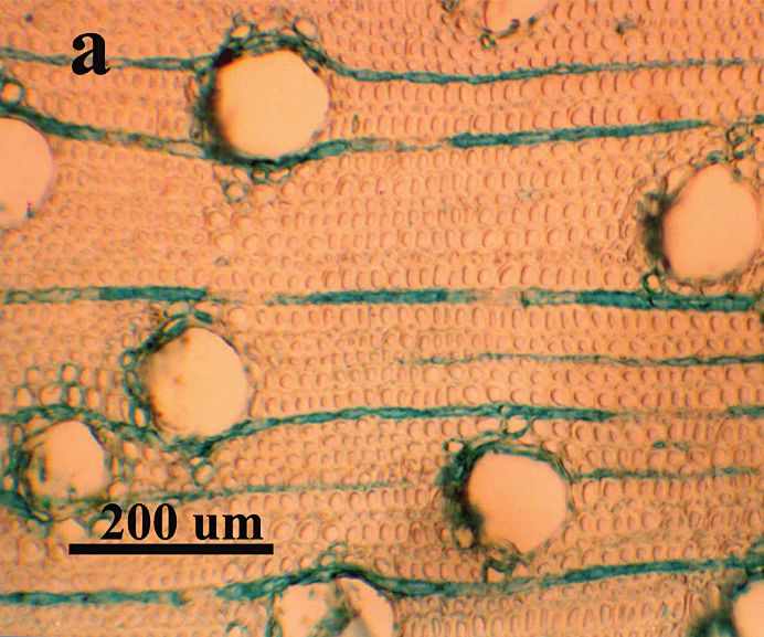
204                                                         IAWA Journal, Vol. 26 (2), 2005

ability of solid wood from these species in manufacturing processes. Tension wood is
a reaction wood in hardwoods that can produce high transverse and longitudinal shrink-
age in solid wood and very high growth stresses. These stresses present difficulties dur-
ing primary processing (Bekele 1995; Washusen et al. 2000). The study of tension
wood and the factors controlling its formation will support efforts to reduce its occur-
rence and severity and thereby improve processing efficiencies. Traditionally, tension
wood occurrence has been confirmed through the anatomical identification of unligni-
fied gelatinous layers in the fibre wall (Wardrop & Dadswell 1948, 1955). Indirectly
it has also been identified through shrinkage during drying of small wood samples or
by direct measurement of growth strain at the stem periphery of trees and logs. These
methods are tedious and are a difficult way of identifying tension wood occurrence and
severity (Washusen 2000; Washusen et al. 2003).
    As part of ongoing studies to assess the effectiveness of SilviScan 2 in tension wood
detection, and broader studies of gene expression in developing xylem, the micro-
structure of Eucalyptus grandis and E. globulus branch wood was examined by X-ray
diffractometry on SilviScan-2 and by conventional microtechniques. Recent research at
CSIRO-FFP in Eucalyptus globulus found that cellulose crystallite width, estimated from
the intensity profiles of X-ray diffraction patterns produced by SilviScan-2, was posi-
tively associated with tangential shrinkage in solid wood (Washusen & Evans 2001a)
and gelatinous fibres in tension wood (Washusen & Evans 2001b). While this associa-
tion with tension wood is not new (Goto et al. 1975; Nishimura et al. 1981; Blaho et al.
1994), the results suggest that the width of cellulose crystallites may be a definitive meas-
ure of tension wood severity and it may be useful to develop its measurement on auto-
mated systems such as SilviScan-2, which incorporates an X-ray diffractometer (Evans
1994, 1999; Evans et al. 1995). Originally developed for the rapid characterization of
pulpwood, SilviScan-2 is more recently finding application in the solid wood processing
industries. The ability to rapidly measure crystallite width may allow routine identi-
fication of tension wood and associated high growth stresses. In this study the experi-
mental measurements of crystallite width currently produced by SilviScan-2 are as-
sessed for their potential to detect tension wood in branch samples where tension wood
commonly forms on the upper side. In addition, microfibril angle is assessed as another
possible indicator of tension wood.

                            MATERIALS AND METHODS
The wood samples
    The samples were taken from single branches that were oriented at an angle of ap-
proximately 80° to the vertical main stem of two 9-year-old trees of Eucalyptus grandis
and E. globulus. These samples were collected as part of a wider molecular study, that
is reported elsewhere, comparing gene expression in developing xylem for upper and
lower branches. Discs 25 mm thick were cut from the branches approximately 20 cm
from the main stem and marked to record the location of the top and bottom of the
branch (in preliminary work, discs at this location taken from branches at a large angle

                                                               Downloaded from Brill.com02/27/2021 11:57:08AM
                                                                                                via free access
Washusen, Evans & Southerton — Eucalyptus branch wood                                         205

to the main stem contained well-defined tension wood zones in both species). The discs
were placed in ethanol in three steps to remove water, each step taking approximately
one week. The discs were then dried at room temperature to about 8% moisture content.
This procedure prevents cellular collapse on drying. A diametral strip 10 mm tangenti-
ally × 25 mm radially running through the pith, was cut from each disc. The strip was
then cut to produce 2 matching diametral strips. One strip was used for X-ray diffraction
analysis and the other was used for anatomical analysis by light microscopy.

X-ray diffraction
   The strips for SilviScan-2 were mounted on wooden sample holders with PVA glue
and trimmed to a thickness of 2 mm in the tangential direction and 7 mm in the longi-
tudinal (fibre axis) direction using a twin-blade saw.
   X-ray diffraction patterns were obtained using SilviScan-2 over 7–30 seconds on
a CCD area detector. The SilviScan-2 system was set up with a rotating copper anode
operating at 45 kV and 15 mA and a focussing capillary giving a spot size of approxi-
mately 200 μm at the sample. Diffraction patterns were acquired at 500 μm intervals
and with a 30 second exposure. At each position along the sample strip, MFA and crys-
tallite width were determined.
   The 002 peak in the diffraction patterns contained information on both MFA and crys-
tallite width. According to the Scherrer formula (Cutter & Murphey 1972), crystallite
width is inversely proportional to the width, at half maximum intensity, of the 002 peak
in the 2θ (radial) direction. Software has been developed on SilviScan-2 for calculat-
ing crystallite width using the Scherrer formula, and these measurements have been
independently validated from manual measurements taken from Eucalyptus globulus
tension wood and normal wood samples (Washusen & Evans 2002).

Microtechniques
   The matching diametral strips were saturated in water and 12 μm thick transverse sec-
tions cut on an American Optical sliding microtome along the entire transverse face of
each strip on the face closest to the sample used for SilviScan analysis. The sections were
stained with 1% aqueous alcian blue and examined microscopically to identify the loca-
tion of tension wood. Alcian blue stains for insoluble carbohydrates (Gurr 1960; Gahan
1984) and hence stains blue the cellulose in the unlignified layers of gelatinous fibres
that are characteristic of tension wood.
   In addition, ten successive radial/longitudinal and tangential/longitudinal sections,
20 μm thick, were cut from regions where unusually large MFA was recorded at the
bottom of both branch samples. Similar sections were taken from the top of the branches
for comparison. The sections were taken from wood left after the 2 mm strip was cut
on the twin blade saw. Fibre orientation, pit angles, and cracks in fibre walls were
examined microscopically in these sections without staining. Photomicrographs were
taken of 12 μm sections that typified the fibre alignment, after staining for lignin with
1% aqueous crystal violet (Conn & Darrow 1948).

                                                               Downloaded from Brill.com02/27/2021 11:57:08AM
                                                                                                via free access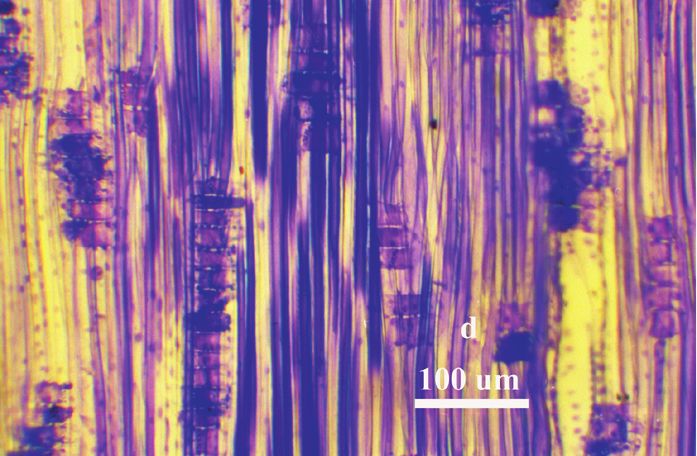
206 IAWA Journal, Vol. 26 (2), 2005
RESULTS AND DISCUSSION
The profiles of microfibril angle (MFA) and cellulose crystallite width for Eucalyptus
grandis and E. globulus are shown in Figure 1a & b respectively. These traces are
remarkably similar in many characteristics with very high MFA at the bottom of both
branch samples and similar trends in crystallite width.
(a) Eucalyptus grandis
80 3.6
Tension wood
70 bands 3.4
Microfibril angle (degrees)
Crystallite width (nm)
60 3.2
50 3.0
40 2.8
Pith
30 2.6
20 2.4
10 2.2
Lower side Upper side
0 2.0
0 5 10 15 20 25 30 35 40
Distance (mm)
(b) Eucalyptus globulus
60 4.0
Tension wood bands
3.8
Microfibril angle (degrees)
50
3.6
Crystallite width (nm)
40 3.4
3.2
30 3.0
Pith 2.8
20
2.6
10 2.4
2.2
Lower side Upper side
0 2.0
0 5 10 15 20 25 30 35 40 45 50 55 60
Distance (mm)
Fig. 1. Plots of microfibril angle (:) and cellulose crystallite width (-) through a Eucalyptus
grandis branch (a) and Eucalyptus globulus branch (b).
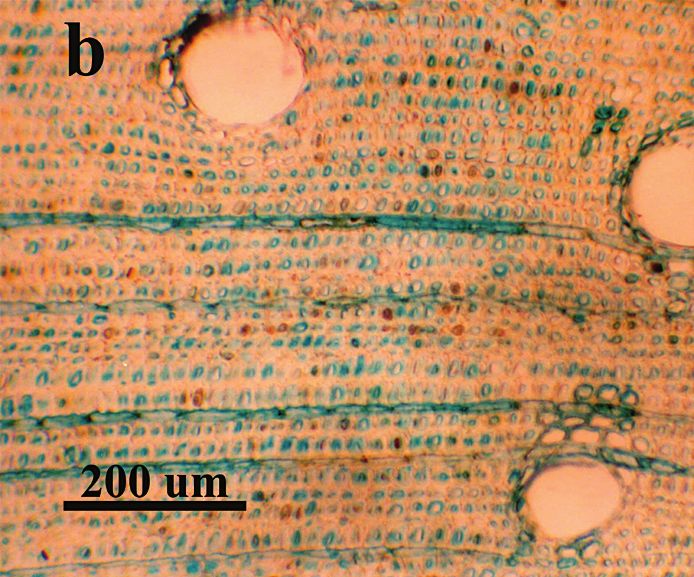
Cellulose crystallite width
The low crystallite width was found to coincide with normal wood (Fig. 2a) and
the high peaks coincided with well-developed tension wood bands (shown in part in
Fig. 2b) on the upper side of the branches. These tension wood zones were very similar
anatomically in both species, with numerous gelatinous fibres and thick gelatinous lay-
ers (Fig. 2b) and thin normal walls that suggested an S1 +G secondary wall structure.
Downloaded from Brill.com02/27/2021 11:57:08AM
via free access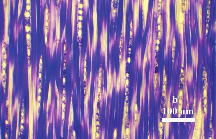
Washusen, Evans & Southerton — Eucalyptus branch wood                                         207

In normal wood zones, the crystallite width ranged from approximately 2.7–3.0 nm
(E. grandis) and 2.8–3.1 nm (E. globulus) and in tension wood zones from 3.0–3.5 nm
(E. grandis) and 3.1–3.8 nm (E. globulus). The range in crystallite width for tension
wood is similar but not identical to the range found by Washusen and Evans (2001b),
where crystallite width was calculated manually from measurements of the width of
the 002 peak. This may be partly due to the difficulty in quantifying tension wood
severity by histochemical methods, or even identification of tension wood that does
not stain well with lignin stains. In the earlier work cited above several fibres stained
as if they were normally lignified but displayed many of the characteristics of tension
wood, such as distortion of fibres in the transverse plane, little or no visible lumen,
cracks in the outer wall and/or distortion or even separation of the bulk of the secondary
wall from the inner layers. We suggest that the small differences between both studies
are probably due to differences in the way the width of the peak was measured. The
peak width determined by SilviScan-2 is measured at half of the total peak height. In
earlier experimental work the width of the 002 peak was measured at half the height
of the peak above the trough between the 101 and 002 reflections. This method gives
a smaller peak width and as there is an inverse relationship between peak width and
crystallite width (Cutter & Murphey 1972), larger crystallite width measurements will
be produced. Even given this difference, good correlations have been observed between
data measured by the two methods (Washusen & Evans 2002).

MFA in tension wood zones
   As expected from earlier work with tension wood by Wardrop and Dadswell (1955)
and numerous other researchers, the tension wood zones aligned with the zones with
lowest MFA (Fig. 1a & b), and the MFA values could be used to differentiate tension
wood. This is in contrast to work by Washusen et al. (2001) in stem wood of E. glo-
bulus where the MFA data produced by SilviScan-2 could not differentiate tension wood
because of low MFA in normal wood zones. This suggests that there are greater micro-
structural differences between tension wood and normal wood of these branch samples
than is the case in stem wood.

MFA on the lower side of branches
   The very high MFA recorded on the lower side of both branch samples was unex-
pected. High MFA has also been observed by the authors (unpublished data) in wood
from the lower side of Eucalyptus nitens branches, and is typical of compression wood
zones in softwoods (unpublished data). The high MFA, as estimated by X-ray diffrac-
tometry, cannot be explained by the buckled pattern of fibres observed in tangential sec-
tions and occasionally in the radial sections (Fig. 3a & c). As SilviScan-2 calculates
MFA from the standard deviation of the intensity profile of the 002 peak, dispersion in
fibre orientation may add to the dispersion of the 002 diffraction peak, thereby causing
overestimates of MFA. However, based on an examination of fibre alignment in the
lower side of the branch (Fig. 3a & c), it appears unlikely that the effect of local mis-

                                                               Downloaded from Brill.com02/27/2021 11:57:08AM
                                                                                                via free access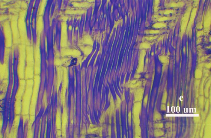
208                                                            IAWA Journal, Vol. 26 (2), 2005

Fig. 2. Eucalyptus grandis transverse sections (20 µm thick) stained with alcian blue. – a: Normal
wood on the lower side of the branch. – b: Tension wood band from the top of the branch. Note
that the gelatinous layers in the tension wood sample are stained blue and appear dark.

                                                                   Downloaded from Brill.com02/27/2021 11:57:08AM
                                                                                                    via free access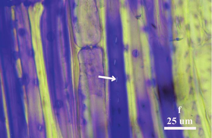
Washusen, Evans & Southerton — Eucalyptus branch wood                                                 209

alignment of fibres would result in an increase in MFA of more than 3 degrees. The
effect is likely to be small because the variation in fibre axis orientation is less than
15 degrees. In the upper (tension) side of the branch samples the fibres were relatively
straight (Fig. 3b & d). Evidence from the pit angles (Fig. 3e) and cracks and striations
on fibre walls indicated that the MFA was indeed large and in some zones approached
45° (Fig. 3e) on the lower side. On the upper side of the branches similar evidence sup-
ported the X-ray diffractometric data; the MFA was indeed very low (Fig. 3f). Figure 3f
also shows that fibres with normally lignified walls may have very low MFA. In this
case the indicated pit is aligned almost axially, indicating a low MFA in this part of
the fibre.

                                         CONCLUSIONS

Tension wood zones confirmed by histochemical assessment were found to occur in
wood with cellulose crystallite widths above 3.0 and 3.1 nm in Eucalyptus grandis and
E. globulus branch samples, respectively. The precise matching of crystallite width data
with tension wood occurrence indicates that the experimental measurements of crystal-
lite width produced by SilviScan-2 are capable of accurately locating tension wood
zones in both species. Further validation work in these two species and expansion to
other species is warranted in an attempt to establish the universality of crystallite width
measurement as a tension wood detection method. This work should be conducted in
other branch samples where tension wood can be conveniently located, and expanded
to stem wood where tension wood has formed.

The very large MFA in opposite wood, which regularly exceeded 40° on the lower side
of both branch samples, was surprising and not often seen in SilviScan-2 data. However,
the large MFA was confirmed by evidence from pit angles and the cracks and striations in
fibre walls. The high MFA is similar to that often recorded for compression wood zones
in softwoods and suggests that it might be a response to high compressive stresses that
develop on the lower side of branches. Such wood may therefore provide a complimen-
tary structural role to the tension wood that formed on the upper side of the branches.
The fibre misalignment and buckling may also be a response to the very high compres-
sive stress.

←
Fig. 3. Examples of tangential and radial microtome sections of Eucalyptus grandis branch wood
stained with crystal violet. – a: Tangential section from the lower side of the branch displaying
the typical irregular alignment of fibres. – b: Tangential section from the upper side showing
normal fibre alignment. – c: Radial section from the lower side showing buckling of some fibres. –
d: Radial section from the upper side showing normal fibres. – e: Tangential section from the
lower side showing irregular alignment of some fibres and the oblique angle of the pits indicat-
ing a high microfibril angle. – f: Tangential section from the upper side showing an almost axial
alignment of the pits in the fibres indicating a very low microfibril angle; the staining with crystal
violet indicates that these are normal lignified fibres with low microfibril angle.

                                                                       Downloaded from Brill.com02/27/2021 11:57:08AM
                                                                                                        via free access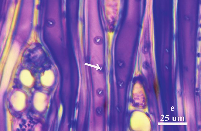
210                                                            IAWA Journal, Vol. 26 (2), 2005

                                        REFERENCES
Bekele, T. 1995. Degradation of boards of Eucalyptus globulus Labill. and Eucalyptus camaldu-
   lensis Dehnh. during air drying. Holz Roh- u. Werkstoff 53: 407–412.
Blaho, J., M. Vozar & J. Sindler. 1994. Some properties of reaction wood and normal wood of
   beech (Fagus sylvatica L.). Zbornik Vedeckych Prac Drevarskej Fakulty Technickej Uni-
   verzity vo Zvolene: 297–306.
Conn, H. J. & M. A. Darrow. 1948. Staining procedures used by the Biological Stain Commis-
   sion. Parts II and III edited by H. J. Conn & M. J. Darrow: pp. 181–194. The Williams and
   Wilkins Company, Baltimore.
Cutter, B. E. & W. K. Murphey. 1972. X-ray measurement of crystallite size in wood. Wood and
   Fiber 4: 43– 44.
Evans, R. 1994. Rapid measurement of the transverse dimensions of tracheids in radial wood sec-
   tions from Pinus radiata. Holzforschung. 48: 168–173.
Evans, R. 1999. A variance approach to the X-ray diffractometric estimation of microfibril angle
   in wood. Appita J. 52: 283–294.
Evans, R., G. Downes, R. Menz & S. Stringer. 1995. Rapid measurement of variation in tracheid
   dimensions in a radiata pine tree. Appita J. 48: 134–138.
Gahan, P. B. 1984. Plant Histochemistry and Cytochemistry: pp. 301. Academic Press.
Goto, T., H. Harada & H. Saiki. 1975. Cross-sectional view of microfibrils in gelatinous layer of
   poplar tension wood (Populus euramericana). Mokuzai Gakkaishi. 21: 537–542.
Gurr, E. 1960. Encyclopaedia of microscopic stains: 19–393. Leonard Hill (Books) Ltd, London.
Nishimura, H., T. Okano & I. Asano. 1981. Fine structure of wood cell walls. II. Crystallite size
   and several peak positions of X-ray diagram of cellulose I. Mokuzai Gakkaishi. 27: 709–715.
Wardrop, A.B. & H.E. Dadswell. 1948. The nature of reaction wood. I. The structure and proper-
   ties of tension wood fibres. Austral. J. Sci. Res. B 1: 3.
Wardrop, A. B. & H. E. Dadswell. 1955. The nature of reaction wood. IV. Variation in cell wall
   organisation of tension wood fibres. Austral. J. Bot. 3: 177–189.
Washusen, R. 2000. The occurrence and characteristics of tension wood and associated wood
   properties in Eucalyptus globulus Labill. PhD Thesis. Dept. of Forestry, the University of Mel-
   bourne. pp 256.
Washusen, R., P. Ades, R. Evans, J. Ilic & P. Vinden. 2001. Relationships between density, shrink-
   age, extractives content and microfibril angle in tension wood from three provenances of
   10-year-old Eucalyptus globulus Labill. Holzforschung 55: 176–182.
Washusen, R., P. Blakemore, R. Northway, P. Vinden & G. Waugh. 2000. Recovery of dried ap-
   pearance grade timber from Eucalyptus globulus Labill. grown in plantations in medium
   rainfall areas of the southern Murray-Darling Basin. Austral. For. 63: 195–201.
Washusen, R. & R. Evans. 2001a. Prediction of wood tangential shrinkage from cellulose crystal-
   lite width in one 11-year-old tree of Eucalyptus globulus Labill. Austral. For. 64: 123–126.
Washusen, R. & R. Evans. 2001b. The association between cellulose crystallite width and tension
   wood occurrence in Eucalyptus globulus Labill. IAWA J. 22: 235–243.
Washusen, R. & R. Evans. 2002. The measurement of cellulose crystallite width by X-ray diffrac-
   tion on SilviScan-2 and its possible application in eucalypt and acacia species. In, Wood
   Structure and Properties ʻ02. In: J. Kúdela & S. Kurjatko (eds.), Wood Structure and Proper-
   ties ʼ02: 7–12. Arbora Publishers, Zvolen, Slovakia.
Washusen, R., J. Ilic & G. Waugh. 2003. The relationship between longitudinal growth strain, tree
   form and tension wood at the stem periphery of 10 to 11 year-old Eucalyptus globulus Labill.
   Holzforschung 57: 308 –316.

                                                                   Downloaded from Brill.com02/27/2021 11:57:08AM
                                                                                                    via free access
You can also read