Accuracy Verication of 4D-CT Analysis of Knee Joint Movements: A Pilot Study Using a Knee Joint Model and Motion-capture System
←
→
Page content transcription
If your browser does not render page correctly, please read the page content below
Accuracy Verification of 4D-CT Analysis of Knee Joint
Movements: A Pilot Study Using a Knee Joint Model and
Motion-capture System
Takuya Adachi
Tokyo Medical and Dental University: Tokyo Ika Shika Daigaku
Yuki Kato ( nusportskato@gmail.com )
Kameda Medical Center https://orcid.org/0000-0003-1201-6176
Daii Kiyotomo
Taijin Nakashima, Co. Ltd.
Katsushige Kawamukai
Kameda Medical Center
Yoichi Machida
Kameda Medical Center
Research Article
Keywords: 4D-CT, image analysis, motion analysis, joint kinematics, computed tomography
Posted Date: September 27th, 2021
DOI: https://doi.org/10.21203/rs.3.rs-923912/v1
License: This work is licensed under a Creative Commons Attribution 4.0 International License. Read Full
License
Page 1/14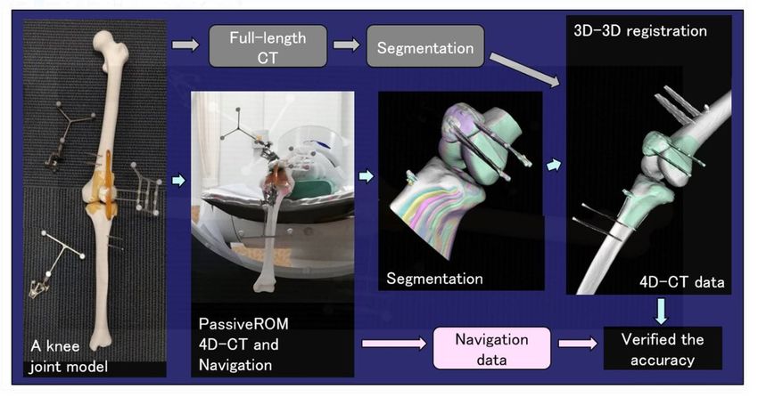
Abstract
Background

Four-dimensional CT(4D-CT) is an advanced imaging method with the ability to acquire kinematic and three-
dimensional morphological information. Although its use for analysis of the six degrees of freedom in the knee is
expected, its accuracy has not been reported. This study aimed to use the optical motion-capture method to verify the
accuracy of 4D-CT analysis of knee joint movement.

Methods

One static CT and three 4D-CT examinations of the knee joint model were obtained. The knee joint model was
passively moved in the CT gantry during 4D-CT acquisitions. 4D-CT and static CT examinations were matched to
perform 3D-3D registration. An optical motion-capture system recorded the position-posture of the knee joint model
simultaneously with the 4D-CT acquisitions. These results were used as the correct answer value, the position-posture
measurements using 4D-CT were compared to these values, and the accuracy of the 4D-CT analysis of knee joint
movements was quantitatively assessed.

Results

The position-posture measurements obtained from 4D-CT showed similar tendency to those obtained from the motion-
capture system. In the femorotibial joint, the difference in the spatial orientation between the two measurements was
0.7 mm in the X direction, 0.9 mm in the Y direction, and 2.8 mm in the Z direction. The difference in angle was 1.9° in
the varus/valgus direction, 1.1° in the internal/external rotation, and 1.8° in extension/flexion. In the patellofemoral
joint, the difference between the two measurements was 0.9 mm in the X direction, 1.3 mm in the Y direction, and 1.2
mm in the Z direction. The difference in angle was 0.9° for varus/valgus, 1.1° for internal/external rotation, and 1.3° for
extension / flexion.

Conclusions

4D-CT with 3D-3D registration could record the position-posture of knee joint movements with an error of less than 3
mm and less than 2° when compared with the highly accurate motion-capture system. Knee joint movement analysis
using 4D-CT with 3D-3D registration showed excellent accuracy for in vivo applications.

Background
The prevalence of locomotor disorders is expected to increase with the aging of the global population. Elderly people
with locomotor disorders may experience joint pain and require nursing care support, and locomotor disorders are
reported to account for 12% of the "bedridden" cases. Knee osteoarthritis is a common locomotor disorder estimated to
affect 24 million individuals, and its prevention and treatment are of substantial importance [1]. Surgical treatments
such as total knee arthroplasty (TKA) have been performed for advanced knee osteoarthritis, and excellent results
have been reported. However, some reports have also described TKA failure caused by infection, loosening,
malalignment, patellofemoral issues, and instability [2–4].

Kinetically abnormal joint movement is involved in the progression of osteoarthritis and articular cartilage damage [5].
Therefore, accurate analysis of the six degrees of freedom of the knee joint is important for tracking the onset and
progression of osteoarthritis and for assessing kinematic changes after medical interventions such as surgeries and
rehabilitation. Several methods for evaluating dynamic knee joint movement have been reported to date, including the

                                                        Page 2/14
motion-capture method and the 2D-3D matching method. Motion-capture systems that measure "positions of human
bodies, animals, and objects" and "time-series movements of joints" have recently gained acceptance. These systems
are classified into the “optical type,” which is widely used, and the “non-optical type,” which include mechanical
motion-capture systems and magnetic systems. The principles of the optical motion-capture method were proposed
by Rashid et al. [6 33] in 1980. In this method, reflective markers are attached to an object, and its coordinates are
measured using a camera. Because of its high accuracy in detection of the position and degree of free movement of
objects, attempts have been made to use this method to evaluate gross joint movement. However, this method may
not necessarily show a high accuracy when applied to the knee joint, since there is a gap between the movements of
bone and soft tissues (skin, subcutaneous fat, and muscle) [7–9]. Implantation of beads in the bone to reduce this gap
has been attempted, although it is an invasive evaluation [10, 11].

The 2D-3D matching method is a technique for superimposing a projected image (2D data) created from 3D-CT (3D
data) on an X-ray fluoroscopic image (2D data). This enables measurement of distances and angles in three axial
directions, and it has been used in computer-assisted navigation systems for analysis of joint dynamics. This method
enables highly accurate measurement of knee joint movement, especially for biplane analysis [10, 12–14]. However,
evaluation of the patella with a few morphological features is difficult [15 11], necessitating an additional CT scan to
obtain three-dimensional morphological information [15].

Recent improvements in the multi-row structure of CT systems have enabled acquisition of a wide range at high speed.
Four-dimensional CT (4D-CT) adds a time axis to three-dimensional data and allows continuous CT imaging on a time-
series basis. This method has widespread clinical application for assessments such as evaluation of blood flow
dynamics [16–19]. In the field of orthopedic surgery, this method is expected to improve evaluations of joint dynamics
and functions [20]. Unlike the 2D-3D matching method, 4D-CT can acquire three-dimensional morphological
information as well as kinematic information. However, there are concerns about the accuracy of joint dynamics
evaluations performed by 4D-CT due to the presence of motion artifacts and partial-volume effects [21]. In response to
this concern, Oki et al. [22] reported a dynamic evaluation method using 3D-3D registration, which superimposes 3D-
CT images created from static lower-limb full-length CT onto 4D-CT. However, no studies have been conducted to verify
the position-posture evaluation accuracy of knee joint movement using 4D-CT.

We hypothesized that 4D-CT using 3D-3D registration would enable highly accurate analysis of knee joint movement.
Thus, the purpose of this study was to use the optical motion-capture method to verify whether high-precision analysis
of knee joint movement is possible with 4D-CT using 3D-3D registration.

Methods
A knee-joint model was used as the subject. The model was passively moved inside the gantry of the CT device, and
4D-CT data were obtained, which were matched with the static CT data of the entire knee joint model to perform 3D-3D
registration. An optical motion-capture system was used to verify the accuracy of 4D-CT data with 3D-3D registration
(Fig. 1).

Bone model creation
A human knee skeleton model, including the femur, tibia, and patella, was manufactured by the laminate-shaping
method using ZPrinter150 (3D Systems, Inc., California, USA), yielding the same shape as “SAWBONES SKU: 1148-3”
(Pacific Research Laboratories, Inc. Sawbones, WA, USA). Cushioning materials with a thickness of approximately 5
mm were taped to the articular surface of the tibial model and the patellofemoral joint of the femur model to mimic the
articular cartilage. Rubber bands were attached to both ends (distal and proximal) of the patella model, with the distal
                                                       Page 3/14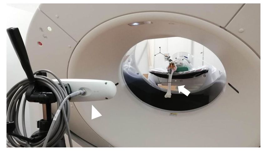
rubber band fixed to the anterior surface of the femoral model and the proximal rubber band to the anterior surface of
the tibial model. Using this approach, the three bone models that constituted the knee joint were connected. Tracker
pins with a diameter of 3 mm were placed in each bone model to attach the tracker used in the navigation system
(Fig. 2).

CT acquisition
A 320-slice multidetector CT (Aquilion One, Canon Medical Systems, Otawara, Japan) was used for CT imaging.
Table 1 shows the imaging parameters for static CT images and 4D-CT images. First, a static CT image of the entire
knee joint model was taken in the extension position. Next, the femoral side was fixed to the table such that the center
of knee joint movement was located at the center of the CT imaging range. With the CT gantry tilted at 30°, 4D-CT
scans were obtained with a rotation time of 10.5 s while the knee joint model was passively flexed from 0° to 90° by an
experimenter. A total of 1.5 cycles of flexion-extension movement were performed in approximately 10.5 s, with the
obtained 4D-CT data consisting of 20 volume scans. During 4D-CT imaging, the position-posture of the knee joint
model was concurrently recorded by the navigation system (Fig. 3). The entire process of CT imaging as described
above was repeated 3 times.

                                                       Table 1
                            Scanning parameters for static CT and 4D-CT data acquisitions
           Gantry       Section       Range     Tube        Effective    Rotation    Acquisition     CT         Dose-
           tilt         thickness     (mm)      potential   mAs          time (s)    interval        Dose       length
           (degrees)    (mm)                    (kV)        (mAs)                                    Index      product
                                                                                                     Volume     (mGy-
                                                                                                     (mGy)      cm)

 Static    0            1.0×40        1000      120         9            0.5         NA              0.7        49

 4D-       30           0.5×160       160       80          87           0.5         Continuous      31.0       495.6
 CT

Surface reconstruction and reference axis
The 4D-CT images in DICOM format were imported into a 3D model construction software (N-View, Teijin-Nakashima
Medical. Co., Ltd. Okayama, Japan), and 3D models of the femur, tibia, and patella, which will be referred to as the “4D-
CT model” hereafter, were constructed for each of the 20 volume scan acquisitions. Similarly, 3D models of the femur,
tibia, and patella, which will be referred to as the “static CT model” hereafter, were constructed from the static CT
images along the entire length of the lower limbs of the knee joint model. Simultaneously, a reference axis was defined
on the static CT model (Figs. 4). In the 4D-CT model, the coordinate system was set for each volume scan based on
the reference axis.

3D-3D registration
The imaging range of 4D-CT can be as small as 16 cm wide. The center of the femoral head and the center of the
ankle joint, which are the reference points on the functional axis, are outside the imaging range of 4D-CT. In addition,
the reference axis for evaluating the position-posture needs to be re-established for each volume scan. Position-
posture evaluation with the 4D-CT model alone can be inaccurate for these two reasons. Therefore, in this study, the
reference axis defined in the static CT model was applied to the 4D-CT model by surface-matching the two models in
order to calculate the accurate position-posture of the 4D-CT model. Surface matching was completed using a
software for editing polygon meshes (GOM Inspect 2018, GOM GmbH, Braunschweig, Germany).

                                                        Page 4/14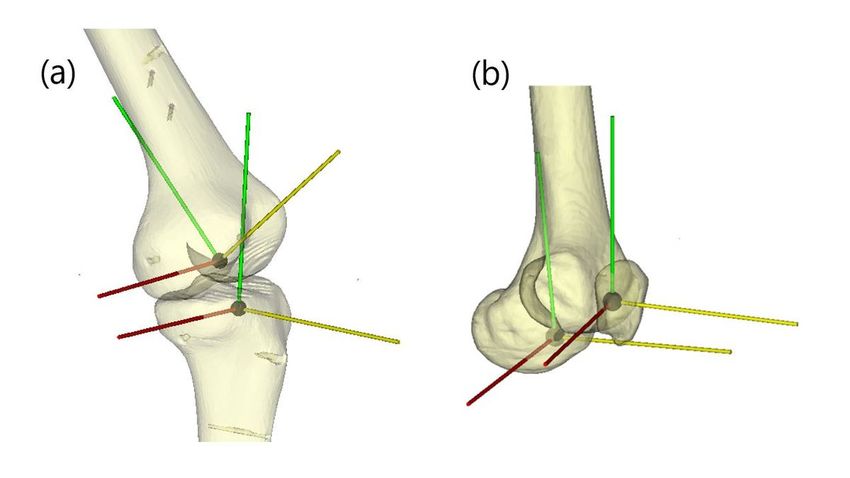
Precision validation
For each volume scan, the position-posture expressed in the femur model coordinate system was calculated with
reference to the tibia model coordinate system as the “FT position-posture.” Similarly, for each volume scan, the
position-posture expressed in the femur coordinate system based on the coordinate system of the patella model was
also calculated as the “PF position-posture.” The position of each volume scan was determined by determining the
positional relationship between the origins of the coordinate system between volume scans, and the posture was
obtained by angle-resolving the "distortion" between the coordinate systems.

The position-posture measurement of the bone model by the CT-based Navigation System (N-navi, Teijin Nakashima,
Co., Ltd.) was performed in synchronization with 4D-CT imaging. For measurements with the navigation system, the
same CT model and alignment definition as those adapted for 3D-3D registration were used. This measurement of the
position and orientation of the simulated bone by the navigation system facilitated comparison of the position and
orientation calculated by 3D-3D registration with the same criteria. Since the navigation system used in this study had
an error of 0.6 mm and within 1 deg according to a previous study [23], the position and orientation measurement by
this navigation system was used as the correct answer value. In each 4D-CT volume scan, the difference in position
and posture (coordinates, varus/valgus, internal rotation/external rotation, extension/flexion) from the navigation
system was calculated. The same analysis was performed on each of the three acquisitions of CT data. The accuracy
of position-posture evaluation by 4D-CT was verified by calculating the average of absolute value differences.

Results
Figure 5 shows one of the three temporal position-posture measurements of 4D-CT and the navigation system for the
tibiofemoral joint (FT joint) (Fig. 5-a) and patellofemoral joint (PF joint) (Fig. 5-b). Figure 6 shows the difference in
position-posture measurements between 4D-CT and the navigation system for the FT joint (Fig. 6-a) and PF joint
(Fig. 6-b).

The position-posture obtained from 4D-CT showed similar tendency as that obtained from the navigation system. In
the FT joint, the difference in spatial orientation between the two measurements was 0.7 mm in the X direction, 0.9
mm in the Y direction, and 2.8 mm in the Z direction. The difference in angle was 1.9° in the varus/valgus direction,
1.1° in the internal/external rotation, and 1.8° in the extension/flexion. (Fig. 6-a) In the PF joint, the difference between
the two measurements was 0.9 mm in the X direction, 1.3 mm in the Y direction, and 1.2 mm in the Z direction. The
difference in angle was 0.9° for varus/valgus, 1.1° for internal/external rotation, and 1.3° for extension/flexion. (Fig. 6-
b)

Since the subject itself moved while the gantry rotated, the 4D-CT data exhibited a partial-volume effect and motion
artifacts (Appendix 1). Therefore, some artifacts were included in the bone model constructed by the binarization
method from this 4D-CT image. In particular, artifacts were most noticeable in the middle of the flexion and extension
movements, which had a high movement speed, and the afterimage of movements was confirmed in the bone axis of
the tibia, which showed the largest movement (Fig. 7).

Discussion
In the present study, knee joint movement of the bone model was analyzed by matching 4D-CT and static CT scans of
the entire length of the lower limbs by using 3D-3D registration. In order to examine the analysis accuracy, this method
was compared with the navigation system using high-precision optical motion capture. The findings revealed that this
was a highly accurate method for evaluating knee joint movements, with an error of less than 3 mm and less than 2°.
                                                          Page 5/14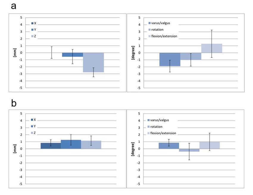
To our knowledge, reports on the accuracy of 4D-CT have been primarily focused on radiotherapy [24–27]. Lee et al.
validated the accuracy of 4D cone-beam CT for radiotherapy by using a phantom and reported that the error was less
than 2 mm when the respiratory movement was up to 30 mm [21]. The present study is the first to analyze the
accuracy of 4D-CT in the joint area, and it showed that this technique showed the same accuracy as it did in the
evaluation of respiratory movement.

Several reports in the field of radiotherapy have referred to artifacts in 4D-CT, demonstrating that artifacts affect lung
function imaging and tumor volume assessment [24–27]. A similar problem may affect 4D-CT of the bone joint area
for evaluating joint movement. In this study, highly accurate evaluation was possible by performing 3D-3D matching.
However, it is unclear whether joint motion was evaluated correctly in the previous reports that did not perform
registration with static CT.

In assessments using 4D-CT, the imaging range is limited to the area around the knee joint, making the alignment of
the entire lower limbs unclear. In addition, the reference axis for evaluating the position and orientation must be
provided for each volume scan, which may introduce errors. To avoid these problems, we measured knee joint
movement using 3D-3D registration, matching the static CT model of the full length of the lower limbs with 4D-CT in
accordance with the method described by Oki et al. [22 14]. Conventional methods such as 2D-3D matching have been
considered to be particularly difficult to analyze the patella in the dynamics of the knee joint [12]. However, 4D-CT
enabled the position and posture of the patella to be determined from three-dimensional data, allowing measurements
of the patellofemoral joint with the same high accuracy as the femoral tibial joint.

Various methods have been used for analysis of knee joint dynamics, and the accuracy of these methods has also
been reported. However, while this study investigated the accuracy of analyzing passive ROM movement in the non-
weight-bearing position, previous reports performed dynamic analysis during walking and running. Therefore,
comparisons between the present and previous studies are difficult. In dynamic analysis using body surface markers,
the deviation from the bone movement due to the movement of the skin is a major concern. Sati et al. [28 51] reported
a 2–17 mm gap between skin and bone in medial and lateral femoral condyles. Therefore, high accuracy cannot be
expected using this method. Another common method is to attach external markers directly to the bone using
percutaneous pins. However, Holden et al. [29 52] reported an error of up to 10 mm and 8° using this method. Accuracy
analyses using fluoroscopy have also been performed. Hoff et al. [13 7] achieved very high accuracy of 2.25 mm and
less than 0.035°. However, this analysis method can only be applied to the knee in which the implant is inserted. The
analysis of knee joint movement by 4D-CT in this study can be applied to patients who have not undergone knee joint
surgery to receive implants. 4D-CT can be expected to yield high accuracy in evaluation of actual patients.

The matching error between the 4D-CT and CT models can be explained by the fact that the 4D-CT model includes the
effect of movement, and that the navigation system used for reference also contains an error. Thus, the accuracy of
analysis by 4D-CT can be improved by reducing the motion speed of knee joint movement.

This study had several limitations. First, this study involved experimental verification using a bone model, which
evaluated the dynamics of passive motion in the non-weight-bearing position. Verification with actual clinical patients
can be expected in future studies. However, conventional CT equipment only allows participants to be evaluated in the
non-weight-bearing position, which is a major disadvantage in any study. Upright CT, which allows a patient to stand,
has been recently developed at the research level. While evaluations in the weight-bearing position are expected to be
facilitated by this new CT technique, clinical application of the new technology will take more time [30 45]. Therefore, it
is meaningful to pursue a highly accurate joint dynamics evaluation method in a widely used device. Second, the
navigation system using optical motion capture contained inherent errors, which may have affected the analysis
accuracy of this study. This navigation system, which is also used in surgery, has been used in many studies
                                                         Page 6/14
analyzing knee joint motion [31 54, 32 53] and was shown to be extremely accurate, with an error of 0.6° and within 1
mm, indicating only a minimal effect on the scale of knee joint movement [23]. However, there may be problems when
using this technique for small joints, because small errors can have a relatively large effect in small joints when
compared to large joints. Third, both 4D-CT and general CT examinations are associated with the risk of radiation
exposure. To avoid this risk, patients’ gonads should be protected and possible measures should be taken to reduce
radiation exposure when the patients undergo CT examinations. Further studies should be made using an appropriate
4D-CT imaging protocol.
Conclusion
In this study, the accuracy of knee joint analysis using 4D-CT with 3D-3D registration was determined using the optical
motion-capture system. This method showed excellent accuracy and could be used in vivo applications.
Abbreviations
TKA, total knee arthroplasty; 4D-CT, four-dimensional CT; FT, femorotibial; PF, patellofemoral; ROM, range of motion
Declarations
Ethics approval and consent to participate: Not applicable
Consent for publication: Not applicable
Availability of data and materials: Not applicable
Competing interests: Not applicable
Funding: This research was carried out in collaboration with Teijin Nakashima Co., Ltd.
Authors' contributions:
Contributed equally to this work: Yuki Kato and Takuya Adachi.
Yuki Kato and Takuya Adachi were involved in study design and data interpretation.
Yuki Kato, Takuya Adachi, and Daii Kiyotomo were involved in the data analysis.
Yuki Kato mainly performed the experimental procedures.
Youichi Machida and Katsushige Kawamukai supported the experimental procedures.
All authors critically revised the report, commented on drafts of the manuscript, and approved the final report.
Acknowledgements: The authors would like to thank Mrs. Yuko Yanai for the careful English proofreading. This work
was supported by a grant from Teijin Nakashima, Co., Ltd.
References
1. Yoshimura N, Muraki S, Oka H, Mabuchi A, En-Yo Y, Yoshida M, et al. Prevalence of knee osteoarthritis, lumbar
spondylosis, and osteoporosis in Japanese men and women: the research on osteoarthritis/osteoporosis against
Page 7/14disability study. J Bone Miner Metab. 2009;27:620-8.
2. Anderson JG, Wixson RL, Tsai D, Stulberg SD, Chang RW. Functional outcome and patient satisfaction in total
knee patients over the age of 75. J Arthroplasty. 1996;11:831-40.
3. Bourne RB, Chesworth BM, Davis AM, Mahomed NN, Charron KD. Patient satisfaction after total knee
arthroplasty: who is satisfied and who is not? Clin Orthop Relat Res. 2010;468:57-63.
4. Pitta M, Esposito CI, Li Z, Lee YY, Wright TM, Padgett DE. Failure after modern total knee arthroplasty: A
prospective study of 18,065 knees. J Arthroplasty. 2018;33:407-14.
5. Andriacchi TP, Koo S, Scanlan SF. Gait mechanics influence healthy cartilage morphology and osteoarthritis of
the knee. J Bone Joint Surg Am. 2009;91 Suppl 1:95-101.
6. Rashid RF. Towards a system for the interpretation of moving light displays. IEEE Trans Pattern Anal Mach
Intell. 1980;2:574-81.
7. Cappozzo A. Three-dimensional analysis of human walking: Experimental methods and associated artifacts.
Hum Mov Sci. 1991;10:589-602.
8. Cappozzo A, Cappello A, Della Croce U, Pensalfini F. Surface-marker cluster design criteria for 3-D bone
movement reconstruction. IEEE Trans Biomed Eng. 1997;44:1165-74.
9. Reinschmidt C, van den Bogert AJ, Lundberg A, Nigg BM, Murphy N, Stacoff A, et al. Tibiofemoral and
tibiocalcaneal motion during walking: external vs. skeletal markers. Gait Posture. 1997;6:98-109.
10. Anderst W, Zauel R, Bishop J, Demps E, Tashman S. Validation of three-dimensional model-based tibio-femoral
tracking during running. Med Eng Phys. 2009;31:10-6.
11. Inoue T, Abe N, Fujiwara K, Hashizume H, Nakajima Y, Sugita N, et al. Validation of 3-dimensional postoperative
evaluation technique for total knee arthroplasty. J Jpn Soc Comput Aid Surg. 2016;18:71-9.
12. Fregly BJ, Rahman HA, Banks SA. Theoretical accuracy of model-based shape matching for measuring natural
knee kinematics with single-plane fluoroscopy. J Biomech Eng. 2005;127:692-9.
13. Hoff WA, Komistek RD, Dennis DA, Gabriel SM, Walker SA. Three-dimensional determination of femoral-tibial
contact positions under in vivo conditions using fluoroscopy. Clin Biomech (Bristol, Avon). 1998;13:455-72.
14. Li G, Kozanek M, Hosseini A, Liu F, Van de Velde SK, Rubash HE. New fluoroscopic imaging technique for
investigation of 6DOF knee kinematics during treadmill gait. J Orthop Surg Res. 2009;4:6.
15. Ishimaru M, Shiraishi Y, Ikebe S, Higaki H, Hino K, Onishi Y, et al. Three-dimensional motion analysis of the
patellar component in total knee arthroplasty by the image matching method using image correlations. J Orthop Res.
2014;32:619-26.
16. Apfaltrer G, Lavra F, Schoepf UJ, Scarabello M, Yamada R, van Assen M, et al. Quantitative analysis of dynamic
computed tomography angiography for the detection of endoleaks after abdominal aorta aneurysm endovascular
repair: A feasibility study. PLOS ONE. 2021;16:e0245134.
Page 8/1417. Cancelliere NM, Najafi M, Brina O, Bouillot P, Vargas MI, Lovblad KO, et al. 4D-CT angiography versus 3D-
rotational angiography as the imaging modality for computational fluid dynamics of cerebral aneurysms. J
Neurointerv Surg. 2020;12:626-30.
18. Kortman HG, Smit EJ, Oei MT, Manniesing R, Prokop M, Meijer FJ. 4D-CTA in neurovascular disease: a review.
AJNR Am J Neuroradiol. 2015;36:1026-33.
19. Siow T, Lim S. Correlating lung tumour location and motion with respiration using 4D CT scans. J Radiother
Pract 2021;20:17-21.
20. Demehri S, Thawait GK, Williams AA, Kompel A, Elias JJ, Carrino JA, et al. Imaging characteristics of
contralateral asymptomatic patellofemoral joints in patients with unilateral instability. Radiology. 2014;273:821-30.
21. Lee S, Yan G, Lu B, Kahler D, Li JG, Sanjiv SS. Impact of scanning parameters and breathing patterns on image
quality and accuracy of tumor motion reconstruction in 4D CBCT: a phantom study. J Appl Clin Med Phys.
2015;16:195-212.
22. Oki S, Kaneda K, Yamada Y, Yamada M, Morishige Y, Harato K, et al. Four-dimensional CT analysis using
sequential 3D-3D registration. J Vis Exp. 2019;23:e59857 .
23. Yokoyama Y, Abe N, Fujiwara K, Suzuki M, Nakajima Y, Sugita N, et al. A new navigation system for minimally
invasive total knee arthroplasty. Acta Med Okayama. 2013;67:351-8.
24. Louie AV, Rodrigues G, Olsthoorn J, Palma D, Yu E, Yaremko B, et al. Inter-observer and intra-observer reliability
for lung cancer target volume delineation in the 4D-CT era. Radiother Oncol. 2010;95:166-71.
25. Nakamura M, Narita Y, Sawada A, Matsugi K, Nakata M, Matsuo Y, et al. Impact of motion velocity on four-
dimensional target volumes: a phantom study. Med Phys. 2009;36:1610-7.
26. Persson GF, Nygaard DE, Brink C, Jahn JW, Munck af Rosenschold P, Specht L, et al. Deviations in delineated
GTV caused by artefacts in 4DCT. Radiother Oncol. 2010;96:61-6.
27. Yamamoto T, Langner U, Loo BW, Jr., Shen J, Keall PJ. Retrospective analysis of artifacts in four-dimensional
CT images of 50 abdominal and thoracic radiotherapy patients. Int J Radiat Oncol Biol Phys. 2008;72:1250-8.
28. Sati M, de Guise JA, Larouche S, Drouin G. Quantitative assessment of skin-bone movement at the knee. Knee.
1996;3:121-38.
29. Holden JP, Orsini JA, Stanhope SJ.Estimates of skeletal motion: movement of surface-mounted targets relative
to bone during gait.: Engineering Research Center, University of the District of Columbia; 1994.
30. Jinzaki M. Development of Upright CT and Its Initial Evaluation: Effect of Gravity on Human Body and
Potential Clinical Application 2020. Singapore: Springer Singapore; 2020: 273-9.
31. Pearle AD, Solomon DJ, Wanich T, Moreau-Gaudry A, Granchi CC, Wickiewicz TL, et al. Reliability of navigated
knee stability examination: a cadaveric evaluation. Am J Sports Med. 2007;35:1315-20.
32. Robinson J, Carrat L, Granchi C, Colombet P. Influence of anterior cruciate ligament bundles on knee kinematics:
clinical assessment using computer-assisted navigation. Am J Sports Med. 2007;35:2006-13.
Page 9/14Figures
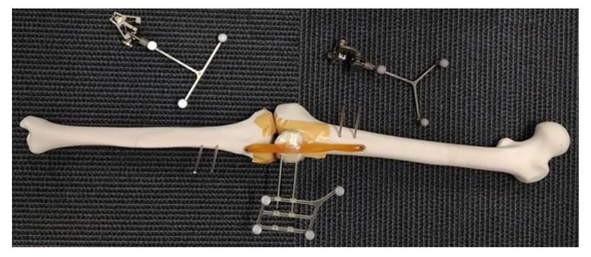
Figure 1
Process flow from CT acquisition to dynamic analysis.
Figure 2
Human knee skeletal bone model. The bone models were connected with rubber bands, and two tracker pins were
attached to each bone model for the navigation system.
Page 10/14Figure 3
Knee joint model in the CT gantry (arrow) and the navigation system (arrowhead).
Figure 4
Page 11/14(a) Local coordinate systems of the femur and tibia, (b) local coordinate systems of the femur and patella.
Figure 5
Changes in the position-posture measured by 4D-CT (blue line) and the navigation system (red line) in the FT joint (Fig
5-a) and the PF joint (Fig 5-b). 4D-CT and the navigation system showed similar movement patterns in both FT and PF
joints.
Page 12/14Figure 6
Differences between 4D-CT and the navigation system in the FT joint (a) and the PF joint (b). Differences are shown as
relative values, indicating errors of up to 2.8 mm and 1.8° for the FT joint, and up to 1.3 mm and 1.8° for the PF joint.
Thus, 4D-CT could measure the position and orientation with high accuracy.
Page 13/14Figure 7
3D-CT model reconstructed from 4D-CT data. Some motion artifacts (arrows) occurred in the tibia and fibula during
the mid-term flexion and extension movements (Fig. 7-a), where the movement speed was high. In contrast, when the
movement speed was relatively low (that is, when the movement per unit time was small), no artifact was observed
(Fig. 7-b).
Page 14/14You can also read