Advanced Optical Imaging - IBS Conference on ABSTRACT BOOK
←
→
Page content transcription
If your browser does not render page correctly, please read the page content below
IBS Conference on
Advanced Optical Imaging
June 19th ~ 21st , 2019
Hana Square Auditorium,
Korea University, Seoul, Korea

ABSTRACT BOOK

ibs-conference.org/2019/aoi
Korea University, Seoul,
    South Korea

                                                      CONTENTS

    COMMITTEE MEMBERS                                 COMMITTEE MEMBERS                             ••••••••••••••••••••••••••••••••••••••••••••••••••
                                                                                                                                                         02

                                                      PROGRAM      •••••••••••••••••••••••••••••••••••••••••••••••••••••••••••••••••••••••••••••••••••
                                                                                                                                                         04

    Organizers                                        DAY 1
                                                      ORAL SESSION 1-3                 •••••••••••••••••••••••••••••••••••••••••••••••••••••••••••••••
                                                                                                                                                         07
    Cho, Minhaeng
    Director, IBS Center for Molecular Spectroscopy   DAY 2
    and Dynamics, Korea University
                                                      ORAL SESSION 4              ••••••••••••••••••••••••••••••••••••••••••••••••••••••••••••••••••••
                                                                                                                                                         33
    Choi, Wonshik
    Associate Director, IBS Center for Molecular      DAY 3
    Spectroscopy and Dynamics, Korea University
                                                      ORAL SESSION 5-6                 •••••••••••••••••••••••••••••••••••••••••••••••••••••••••••••••
                                                                                                                                                         43
    Hong, Seok-Cheol
    Professor, IBS Center for Molecular               POSTER   •••••••••••••••••••••••••••••••••••••••••••••••••••••••••••••••••••••••••••••••••••••••
                                                                                                                                                         61
    Spectroscopy and Dynamics, Korea University

2                                                                                                                                                             3
PROGRAM                                                                                                                                        2019 IBS Conference on Advanced Optical Imaging

    PROGRAM                                                                                    DAY                                                                   Thursday, June 20, 2019
                                                                                                     Oral Session 4   Single-Molecule Imaging II

                                                                                                      09:20 - 09:30   Chair

                                                                                                      09:30 - 10:10   Vahid Sandoghdar

                                                                                                      10:10 - 10:50   Seok-Cheol Hong

                                                                                                      10:50 - 11:30   Joshua C. Vaughan

                                                                                                      11:30 - 13:00   Lunch

                                                                                                      13:00 - 14:30   Poster Session

                                                                                                      14:30 - 19:00   Palace Tour for Invited Speakers

    DAY                                                             Wednesday, June 19, 2019          19:00 -         Dinner for Invited Speakers
               08:30 - 09:10   Registration

               09:10 - 09:20   Opening Remarks

              Oral Session 1   Coherent Raman Microscopy and Quantum Imaging

               09:20 - 09:30   Chair

               09:30 - 10:10   Marcus T. Cicerone

               10:10 - 10:50   Minbiao Ji
                                                                                               DAY                                                                       Friday, June 21, 2019
               10:50 - 11:30   Dan Oron                                                              Oral Session 5   Imaging with Nanodiamond Probes

               11:30 - 11:50   Conference Photo                                                       09:20 - 09:30   Chair

               11:50 - 13:00   Lunch                                                                  09:30 - 10:10   Francois Marquier

              Oral Session 2   Single-Molecule Imaging I                                              10:10 - 10:50   Keir C. Neuman

               13:00 - 13:10   Chair                                                                  10:50 - 11:30   Romana Schirhagl

               13:10 - 13:50   Haw Yang                                                               11:30 - 13:00   Lunch

               13:50 - 14:30   Hanbin Mao                                                            Oral Session 6   Adaptive Optics Microscopy

               14:30 - 15:10   Claus Seidel                                                           13:00 - 13:10   Chair

               15:10 - 15:30   Coffee Break                                                           13:10 - 13:50   Martin Booth

              Oral Session 3   Deep-Tissue Imaging                                                    13:50 - 14:30   Steven Graham Adie

               15:30 - 15:40   Chair                                                                  14:30 - 15:10   Wonshik Choi

               15:40 - 16:20   Changhuei Yang                                                         15:10 - 15:30   Award Ceremony

               16:20 - 17:00   Vivek Srinivasan                                                       15:30 - 15:40   Closing Remarks

               17:00 - 17:40   Peter So                                                               15:40 - 17:40   IBS CMSD Lab Tour

               17:40 -         Banquet                                                                17:40 -         After Conference Event

4                                                                                                                                                                                                    5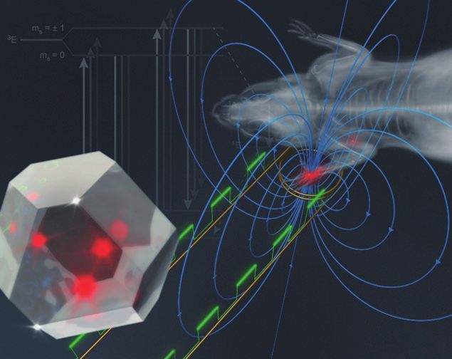
2019 IBS Conference on Advanced Optical Imaging

    IBS Conference on
    Advanced Optical Imaging
    June 19th ~ 21st , 2019

                                                                   DAY
    Korea University, Seoul, Korea

    ibs-conference.org/2019/aoi

                                            Wednesday, June 19, 2019

                                        ORAL SESSION 1

                                        ORAL SESSION 2

                                        ORAL SESSION 3

6                                                                                      7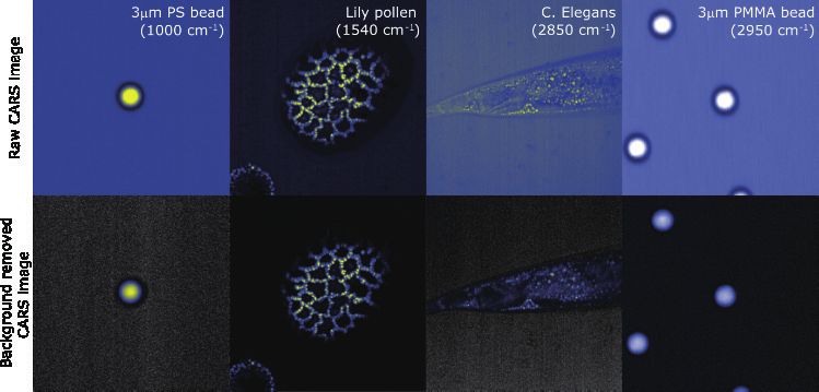
ORAL SESSION
    Coherent Raman Microscopy and Quantum Imaging

                        Speakers

                  Marcus T. Cicerone
                        Georgia Tech

                       Minbiao Ji
                      Fudan University

                        Dan Oron
                Weizmann Institute of Science

8                                                   9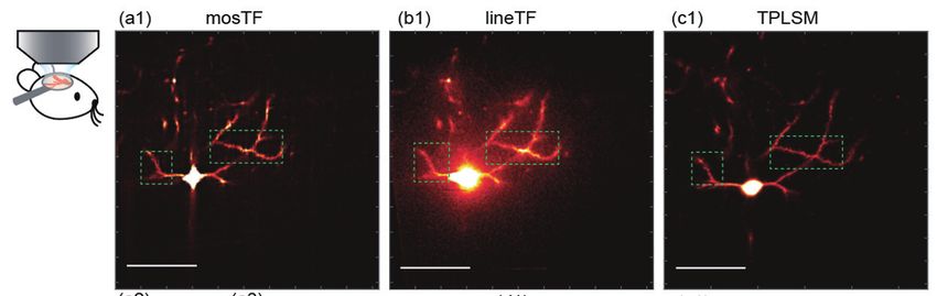
ORAL SESSION 1                                                                                                                                                                                                  2019 IBS Conference on Advanced Optical Imaging

       Abstract                                                                                                                            Abstract

      Discovering Biology with Broadband Coherent Raman Microscopy                                                                      Label-free Histology with Stimulated Raman Scattering Microscopy
                                                       Marcus T. Cicerone                                                                                                                      Minbiao Ji
                 Department of Chemistry and Biochemistry, Georgia Institute of Technology, Georgia 30332, USA                                                     Department of Physics, Fudan University, Shanghai 200433, China

        Spectroscopic coherent Raman imaging (CRI) methods allow label-free, chemically specific imaging                                   Stimulated Raman scattering (SRS) microscopy is an emerging label-free imaging technique. It is a
     of materials and biological systems, and are opening up many exciting possibilities for understanding                              nonlinear version of Raman scattering, with the advantages of high chemical selectivity and rapid imaging.
     phenomena in these systems. When we first introduced spectroscopic coherent Raman imaging in 2004[1], we                           We have applied SRS microcopy for brain tumor detection on both in vivo mouse models and ex vivo human
     could acquire spectra only from bulk polymers and liquids. Now, after many years of development, we easily                         surgical specimens, based on the spectral differences between normal and tumor tissues. We further developed
     acquire label-free micrographs of biological cells and tissues at 3.5 ms for each spectral image pixel[2]. Figure                  dual-phase parallel imaging technique for real-time two color SRS imaging for rapid histology. Our recent
     1 provides an example of a murine pancreas section, showing major structural features, although much more                          efforts in combining SRS with deep-learning networks may provide smart intraoperative virtual histology for
     spectral image information is available. Initially it was not clear that acquiring such spectra would be possible,                 solid tumors.
     but a recognition that an intrinsic and strong nonresonant signal could be used to enhance weak resonant
     signals[3] made it possible to obtain these signals above detector noise. Further improvements in signal                           References
     generation and spectral retrieval algorithms[4], now provide for highly robust and rapid spectral imaging. I                       [1] Ji, M.*; Arbel, M.; Zhang, L.; Freudiger, W. C.; Hou, S. S.; Lin, D.; Yang, X.; Bacskai, J. B.*; Xie, X. S.*, “Label-free
     will briefly introduce broadband spectroscopic coherent anti-Stokes Raman scattering (BCARS) microscopy,                           imaging of amyloid plaques in Alzheimer’s Disease with stimulated Raman scattering microscopy”. Science Advances 2018,
     discussing some of the key concepts that make this method practical. I will also present selected application                      4(11), eaat7715.
     examples from studies on C. elegans metabolism and human cytomegalovirus infection that highlight the                              [2] He, R.; Xu, Y.; Zhang, L.; Ma, S.; Wang, X.; Ye, D.; Ji, M.*, “Dual-phase stimulated Raman scattering microscopy
     utility of BCARS for discovering new biology through in-depth characterization of highly complex biological                        for real-time two-color imaging”. Optica 2017, 4 (1), 44-47. (Cover story)
     systems.                                                                                                                           [3] Ji, M., S. Lewis, X. Sunney Xie, et al. “Detection of human brain tumor infiltration with quantitative stimulated Ra-
                                                                                                                                        man scattering microscopy.” Science Translational Medicine 2015, 7, 309ra163
                                                                                                                                        [4] Ji, M., D. A. Orringer, C. W. Freudiger, X. Sunney Xie, et al. (2013). “Rapid, label-free detection of brain tumors
                                                                                                                                        with stimulated Raman scattering microscopy.” Science Translational Medicine 2013, 5, 201ra119. (Cover story)
                                                                                                                                        [5] Ji, M., M. Odelius, K. J. Gaffney. (2010). “Large Angular Jump Mechanism Observed for Hydrogen Bond Exchange
                                                                                                                                        in Aqueous Perchlorate Solution.” Science 2010, 328(5981): 1003-1005.

                                                                   Figure 1. M
                                                                              urine pancreas with contrast from Raman spectra
                                                                             (e.g., shown at right) obtained at each pixel in 3.5
                                                                             ms. Contrast: Cell nuclei (blue), collagen (red), and
                                                                             arterial wall (green).

     References
     [1] T. W. Kee and M. T. Cicerone, “Simple approach to one-laser, broadband coherent anti-stokes raman scattering microscopy,”
     Optics Letters 29 , 2701–2703 (2004).
     [2] C. H. Camp Jr., Y. J. Lee, J. M. Heddleston, C. M. Hartshorn, A. R. H. Walker, J. N. Rich, J. D. Lathia, and M. T. Cicerone,
     “High-speed coherent Raman fingerprint imaging of biological tissues,” Nature photonics 8 , 627–634 (2014).
     [3] Y. Liu, Y. J. Lee, and M. T. Cicerone, “Broadband CARS spectral phase retrieval using a time-domain KramersKronig
     transform,” Optics Letters 34 , 1363–1365 (2009).
     [4] C. H. Camp Jr., Y. J. Lee, and M. T. Cicerone, “Quantitative, comparable coherent anti-Stokes Raman scattering (CARS)
     spectroscopy: correcting errors in phase retrieval,” Journal of Raman Spectroscopy 47, 408–415 (2015).

10                                                                                                                                                                                                                                                                      11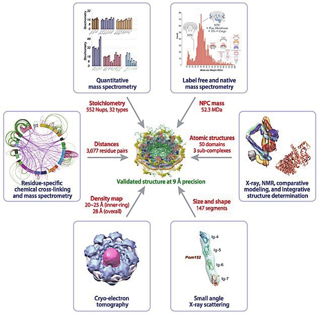
ORAL SESSION 1                                                                                                                                                                                     2019 IBS Conference on Advanced Optical Imaging

       Abstract

                     Quantum Enhanced Superresolution Microscopy                                                               References
                                                                                                                               [1] O. Schwartz, et al., “Superresolution microscopy with quantum emitters,” Nano letters, 13, 5832–5836 (2013).
                                                          Dan Oron                                                             [2] Y. Israel et al., “Quantum correlation enhanced super-resolution localization microscopy enabled by a fiber bundle
                Department of Physics of Complex Systems, Weizmann Institute of Science, Rehovot 76100, Israel                 camera”, Nat. Commun. 8, 14786 (2017)
                                                                                                                               [3] R. Tenne et al., “Super-resolution enhancement by quantum image scanning microscopy”, Nature Photonics 13, 116
        Far-field optical microscopy beyond the Abbe diffraction limit, making use of nonlinear excitation (e.g.               (2019).
     STED), or temporal fluctuations in fluorescence (PALM, STORM, SOFI) is already a reality. In contrast,
     overcoming the diffraction limit using non-classical properties of light is very difficult to achieve due to the
     requirement of nonlinearity and the fragility of quantum states of light. Here, we experimentally demonstrate
     superresolution microscopy based on quantum properties of light naturally emitted by fluorophores used
     as markers in fluorescence microscopy. Our approach is based on photon antibunching, the tendency of
     fluorophores to emit photons one by one rather than in bursts. Although a distinctively quantum phenomenon,
     antibunching is readily observed in most common fluorophores even at room temperature.
        This nonclassical resource can be utilized directly to enhance the imaging resolution, since the non-classical
     far-field intensity correlations induced by antibunching carry high spatial frequency information on the spatial
     distribution of emitters[1]. Detecting photon statistics simultaneously in the entire field of view, we were able
     to detect non-classical correlations of the second and third order, and reconstructed images with resolution
     significantly beyond the diffraction limit.
        Alternatively, we demonstrate the utilization of antibunching for augmenting the capabilities of other
     commonly used superresolution techniques, such as localization-based superresolution imaging [2] or image-
     scanning confocal microscopy[3]. For this end, we use a novel detector comprised of an array of single photon
     detectors connected to a densely packed fiber bundle, acting as a low-noise single photon sensitive camera, and
     enabling the measurement of photon correlations. These features allow us to enhance the spatial resolution with
     which multiple emitters can be imaged and localized compared with techniques that rely on an integrated photon
     count using CCD cameras. An example for resolution enhancement by quantum image scanning confocal
     microscopy is given in Fig. 1
        Finally, new modalities for harnessing quantum photon statistics for super-resolved imaging will be
     discussed.

     Figure 1. I mages of a microtubule-labeled 3T3 cell stained with quantum dots by: confocal (left), image-scanning con-
               focal (center) and quantum image scanning confocal (right). Scale bar is 0.5μm

12                                                                                                                                                                                                                                                        13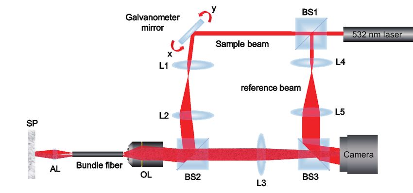
ORAL SESSION 1 | MEMO   2019 IBS Conference on Advanced Optical Imaging

14                                                                             15
ORAL SESSION
          Single-Molecule Imaging I

                   Speakers

                    Haw Yang
                Princeton University

                  Hanbin Mao
                Kent State University

                  Claus Seidel
        Heinrich-Heine-Universität Düsseldorf

16                                              17
ORAL SESSION 2                                                                                                                                                                             2019 IBS Conference on Advanced Optical Imaging

       Abstract                                                                                                             Abstract

                              Real-Time 3D Single-Particle Tracking:                                                                           3-D Imaging of Bimolecular Structures
                               Spectroscopy, Imaging, and Control                                                                        at the Single Molecular Level by Optical Tweezers
                                                         Haw Yang                                                                                                         Hanbin Mao
                            Department of Chemistry, Princeton University, New Jersey 08544, USA                                            Department of Chemistry Biochemistry, Kent State University, Ohio 44240, USA

        Real-time 3D single-particle tracking spectroscopy is an experimental technique that allows one to follow            Bimolecular structures can be resolved by conventional techniques such as X-ray crystallography and
     a nanoprobe as its moves in three-dimensional space, either by diffusion or by active locomotion[1]. The             NMR. These methods are able to resolve structures at atomic level. However, homogeneous samples are often
     technique keeps the nanoprobe at the center of the microscope objective focus at all times by moving the             required in these approaches. In NMR, relatively high concentration of molecules is needed whereas solvent
     sample stage to counter any movements that the probe exhibits. Effectively, the apparatus transforms the             conditions are important to resolve the structure. These conditions are difficult to achieve experimentally. For
     experimental coordinate from a lab-based frame to the probe-based frame[2-4]. This way, it becomes possible to       example, many biomolecules only assume active conformations in viscous solutions that contain many other
     do time-dependent experiments on the probe or on the molecule tethered to the probe as if the particle or the        macromolecules (the so-called molecular crowded condition). In our lab, we have developed a new method
     molecule is immobilized. This presentation will discuss its principles[5-7], capabilities[8-10], and applications—   to image 3D structures at the single-molecule level. Using click chemistry, we attached two pulling handles
     including (1) single-particle spectroscopy, (2) 3D multi-resolution imaging of individual virus-like nano            to two residues of a biomolecule, DNA G-quadruplex[1]. The other end of each pulling handle is tethered
     particles interacting with a live cell[11], and (3) steering of self-propelled micro-swimmers and its implications   to an optically trapped bead. Upon moving the two trapped beads apart, we increased the tension in the
     in bacterial locomotion[12].                                                                                         pulling handles, which mechanically unfolded the G-quadruplex attached in between[2] Each unfolding event
                                                                                                                          allowed us to retrieve the distance between the two pulling residues. By repeating this mechanical unfolding
     References                                                                                                           process between different pairs of pulling residues, we were able to obtain an array of distances among
     [1] H. Cang, C. M. Wong, C. S. Xu, A. H. Rizvi, and H. Yang, Appl. Phys. Lett. 88 (2006) 223901.                     residue pairs. After comparing this profile with those measured from the known X-ray or NMR structures, we
     [2] H. Cang, C. S. Xu, and H. Yang, Chem. Phys. Lett. 457 (2008) 285.                                                were able to identify the best matching structure of the G-quadruplex. This method allows to quickly image
     [3] D. Montiel and H. Yang, Laser & Photonics Reviews 3 (2010) 374.                                                  specific structure of a biomolecule under conditions (low concentration and complex solvent)3 not amenable
     [4] H. Yang, Curr. Opin. Chem. Biol. 14 (2010) 3.                                                                    to conventional structural determination approaches. In addition, this method can reveal the folding and
     [5] H. Yang, J. Chem. Phys. 129 (2008) 074701.                                                                       unfolding kinetics of the biomolecule, as well as its mechanical properties[4].
     [6] D. Montiel, H. Cang, and H. Yang, J. Phys. Chem. B 110 (2006) 19763.
     [7] K. Welsher and H. Yang, Faraday Discuss. 184 (2015) 359.
     [8] H. Cang, C. S. Xu, D. Montiel, and H. Yang, Opt. Lett. 32 (2007) 2729.
     [9] H. Cang, D. Montiel, C. S. Xu, and H. Yang, J. Chem. Phys. 129 (2008) 044503.
     [10] C. S. Xu, H. Cang, D. Montiel, and H. Yang, J. Phys. Chem. C 111 (2007) 32.
     [11] K. Welsher and H. Yang, Nat. Nanotechnol. 9 (2014) 198.
     [12] B. Qian, D. Montiel, A. Bregulla, F. Cichos, and H. Yang, Chem. Sci. 4 (2013) 1420.

18                                                                                                                                                                                                                                                19
ORAL SESSION 2 2019 IBS Conference on Advanced Optical Imaging
Abstract
References Quantitative FRET Image Spectroscopy and Integrative
[1] Zhongbo Yu, Deepak Koirala, Yunxi Cui, Leah F. Easterling, Yuan Zhao, and Hanbin Mao. “Click-Chemistry As-
sisted Single-Molecule Fingerprinting Reveals a 3D Biomolecular Folding Funnel”. Journal of the American Chemical Modeling Unravel the Structure and Functional
Society, 2012, 134, 12338-12341. Dynamics of Biomolecular Systems
[2] Deepak Koirala, Soma Dhakal, Beth Ashbridge, Yuta Sannohe, Raphine Rodriguez, Hiroshi Sugiyama, Shankar Bal-
asubramanian, Hanbin Mao, “A Single-Molecule Platform for Investigation of Interactions between G-quadruplexes and
Claus A.M. Seidel
Small-Molecule Ligands”, Nature Chemistry, 2011, 3, 782-787
Chair for Molecular Physical Chemistry, Heinrich Heine University Duesseldorf, Universitaetsstr 1,
[3] Soma Dhakal, Yunxi Cui, Deepak Koirala, Chiran Ghimire, Saurabh Kushwaha, Zhongbo Yu, Philip M. Yangyuoru,
and Hanbin Mao. “Structural and Mechanical Properties of Individual Human Telomeric G-quadruplexes in Molecularly 40225 Duesseldorf, Germany.
Crowded Solutions”. Nucleic Acids Research, 2013, 41, 3915-3923.
[4] Sagun Jonchhe, Chiran Ghimire, Yunxi Cui, Shogo Sasaki, Mason McCool, Soyoung Park, Keisuke Iida, Kazuo Na- In view of the current progress in super-resolution microscopy, the merits of studying the Förster resonance
gasawa, Hiroshi Sugiyama and Hanbin Mao. “Binding of a Telomestatin Derivative Changes Mechanical Anisotropy of energy transfer (FRET) between an excited donor and acceptor fluorophore in combination with confocal
Human Telomeric G-quadruplex”. Angewandte Chemie International Edition, 2019, 58, 877-881. or stimulated emission depletion (STED) microscopy enables one to reach molecular resolution below
10 nm for studying protein complexes, while simultaneously providing spatial and temporal information
of the cellular context. For this, we established a complete workflow from FRET microscopy in living
cells over FRET-specific data analysis to integrative structural models. (I) We introduced multi-parameter
fluorescence detection (MFD)[1] and multi-parameter fluorescence image spectroscopy (MFIS)[2] to register
all eight characteristic fluorescence parameters so that we gain the complete fluorescence information on
biomolecules in a single measurement. This allows us to study the formation of homo- and hetero-complexes
by homo- and hetero-FRET in live cells simultaneously. (II) We developed a refined analysis of MFIS-
FRET data with a significant noise reduction to infer the structural properties, molecular stoichiometry
and interaction affinities of molecular complexes in living cells by applying detailed models to resolve
the corresponding FRET-parameters (i.e. distances) and species fractions. (III) Using FRET restraints
and computer simulations, we established an automated workflow to generate integrative structural
models of biomolecular assemblies that can be deposited in the new protein data bank, PDB-dev[3, 4].
By combining MFIS and STED-MFIS in living cells with single-molecule FRET studies in vitro, we
studied Guanylate binding proteins (GBPs) that undergo a conformational transition for GTP-controlled
oligomerization to exert their function as part of the innate immune system of mammalian cells - attacking
intra-cellular parasites by disrupting their membranes. We identified GBP’s intrinsic flexibility, a GTP-
triggered association of the GTPase-domains and an assembly-dependent GTP-hydrolysis as functional design
principles that control their reversible oligomerization in polar assemblies and the subsequent formation of
condensates[5]. A further example for functional microscopy of membrane proteins by FRET based MFIS with
molecular resolution is the structural characterization of GPRs in cells[6].
References
[1] Widengren et al.; Anal. Chem. 78, 2039-2050 (2006).
[2] Weidtkamp-Peters et al.; Photochem. Photobiol. Sci. 8, 470-480 (2009).
[3] Kalinin et al.; Nat. Methods 9, 1218-1225 (2012).
[4] Dimura et al.; Curr. Opin. Struct. Biol. 40, 163–185 (2016).
[5] Kravets et. al.; eLife 5, e11479 (2016).
[6] Greife et al.; Sci. Rep. 6, 36792 (2016).
20 21ORAL SESSION 2 | MEMO 2019 IBS Conference on Advanced Optical Imaging 22 23
ORAL SESSION
            Deep-Tissue Imaging

                  Speakers

              Changhuei Yang
        California Institute of Technology

              Vivek Srinivasan
          University of California, Davis

                   Peter So
                       MIT

24                                           25
ORAL SESSION 3 2019 IBS Conference on Advanced Optical Imaging
Abstract Abstract
Wavefront Shaping – the Threading of Light through Scattering Media Interferometric Imaging and Sensing of the Brain:
From Micro to Macro
Changhuei Yang
Department of Electrical Engineering, California Institute of Technology, California, USA
Vivek J. Srinivasan, Jun Zhu, Oybek Kholiqov, Du Le, and Wenjun Zhou
Department of Biomedical Engineering, UC Davis, California, USA
We appear opaque because our tissues scatter light very strongly. Traditionally, optical imaging and
the focusing of light in biological tissues is confounded by the extreme scattering nature of tissues. I summarize interferometric optical techniques, from micron-scale to centimeter-scale, currently being
Interestingly, optical scattering is time-symmetric and we can exploit optical phase conjugation methods to developed by our laboratory. First, I highlight the diverse set of intrinsic optical scattering and absorption
reverse scattering effects. Over the past decade, my team has worked on wavefront control technologies to contrast mechanisms that can be imaged with Optical Coherence Microscopy in the brain and our efforts and
meaningfully focus light through living tissues for imaging and optogenetic stimulation purposes. I will report results in extending penetration depth to sub-cortical regions in the mouse brain using longer wavelength
on our recent experimental findings. In addition, I will also talk about how the interplay between scattering (1300 nm, 1700 nm, and now even 2100 nm) Optical Coherence Microscopy. Next, I present ongoing
and wavefront control is rich and tangled, with surprising optical opportunities waiting to be uncovered. For work in a novel statistical approach to gate out multiply scattered light and further improve imaging depth.
example, the incorporation of scattering within an optical system can actually improve system performance – Finally, at the macroscopic scale, I demonstrate our novel approach to near-infrared (~850 nm) diffuse
a cloudy piece of plastic can actually be a better optical element than a well made lens. optical measurements of the adult human brain in vivo using interferometry. Using CMOS sensors for
detection, I demonstrate a reduction in cost of optical cerebral blood flow (CBF) measurements by two
orders of magnitude over conventional techniques, as well as, quite possibly, the most brain-specific optical
measurements achieved to date in the adult human brain non-invasively. Then, I show time-of-flight
resolved measurements of flow dynamics in the human brain, illustrating the ability to clearly distinguish
scalp and skull signals from brain signals at a level of detail not achieved by previous near-infrared optical
measurements in adult humans.
26 27ORAL SESSION 3 2019 IBS Conference on Advanced Optical Imaging
Abstract
High Throughput, Wide-Field Multiphoton Microscopy
For Deep Structural Imaging of Neuronal Synapse Remodeling
Peter T. C. So
Department of Mechanical and Biological Engineering, Massachusetts Institute of Technology, Massachusetts 02139, USA
Neuron structural remodeling is closely related to mammalian memory plasticity. Many pioneering studies
in this field were enabled by point scanning multiphoton microscopy. In vivo, high resolution 3D imaging
of the whole dendritic tree requires about 30 minutes. Significant remodeling on the synaptic level has been
observed within a day but we hypothesize that there are important faster dynamics to be explored. To access
events on the time scale of minutes to hours, we are developing high throughput wide-field multiphoton
microscopy based on temporal focusing. While this method has been developed for over a decade, it has been
limited by having modest penetration in tissues due to scattering of excitation and emission photons. We
will explore several approaches to overcome this limitation including three-photon excitation and coupling
structured illumination with computation image recovery.
28 29ORAL SESSION 3 | MEMO 2019 IBS Conference on Advanced Optical Imaging 30 31
2019 IBS Conference on Advanced Optical Imaging

 IBS Conference on
 Advanced Optical Imaging
 June 19th ~ 21st , 2019

                                                                DAY
 Korea University, Seoul, Korea

 ibs-conference.org/2019/aoi

                                            Thursday, June 20, 2019

                                     ORAL SESSION 4

32                                                                                  33
ORAL SESSION
Single-Molecule Imaging II
Speakers
Vahid Sandoghdar
MPI of the Science of Light
Seok-Cheol Hong
IBS CMSD
Joshua C. Vaughan
University of Washington
34 35ORAL SESSION 4 2019 IBS Conference on Advanced Optical Imaging
Abstract Abstract
Microscopy and Tracking of Lipids, Proteins and Viruses Interferometric Scattering Microscopy: Versatile Optical Imaging
via Interferometric Detection of Scattering (iSCAT) Technique for Biomolecules and Cellular Structures
Vahid Sandoghdar Seok-Cheol Hong
Max Planck Institute for the Science of Light, Staudtstr. 2, 91058 Erlangen, Germany Center for Molecular Spectroscopy and Dynamics, Institute for Basic Science, Seoul 02841, Korea
Lipids and proteins are some of the most ubiquitous and important components of a biological cell. In Interferometric scattering microscopy (iSCAT) has been continuously and widely expanding the range
addition, biological nanoparticles such as viruses or exosomes play important roles in the physiology of living of applications since its birth in 2004[1,2,3]. While it shares several interferometry-based advantages with its
systems. Aside from their structure and chemical properties, the dynamics of these entities influences their precedent technique, RICM (Reflection Interference Contrast Microscopy) or IRM (Interference Reflection
function over time scales ranging from sub-nanoseconds to minutes and hours. Thus, it would be extremely Microscopy)[3], this technique has rejuvenated and innovated the field of interference microscopy.
insightful if one could monitor the motion of various subcellular nano-objects with nanometer spatial In our center, we have been developing iSCAT-type techniques [4,5], which are complementary to
resolution over many temporal decades. The workhorse of biological imaging, fluorescence microscopy, fluorescence microscopy by circumventing known limitations by fluorescence-based methods and also
confronts fundamental limits in satisfying this need. Here, we report on the application of interferometric transform iSCAT into highly versatile, sensitive, selective techniques.
scattering (iSCAT) microscopy on small gold nanoparticle labels and on unlabeled proteins or viruses with In my talk, I will briefly overview our coherent efforts towards the goal by studying nanoparticles,
unprecedented combination of spatial and temporal resolution. We discuss several studies including diffusion biomolecules, and cellular structures with iSCAT.
of lipids in model membranes as well as diffusion and transport of membrane proteins and real-time secretion
of proteins from a live cell. References
[1] K. Lindfors, et al, PRL, 93, 37401 (2004)
[2] J. Ortega-Arroyo, et al, PCCP, 14, 15625 (2012)
[3] R. W. Taylor, et al, arXiv: 1812.10765 (2018)
[4] I. B. Lee, et al, ACS Photonics, 5, 797 (2017)
[5] J. S. Park, et al, Chem. Sci., 9, 2690 (2018)
36 37ORAL SESSION 4 2019 IBS Conference on Advanced Optical Imaging
Abstract
Super-Resolution Microscopy Made Simple
Joshua C. Vaughan
Department of Chemistry, University of Washington Seattle, Washington 98195, USA
Two of the most important developments in fluorescence microscopy over the past 1-2 decades are super-
resolution microscopy, for imaging small features beneath the ~250 nm diffraction limit of resolution of
traditional light microscopy, and cleared tissue microscopy, for deep imaging of intact specimens free
from the debilitating effects of scattering that often limit imaging to ≤100 µm thick tissues. With relatively
widespread adoption of these methods by biomedical researchers, super-resolution microscopy and cleared
tissue microscopy have enabled many important discoveries. These methods also pose new challenges
and opportunities. I will describe four highlights from my research group’s recent efforts to develop
simple, accessible tools in this area that include: improved hydrogel-linking chemistry for super-resolution
microscopy via specimen expansion; expansion structured-illumination microscopy; the extension of
expansion to challenging specimens containing rigid structures that tend to resist expansion; and novel small-
molecule analogs of classic histopathology stains for super-resolution and cleared-tissue microscopy[1–3].
Figure 1. E
xpanded biological specimens. A) Dividing PTK1 cell immunolabeled using improved hydro-
gel-linking chemistry. B) Interphase COS-7 cell stained using novel small-molecule analogs of
classic histopathology stains. C) Giardia adhesive disc imaged by expansion structured-illumination
microscopy.
References
[1] Chozinski, T. J. et al. Expansion microscopy with conventional antibodies and fluorescent proteins. Nat.Methods 13,
485–488 (2016).
[2] Halpern, A. R., Alas, G. C. M., Chozinski, T. J., Paredez, A. R. & Vaughan, J. C. Hybrid Structured Illumination Ex-
pansion Microscopy RevealsMicrobial Cytoskeleton Organization. ACS Nano 11, 12677–12686 (2017).
[3] Chozinski, T. J. et al. Volumetric, Nanoscale Optical Imaging of Mouse and Human Kidney via ExpansionMicrosco-
py. Sci. Rep. 8, (2018).
38 39ORAL SESSION 4 | MEMO 2019 IBS Conference on Advanced Optical Imaging 40 41
2019 IBS Conference on Advanced Optical Imaging

 IBS Conference on
 Advanced Optical Imaging
 June 19th ~ 21st , 2019

                                                                DAY
 Korea University, Seoul, Korea

 ibs-conference.org/2019/aoi

                                                 Friday, June 21, 2019

                                     ORAL SESSION 5

                                     ORAL SESSION 6

42                                                                                  43
ORAL SESSION
Imaging with Nanodiamond Probes
Speakers
Francois Marquier
Ecole Normale Supérieure Paris-Saclay
Keir C. Neuman
National Heart, Lung, and Blood Institute
Romana Schirhagl
University of Groningen
44 45ORAL SESSION 5 2019 IBS Conference on Advanced Optical Imaging
Abstract Abstract
Intraneuronal Transport Abnormalities Revealed by Optically Active Fluorescent Nanodiamonds for
Photostable Nanoparticle Tracking In Vitro and In Vivo Biological Imaging
F. Terras1, Simon Haziza1,†, M. Fréteau2, Q.-L. Chou1, G. Allard1, M. Simonneau1, Haksung Jung, Ganesh Shenoy, Kyung‐Jin Cho, Chandrasekhar Mushti, Jennifer Hong,
C. Langevin2, F. Marquier1, and F. Treussart1 Kelly Lane, Burchelle Blackman, Andrew Dittmore, Yasuharu Takagi, Yeonee Seol,
1
Laboratoire Aimé Cotton, CNRS, Univ. Paris-Sud, ENS Paris-Saclay, Université Paris-Saclay, 91405 Orsay, France Paul A. Roche Martin Brechbiel, Rolf Swenson, and Keir C. Neuman*
2
Virologie et Immunologie Moléculaire, INRA, F-78352, Jouy-en-Josas France Laboratory of Single Molecule Biophysics, National Heart, Lung, and Blood Institute, National Institutes of Health,
†
Present affiliation: James H. Clark Center for Biomedical Engineering & Sciences, and CNC Program, Maryland 20892, USA
Stanford University, Stanford, CA 94305, USA.
Fluorescent nanodiamonds (FNDs) are biocompatible particles with indefinite photo-stability that makes
Neurodegenerative diseases such as Alzheimer’s disease (affecting 18% of >75 years old population) them superior imaging probes that could replace commonly used fluorescent probes for a wide range of appli-
involve a large network of genes displaying subtle changes in their expression. Abnormalities in intraneuronal cations. The negatively charged nitrogen-vacancy (NV¯) center is a defect in the diamond lattice consisting of
transport have been linked to genetic risk factors found in patients, suggesting the relevance of measuring this a substitutional nitrogen and a lattice vacancy that form a nearest-neighbor pair. NV¯ centers are fluorescent
key biological process. However, current techniques are not sensitive enough to detect minor abnormalities. sources with remarkable optical properties including quantum efficiency near unity, indefinite photo-stability,
In 2017, we reported a sensitive method to measure changes in intraneuronal endosomal transport induced i.e., no photo-bleaching or blinking, broad excitation spectra, and exquisitely sensitive magnetic field-depen-
by brain disease-related genetic risk factors using fluorescent nanodiamonds (fNDs)[1]. We showed that the dent fluorescence emission. I will describe the production and unique features of FNDs and illustrate their
high brightness, photostability and absence of cytotoxicity allow fNDs to be spontaneously internalized inside unique capabilities with several examples ranging from high-resolution three-dimensional single-molecule
the endosomes neurons in cultures and subsequently tracked with 12 nm spatial and 50 ms time resolutions. tracking to background-free in vivo imaging.
As proof-of-principle, we applied the fND-tracking assay to two transgenic mouse lines that mimic the slight
changes in protein concentration (~30%) found in brains of patients. In both cases, we showed that the fND
assay is sufficiently sensitive to detect these changes trough modifications of transport parameters.
This nanoparticle tracking based-approach applies also to multiphoton imaging, opening the possibility of
transport measurement in vivo. To be able to keep a high framerate while raster scanning the infrared focused
excitation beam in multiphoton microscopy, we have used nanocrystals possessing a large nonlinear second
order optical response. Our first results indicate that the intraneuronal transport measurement can be inferred
from nonlinear microscopy data, opening applications to thicker samples owing to the low background
of multiphoton imaging. In particular, we have been able to track non-linear nanoparticles in axons of the
neuropil of the optical tectum of living zebrafish larvae at 20 frames/s, with a subwavelength precision of
localization of ≈150 nm, and to extract endosomal transport parameters.
Reference
[1] S. Haziza, et al. Nat. Nanotechnol. 12 (2017), 322.
46 47ORAL SESSION 5 2019 IBS Conference on Advanced Optical Imaging
Abstract
Optically Detected NanoMRI in Cells
A.C. Nusantara, F.P. Perona Martinez, M. Chipaux, R. Schirhagl
Groningen University, University Medical Center Groningen, Antonius Deusinglaan1, 9713AW, Groningen, Netherlands
Free radicals play a major role whenever something is wrong in a cell (which is the case in virtually any
disease) but are also part of the natural metabolism or ageing. Despite their relevance we do not know much
about these radicals since they are short lived and very reactive.
To contribute to a better understanding of radical formation we use a new technique called diamond
magnetometry. The method allows nanoscale MRI. It is based on a fluorescent defect in diamond, which
can convert a small magnetic resonance signal into an optical signal. Since optical signals can be read out
more sensitively, this method holds the world record in sensitivity for room temperature magnetic resonance
methods. For investigating radicals in living cells under stress we use nanodiamonds, which are inserted in
the cells. When optically read out they reveal the magnetic state of their surroundings.
48 49ORAL SESSION 5 | MEMO 2019 IBS Conference on Advanced Optical Imaging 50 51
ORAL SESSION
        Adaptive Optics Microscopy

                Speakers

              Martin Booth
              Oxford University

          Steven Graham Adie
              Cornell University

              Wonshik Choi
                 IBS CMSD

52                                   53
ORAL SESSION 6 2019 IBS Conference on Advanced Optical Imaging
Abstract Abstract
Advances in Dynamic Optics for Microscopy Hybrid Adaptive Optics: a New Approach for High Throughput,
Deep, and Volumetric Optical Coherence Microscopy
Martin J. Booth
Department of Engineering Science, University of Oxford, United Kingdom
Steven G. Adie
School in Advanced Optical Technologies,Universität Erlangen-Nürnberg, Germany
Nancy E. and Peter C. Meinig School of Biomedical Engineering, Cornell University, New York 14850, USA
Dynamic optical elements, such as deformable mirrors and spatial light modulators, are used in
Hardware-based wavefront shaping and computational aberration compensation methods have generated
microscopes to enhance imaging capabilities. One of the most common dynamic methods, adaptive
a lot of interest because of their ability to compensate for sample-induced wavefront distortions, and more
optics (AO), has been introduced into microscopes in order to overcome the problems caused by
generally, because they have the potential to address traditional limitations of optical microscopy. In this
specimen-induced wavefront aberrations, restoring image quality. This is particularly important when
talk I will present our recent work on ‘hybrid adaptive optics’ (hyAO) – a new approach that combines
focusing deep into tissue where the cumulative effect of focusing through the refractive index structure
hardware adaptive optics and computational adaptive optics optical coherence microscopy (CAO-OCM).
of the specimen causes significant wavefront distortion. Adaptive optics has been demonstrated in a
HyAO enables the ‘work’ of image formation to be split in new ways between hardware and computation,
range of microscope modalities including conventional widefield microscopes as well as laser scanning
allowing the strengths of one approach to compensate for the weaknesses of the other. Using hyAO we
systems with various applications in biomedical imaging and other areas. Adaptive microscopy has most
have significantly increased the usable depth range of OCM data, and suppressed the effects of multiple
recently been developed for super-resolution microscopes – or nanoscopes – which enable resolutions
scattering and speckle in reconstructed OCM volumes. These capabilities are demonstrated via measurements
smaller than the diffraction limit of light. Other applications of dynamic optics include the control of
in scattering phantoms, and time-lapse volumetric imaging in live 3D cell culture. Our results suggest that
illumination patterns or even the spatiotemporal profile of ultrashort pulses for non-linear microscopy.
hyAO is a promising approach for expanding the spatiotemporal coverage of OCM, and enabling ultra-deep
We will review a range of recent advances in this field, including applications in cell biology,
volumetric microscopy.
neuroscience and other areas. We will also discuss the future of AO in microscopy.
54 55ORAL SESSION 6 2019 IBS Conference on Advanced Optical Imaging
Abstract
Imaging Deep Within Complex Scattering Media by the Recording of
a Time-gated Reflection Matrix
Wonshik Choi
Center for Molecular Spectroscopy and Dynamics, Institute for Basic Science, Seoul 02841, Korea
Optical microscopy suffers from the loss of resolving power when imaging target objects embedded
deep within complex scattering media. As the depth of the object is increased, the signal wave is attenuated
exponentially due to multiple light scattering, and strong scattering noise generated as a consequence obscures
the signal wave. Furthermore, the sample-induced aberrations further attenuate the signal wave in the image
formation step and distort the acquired image. In this talk, I will present imaging methods based on the
recording of a time-gated reflection matrix[1,2], which goes beyond the confocal detection scheme that most of
the existing microscopy relies on. In essence, we record the amplitude and phase of the backscattered waves
not only at the confocal pinhole but also at the points other than the pinhole.
In the image reconstruction process, we made use of the non-confocal signals in a way to enhance the
imaging depth beyond the conventional limit. Specifically, we developed a unique algorithm termed ‘closed-
loop accumulation of single scattering’ (CLASS) that makes the preferable choice of the non-confocal
signals containing the object information and coherently add them to the confocal signals[3]. In this way, we
could identify the sample-induced aberrations in illumination and imaging paths separately without the need
for guide stars and even in the presence of multiple light scattering. We performed in vivo and label-free
volumetric imaging of a living zebrafish as old as 21 dpf and visualized myelinated axons in the entire neural
network by the ideal diffraction-limited spatial resolution[4]. In addition, we realized an optical coherence
imaging through an intact mouse skull that gives rise to the extreme degrees of aberration and scattering.
References
[1] Kang, S. et al., “Imaging deep within a scattering medium using collective accumulation of single-scattered waves,”
Nat. Photon. 2015, 9, 253–258.
[2] Jeong, S. et al., “Focusing of light energy inside a scattering medium by controlling the time-gated multiple light
scattering,” Nat. Photon. 2018, 12, 277–283.
[3] Kang, S. et al., “High-resolution adaptive optical imaging within thick scattering media using closed-loop accumula-
tion of single scattering.” Nat. Commun. 2017, 8, 2157.
[4] Kim, M. et al., “Label-free neuroimaging in vivo using synchronous angular scanning microscopy with single-scat-
tering accumulation algorithm,” Nat. Commun. 2019, accepted for publication.
56 57ORAL SESSION 6 | MEMO 2019 IBS Conference on Advanced Optical Imaging 58 59
2019 IBS Conference on Advanced Optical Imaging

 IBS Conference on
 Advanced Optical Imaging
 June 19th ~ 21st , 2019
 Korea University, Seoul, Korea

 ibs-conference.org/2019/aoi

                                   POSTER SESSION

60                                                                                  61
POSTER SESSION 2019 IBS Conference on Advanced Optical Imaging
Poster Session
No. Name Title
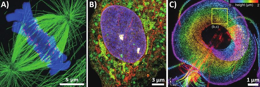
1 Seung Joong Kim Integrative Structure and Functional Anatomy of a Nuclear Pore Complex (NPC)
2 Munkyu Kang Aberration-free and Flexible Bundle-fiber Endomicroscopy
3 Eunsung Seo Experimental Mapping of Near-field Eigenmodes in Sub-wavelength Nanostructures
4 Jungho Moon Real-time Microscopic Recording of Non-repetitive Dynamic Scenes at 5 THz Frame
Wide-field Laser Scanning Microscopy for In Vivo Imaging of Mouse Brain through
5 Hojun Lee
the Intact Skull
6 Ye-Ryoung Lee Dynamics of time-gated reflection eigenchannels
7 Dong-Young Kim Time-gated Iterative Phase Conjugation
Katerina Diffusion Dynamics of Fluorescent Nano-diamonds in Living Cells Detected by
8
Zambochova Fluorescence-combined iSCAT System
9 Sookho Kim Quantitative Detection of DNA Methylation via the B-Z Transition
10 Hyeon-Min Moon Interferometric Scattering Microscopy with Polarization-Selective Detection Scheme
Real-time Observation of the Individual Focal Adhesion Dynamics and Their
11 Jin-Sung Park
Associated Cytoskeletons via iSCAT Microscopy
12 Chanjong Park Mid-IR Photothermal Microscopy
13 Changhyeong Yoon Aberration-corrected and Label-free Scanning Endomicroscopy Using GRIN Lens
14 Hakseok Ko Deep Tissue Space-gated Microscopy via Acousto-optic Interaction
15 Sohee Lim Two-color Resonant CARS Microscopy for an Interfacial Dip-free Detection
Z Sectioning of Intracellular Organelles with Remote-focusing-enabled iSCAT
16 Il-Buem Lee
Microscopy
Selective Suppression of Stimulated Raman Scattering with Another Competing
17 Doyeon Kim
Stimulated Raman Scattering
Bright Ligand-activatable Fluorescent Protein for High-quality Multicolor Live-cell
18 Jiwoong Kwon
Super-resolution Microscopy
19 Minsu Kang Single-molecule Localization Expansion Microscopy
Complimentarily Balanced Real-Time Two-Color Stimulated Raman Scattering
20 Youngjin Choi
Microscopy
21 Hyunmin Kim Transient SHG Microscopy on Atomically Thin 2D Materials
62 63POSTER SESSION 2019 IBS Conference on Advanced Optical Imaging
1
Integrative Structure and Functional Anatomy of References
[1] S.J. Kim*, J. Fernandez-Martinez*, I. Nudelman*, Y. Shi*, W. Zhang* et al. Integrative structure and functional
a Nuclear Pore Complex (NPC) anatomy of a nuclear pore complex. Nature 555, 475-482 (2018).
[2] J. Fernandez-Martinez*, S.J. Kim*, Y. Shi*, P. Upla*, R. Pellarin* et al. Structure and function of the nuclear pore
Seung Joong Kim1,2, Javier Fernandez-Martinez3, Ilona Nudelman3, Yi Shi4, Wenzhu Zhang4,
complex cytoplasmic mRNA export platform. Cell 167 (5), 1215-1228 (2016).
Steven J. Ludtke5, Christopher W. Akey6, Brian T. Chait4, Andrej Sali2, and Michael P. Rout3
1 [3] P. Upla*, S.J. Kim*, P. Sampathkumar*, K. Dutta* et al. Molecular architecture of the major membrane ring
Dept. of Physics and Biological Sciences, Korea Advanced Institute of Science and Technology (KAIST), Daejeon
2 component of the nuclear pore complex, Structure 25 (3), 434-445 (2017).
Dept. of Bioengineering and Therapeutic Sciences, and Pharmaceutical Chemistry, and California Institute for
Quantitative Biosciences, Univ. of California, San Francisco (UCSF), USA
3
Lab. of Cellular and Structural Biology, The Rockefeller University, NY, USA
4
Lab. of Mass Spectrometry and Gaseous Ion Chemistry, The Rockefeller University, NY, USA
5
Dept. of Biochemistry and Molecular Biology, Baylor College of Medicine, Houston, TX, USA
6
Dept. of Physiology and Biophysics, Boston University School of Medicine, Boston, MA, USA
Despite the central role of Nuclear Pore Complexes (NPCs) as gatekeepers of RNA and protein transport
between the cytoplasm and nucleoplasm, their large size and dynamic nature have impeded a full structural
and functional elucidation. Here, we have determined a subnanometer precision structure for the entire
552-protein yeast NPC by satisfying diverse data including stoichiometry, a cryo-electron tomography
map, chemical cross-links, and small angle X-ray scattering (Figure 1)[1-3]. The structure reveals the NPC’s
functional elements in unprecedented detail (Figure 2). The NPC is built of sturdy diagonal columns to which
are attached connector cables, imbuing both strength and flexibility, while tying together all other elements of
the NPC, including membrane-interacting regions and RNA processing platforms. Inwardly-directed anchors
create a high density of transport factor-docking Phe-Gly repeats in the central channel, organized in distinct
functional units. Taken together, this integrative structure allows us to rationalize the architecture, transport
mechanism, and evolutionary origins of the NPC.
Figure 1. I ntegrative structure determination of the NPC Figure 2. Structural dissection of the NPC
64 65POSTER SESSION 2019 IBS Conference on Advanced Optical Imaging
2 3
Aberration-free and Flexible Bundle-fiber Endomicroscopy Experimental Mapping of Near-field Eigenmodes in Sub-wavelength
Nanostructures
Munkyu Kang1,2, Wonjun Choi1,2, Changhyeong Yoon1,2, Sungsoo Woo1,2,
Youngwoon Choi3,*, and Wonshik Choi1,2 Eunsung Seo1,2,+, Young-Ho Jin3,+, Wonjun Choi1,2, Yonghyeon Jo1,2, Kyung-Deok Song1,2,
1
Center for Molecular Spectroscopy and Dynamics, Institute for Basic Science, Seoul 02841, Korea1)
2
Joonmo Ahn1,2, Suyeon Lee4, Q-Han Park2, Myung-Ki Kim3,*, and Wonshik Choi1,2
Department of Physics, Korea University2) 1
Center for Molecular Spectroscopy and Dynamics, Institute for Basic Science, Seoul 02841, Korea
3
School of Biomedical Engineering, Korea University3) 2
Department of Physics, Korea University, Seoul 02841, Korea
3
KU-KIST Graduate School of Converging Science and Technology, Korea University, Seoul 02841, Korea
The bundled fiber is commonly used as a probe of endoscopes due to its small dimension, flexibility 4
Samsung Advanced Institute of Technology, 130, Samsung-ro, Yeongtong-gu, Suwon, Gyeongi-do, 16678, Korea
and relatively large numerical aperture (NA). However, image pixelation and sample-induced aberration
degrade spatial resolving power. T-matrix measurement has been proposed to resolve such problems, but it Near-field scanning optical microscopy (NSOM) is a useful tool for studying sub-diffraction nanostructures.
is vulnerable for the fiber bending. Here we apply our own algorithm, Closed-Loop Accumulation of Single As the size of nanostructures becomes smaller, the ability to observe and manipulate the near-field is getting
Scattering (CLASS)[1], to the fiber bundle system for correcting the core-to-core phase retardation induced by more crucial. In ordinary NSOM imaging, the illumination of light source has not been a major concern since
fiber bending as well as sample-induced aberration. Back-reflection problem from the ultrathin probe was also the spatial resolution is mainly determined by the collection process by the sub-wavelength aperture.
solved since we performed the single-core scanning[2]. We could demonstrate high-resolution and pixelation- We constructed a unique system that integrates far-field wavefront shaping by a spatial light modulator
free images. We are in the process of miniaturizing the endomicroscope by attaching a GRIN lens to the into an NSOM and developed methods to measure a far- to near-field transmission matrix (FNTM). Using the
bundle-fiber to acquire in-vivo biosample images. recorded matrix, we have demonstrated the manipulation of near-field waves and observation of the near-field
eigenmodes generated by the nanostructures.
For the double-slot nanoantenna having the separation of 50 nm, which is about 13 times smaller than
the wavelength of light source and 3 times smaller than the size of NSOM probe, we could obtain an anti-
symmetric transverse mode which has a sharp phase singularity in the middle of the two slot antennas. This
corresponds to the resolving of structures whose separation is smaller than the NSOM aperture. Moreover,
by scanning the NSOM probe over the two-dimensional (2D) surface, we have demonstrated the mapping of
2D near-field eigenmodes for any arbitrary nanostructures. We believe that these studies exploiting the far- to
near-field transmission matrix will open new venues for interrogating the complex nanophotonic structures.
References
[1] Rotenberg, N. & Kuipers, L. “Mapping nanoscale light fields”. Nat Photonics 2014, 8, 919-926.
[2] Kim, M. et al. “Maximal energy transport through disordered media with the implementation of transmission
eigenchannels”. Nat Photonics 2012, 6, 581-585.
[3] Kim, M., Choi, W., Choi, Y., Yoon, C. & Choi, W. “Transmission matrix of a scattering medium and its applications
in biophotonics”. Opt Express 2015, 23, 12648-12668.
[4] Choi, W. et al. “Control of randomly scattered surface plasmon polaritons for multiple-input and multiple-output
Figure 1. E
xperimental setup. BS1~BS3: Beam splitters, L1~L5: Lenses, OL: 10x, 0.25 NA objective lens, AL: Effective
plasmonic switching devices”. Nat Commun 2017, 8.
focal length 1.45 mm aspheric lens. Obtaining complex field images from Mach-Zehnder interferometry
[5] le Feber, B., Rotenberg, N., Beggs, D. M. & Kuipers, L. “Simultaneous measurement of nanoscale electric and
system. Scanning each single core of bundle-fiber by swinging the galvanometer mirror. Fiber is placed at the
magnetic optical fields”. Nat Photonics 2014, 8, 43-46.
Fourier plane of the sample to measure the aberration of core dependent aberration.
References
[1] Kang. S, et al., “High-resolution adaptive optical imaging within thick scattering media using closed-loop
accumulation of single scattering”, Nat. Communications. 8. 2157. (2017).
[2] Yoon. C, et al., “Removal of back-reflection noise at ultrathin imaging probes by the single-core illumination and
wide-field detection”, Scientific Reports. 7, 6524. (2017).
66 67POSTER SESSION 2019 IBS Conference on Advanced Optical Imaging
4 5
Real-time Microscopic Recording of Non-repetitive Wide-field Laser Scanning Microscopy for in Vivo Imaging
Dynamic Scenes at 5 THz Frame of Mouse Brain through The Intact Skull
Jungho Moon, Seok-Chan Yun, and Wonshik Choi Hojun Lee1,2,+, Seokchan Yoon1,2,+, Jin Hee Hong1,2, and Wonshik Choi1,2
Center for Molecular Spectroscopy and Dynamics, Institute for Basic Science, Seoul 02841, Korea 1
Center for Molecular Spectroscopy and Dynamics, Institute for Basic Science, Seoul 02841, Korea
2
Department of Physics, Korea University, Seoul 02855, Korea
Femtoseconds-scale ultrafast imaging has served as an essential tool to directly visualize fast dynamics in
a wide variety of applications in molecular biology, physical chemistry, atomic physics, and fluid dynamics. We present a novel non-fluorescence-based adaptive-optics optical coherence tomography that can
The conventional techniques providing a frame rate up to trillion frames per seconds include a streak cam- eliminate higher-order aberrations even in the regime of strong multiple scattering of light. This adaptive-
era, pump-probe imaging and time of flight system. However, the existing methods are either sacrificing the optics system combines a coherence-gated wavefront sensing and a phase-retrieval algorithm that
number of imaging dimension or demanding many repetitions of the same scene. Recently many interesting independently estimates aberrations along both the illumination and detection pathways. With this technique,
single-shot 2D ultrafast imaging techniques was developed for recording non-repetitive dynamic scenes[1,2,3]. we demonstrate in vivo high-resolution reflectance imaging of myelinated axons in mouse brain through the
Nevertheless, there are still weakness in the number of frames, the number of image pixels, spatial and tempo- intact skull. The imaging configuration relies on the point illumination, which can be easily integrated in
ral resolution. Here, we present a microscopic femtosecond imaging technique that can capture non-repetitive existing multiphoton microscopes. After compensating the measured phase aberration by using a wavefront
dynamic scenes at 200 fs frame interval, 4 mm spatial resolution and a dozen frames. We used an interfero- control device, high-resolution multiphoton images can also be achieved successfully.
metric imaging setup that gives different time delay to multi reference pulses with spatial frequency division.
Our new ultrafast camera scheme, with a potential to achieve a trillions-frame-rate microscopy will have pro- References
found impacts in ultrafast imaging area. [1] Kang, S. et al. Imaging deep within a scattering medium using collective accumulation of single-scattered waves. Nat.
Photonics 9, 253–258 (2015).
References
[2] Kang, S. et al. High-resolution adaptive optical imaging within thick scattering media using closed-loop accumulation
[1] K. Nakagawa, A. Iwasaki, Y. Oishi, R. Horisaki, A. Tsukamoto, A. Nakamura, K. Hirosawa, H. Liao, T. Ushida, K.
of single scattering. Nat. Commun. 8, 2157 (2017).
Goda, F. Kannari & I. Sakuma, “Sequentially timed all-optical mapping photography (STAMP)”, Nature Photonics 2014,
8, 695–700.
[2] Andreas Ehn, Joakim Bood, Zheming Li, Edouard Berrocal, Marcus Aldén and Elias Kristensson, “Single-shot com-
pressed ultrafast photography at one hundred billion frames per second”, Nature,516,74-77.
[3] Andreas Ehn, Joakim Bood, Zheming Li, Edouard Berrocal, Marcus Aldén and Elias Kristensson, “FRAME: femto-
second videography for atomic and molecular dynamics”, Light: Science & Applications 2017,6,e17045.
68 69You can also read