Analysis of remaining motion using one innovative upper airway opening cervical collar and two traditional cervical collars
←
→
Page content transcription
If your browser does not render page correctly, please read the page content below
www.nature.com/scientificreports
OPEN Analysis of remaining motion
using one innovative upper airway
opening cervical collar and two
traditional cervical collars
Matthias K. Jung1, Gregor V. R. von Ehrlich‑Treuenstätt1, Holger Keil2, Paul A. Grützner1,
Niko R. E. Schneider3 & Michael Kreinest1*
The aim of this study was to compare the remaining motion of an immobilized cervical spine using an
innovative cervical collar as well as two traditional cervical collars. The study was performed on eight
fresh human cadavers. The cervical spine was immobilized with one innovative (Lubo Airway Collar)
and two traditional cervical collars (Stifneck and Perfit ACE). The flexion and lateral bending of the
cervical spine were measured using a wireless motion tracker (Xsens). With the Weinman Lubo Airway
Collar attached, the mean remaining flexion was 20.0 ± 9.0°. The mean remaining flexion was lowest
with the Laerdal Stifneck (13.1 ± 6.6°) or Ambu Perfit ACE (10.8 ± 5.8°) applied. Compared to that of
the innovative Weinmann Lubo Airway Collar, the remaining cervical spine flexion was significantly
decreased with the Ambu Perfit ACE. There was no significant difference in lateral bending between
the three examined collars. The most effective immobilization of the cervical spine was achieved when
traditional cervical collars were implemented. However, all tested cervical collars showed remaining
motion of the cervical spine. Thus, alternative immobilization techniques should be considered.
Although evidence of the effectiveness of cervical collars is r are1–3, the use of rigid cervical collars in preclini-
cal trauma care is known to be a common immobilization technique. The importance of immobilization of the
cervical spine is recognized in national and international recommendations for initial trauma care. The applica-
tion of the cervical collar to avoid secondary injuries of the cervical s pine4,5 is recommended as one of the first
emergency measures6,7.
Nevertheless, immobilization of the cervical spine by rigid collars is controversially discussed in the
literature8–12. Cervical spine immobilization with collars can be accompanied by disadvantages, such as pain,
discomfort13,14 or pressure ulceration13,15,16. Furthermore, the application of a cervical collar can also lead to
considerably serious and even life-threatening complications. In particular, restricted respiratory f unction17
and difficult airway management18 have been described, which can quickly lead to severe complications or
even death, especially in trauma p atients7. To ease airway management, an innovative cervical collar has been
developed that should combine cervical spine immobilization and airway protection. This innovative cervical
andible19. While this innovative collar seems to succeed in airway
collar is equipped with a flexible belt for the m
protection, the quality of cervical spine immobilization has not been proven, so far. However, the effectiveness
of a cervical collar depends on its perfect fit20–22.
The aim of this study was to compare the remaining motion of the cervical spine using an innovative cervical
collar as well as two traditional cervical collars on fresh human cadavers.
Materials and methods
Study design. The study was performed on fresh human cadavers. The body donors were given detailed
information before death and had to give their written informed consent to the body donation. After death, the
body was made available for scientific research purposes. Fresh human cadavers were briefly frozen after death.
For the experiments, the bodies were thawed to room temperature. This method enabled the simulation of the
1
BG Trauma Center Ludwigshafen, Clinic for Trauma and Orthopaedic Surgery, University of Heidelberg,
Ludwig‑Guttmann‑Str. 13, 67071 Ludwigshafen on the Rhine, Germany. 2Clinic for Trauma and Orthopaedic
Surgery, Universitätsklinikum Erlangen, Krankenhausstraße 12, 91054 Erlangen, Germany. 3Clinic of
Anesthesiology, University of Heidelberg, Im Neuenheimer Feld 420, 69120 Heidelberg, Germany. *email:
michael.kreinest@bgu-ludwigshafen.de
Scientific Reports | (2021) 11:20619 | https://doi.org/10.1038/s41598-021-00194-w 1
Vol.:(0123456789)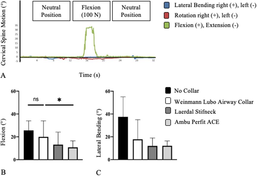
www.nature.com/scientificreports/
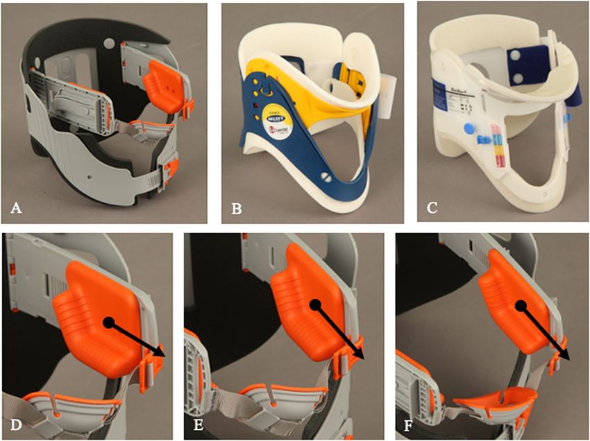
Figure 1. In the present study, three different cervical collars were tested on fresh human cadavers: Weinmann
Lubo Airway Collar (A), Laerdal Stifneck (B) and Ambu Perfit ACE (C). Shown is the innovative mechanism
(D,E,F). The red angle (black dot) pushes the mandible ventrally (black arrow) for opening the upper airway.
joint elasticity and soft tissue situation of a living body. Current biomechanical studies do not describe any sig-
nificant difference in cervical spine motion between fresh human cadavers and p atients4,23,24.
The inclusion criteria for the current study were as follows: (1) existing written informed consent for body
donation for scientific research; (2) no injuries, diseases or operations on the cervical spine; and (3) complete
medical records. The complete medical history and the existing medical data of all examined patients were ana-
lyzed. Patients with diseases such as tumors, thyroid diseases or similar conditions were excluded from the study.
The local ethics committee reviewed and approved the present study (Ethics Committee, Mainz, Rhineland-
Palatinate, Germany, ID: 837.156.16). The study was registered in the German Clinical Trials Register (ID:
DRKS00010499). All experiments were performed in accordance with the relevant guidelines and regulations.
Cervical collars. In the present study, three different cervical collars were tested on fresh human cadavers.
First, the innovative Weinmann Lubo Airway Collar (Weinmann Emergency Medical Technology GmBh and
Co. KG, Hamburg, Germany, Fig. 1A) was tested. This collar was newly developed in 2007 to stabilize the cervi-
cal spine and to avoid supraglottic airway obstruction at the same time (Fig. 1D–F) 19. Second, two traditional
collars were tested, namely, the Laerdal Stifneck (Laerdal Medical GmbH, Puchheim, Germany, Fig. 1B) and the
Ambu Perfit ACE (Ambu GmbH, Bad Nauheim, Germany, Fig. 1C). These traditional rigid collars with one-belt
fixation have been in use nearly worldwide for many decades.
All cervical collars were adjusted according to the manufacturers’ instructions. All different sizes of cervical
collars were available during the study. The three cervical collars (Fig. 1A–C) were applied by one experienced
emergency medical service (EMS) person to fresh human cadavers. Likewise, the repetitive measurements were
performed only by one person experienced in rescue medicine.
Cervical spine motion measurement. In the present study, the remaining flexion and the remaining
lateral bending of the immobilized cervical spine were measured with a wireless human motion tracker system
(Xsens Technologies, Enschede, Netherlands). Compared to other methods, this measurement method with a
motion tracker has been proven25–27 and guarantees exact measurement results28. The endpoints of the measure-
ments were maximal flexion and maximal lateral bending.
In the standardized motion protocol, the rotation was not included separately since combination motions
always occur here. The extension was also not recorded since the fresh human cadavers were lying in the supine
position and thus no extension could be measured (Fig. 2).
Scientific Reports | (2021) 11:20619 | https://doi.org/10.1038/s41598-021-00194-w 2
Vol:.(1234567890)www.nature.com/scientificreports/
Figure 2. A fresh human cadaver is positioned supine on a spine board with a cervical collar; the motion
trackers are fixed to the forehead and to the sternum.
Figure 3. Remaining flexion of the cervical spine if an innovative cervical collar is applied (A). Range of
remaining flexion of the cervical spine (B) and range of remaining lateral bending (C) of the three tested
cervical collars (B,C: Only significant differences are marked).
In the experimental setup, fresh human cadavers were placed on a spineboard (Laerdal BaXstrap, Stavanger,
Norway) in supine position and fixed by a harness fixation system (MIH-Medical Spiderstrap, Georgsmarien-
hütte, Germany). One motion tracker was attached to the forehead, and one motion tracker was attached to
the thorax of the fresh human cadaver (Fig. 2). The 3D data were synchronized every 20 microseconds by the
Xsens recording tool. However, with the help of the motion trackers, rotation and extension were recorded in
the respective measured direction of motion (Fig. 3A).
The head of the fresh human cadaver was passively moved ventrally for flexion (Fig. 3A) and to the right for
lateral bending. The traction force was 100 N. This force is in the range and in the direction as it may be applied
during intubation or patient transport and may therefore initiate or aggravate an assumed spinal i njury3,29,30. The
force was measured with an electronic spring balance (LENI, Fa. Korona, Sundern, Germany).
In the standardized motion protocol, flexion and lateral bending were first analyzed for each fresh human
cadaver without the cervical collar (control group). Subsequently, the motion analysis was performed with the
three cervical collars in place.
Statistical data analysis. In the present study, statistical calculations were performed to determine pos-
sible differences between the three tested cervical collars. With the D’Agostino and Pearson test, the three groups
were tested for normal distribution. The statistical comparison between the four groups was performed using
analysis of variance (ANOVA). A p-value of < 0.05 was considered statistically significant.
Scientific Reports | (2021) 11:20619 | https://doi.org/10.1038/s41598-021-00194-w 3
Vol.:(0123456789)www.nature.com/scientificreports/
No collar Weinmann Lubo airway collar Laerdal Stifneck Ambu Perfit ACE
Flexion, ° (range) 25.6 ± 5.0 (18.0–34.0) 20.0 ± 9.0 (10.0–34.0) 13.1 ± 6.6 (8.0–24.0) 10.8 ± 5.8 (5.0–19.0)
Lateral bending, ° (range) 37.5 ± 8.7 (29.0–55.0) 17.8 ± 10.2 (7.0–35.0) 12.0 ± 4.8 (7.0–19.0) 12.1 ± 4.3 (6.0–17.0)
Table 1. Results of the remaining motion examination of the three cervical collars and the control group.
An exploratory analysis of the measured values was performed. For all measurements, descriptive data with
the mean, standard deviation and ranges are given. The statistical data analysis was performed with GraphPad
PRISM software (Version 8.2.1, San Diego, California, USA).
Results
Study collective. Eight fresh human cadavers were included in the present study. The mean age of the
cadavers at death was 79.0 ± 12.8 years (range: 56–94 years). The study included five female and three male fresh
human cadavers.
Motion measurement of the cervical spine. The highest remaining flexion of the cervical spine with
a cervical collar applied was 34.0° (Fig. 3A). In this case, Weinman Lubo Airway Collar (Fig. 1A) was attached.
Overall, the fresh human cadavers with the Weinman Lubo Airway Collar in place had a mean flexion of
20.0 ± 9.0° (range: 10.0–34.0°; Fig. 3B; Table 1). A lower remaining flexion at the cervical spine could be achieved
using the two traditional cervical collars (Fig. 1B,C). With the Laerdal Stifneck applied, the mean flexion was
13.1 ± 6.6° (range: 8.0–24.0°). The mean flexion was lowest with the Ambu Perfit ACE (10.8 ± 5.8°; range: 5.0–
19.0°; Fig. 3B). Without cervical collar, the fresh human cadavers had a mean cervical flexion of 25 ± 5.0° (range:
18.0°–34.0°, Fig. 3B).
The mean flexion of the cervical spine with the Weinmann Lubo Airway Collar on was almost twice as high
as that with the Ambu Perfit ACE applied. Compared to the Weinmann Lubo Airway Collar, the remaining
cervical spine flexion was significantly (p = 0.0474) decreased with the Ambu Perfit ACE (Fig. 3B). The differ-
ences in remaining cervical spine flexion between the Laerdal Stifneck and the Weinmann Lubo Airway Collar
(p = 0.1655) and between the Laerdal Stifneck and the Ambu Perfit ACE (p = 0.7923) were not significant. There
was no significant difference in cervical flexion with Weinmann Lubo Airway Collar applied or no collar applied
(p = 0.3611). Between the control examination and the Laerdal Stiffneck (p = 0.0049) and the Ambu Perfit ACE
(p = 0.0008) there was a significant reduction in remaining cervical spine flexion.
The mean remaining lateral bending of the Weinmann Lubo Airway Collar was 17.8 ± 10.2° (range:
7.0–35.0°; Table 1). With the Laerdal Stifneck attached, the mean remaining lateral bending was 12.0 ± 4.8°
(range: 7.0–19.0°). The Ambu Perfit ACE allowed for a mean remaining lateral bending of 12.1 ± 4.3° (range:
6.0–17.0°; Figs. 1C and 3C). Without cervical collar, the lateral bending of the cervical spine was 37.5 ± 8.7°
(range: 29.0°–55.0°, Fig. 3C).
There was no significant difference in lateral bending between the three examined collars (Weinmann Lubo
Airway Collar vs. Ambu Perfit ACE: p = 0.2600; Weinmann Lubo Airway Collar vs. Laerdal Stifneck: p = 0.2458;
Laerdal Stifneck vs. Ambu Perfit ACE: p = 0.9993; Fig. 3C). Lateral bending of the cervical spine was significantly
reduced by all three cervical collars (p < 0.0001).
Discussion
The aim of this study was to measure and to evaluate quantitatively the remaining motion of the cervical spine
during immobilization using one innovative upper airway opening cervical collar and two traditional cervical
collars. The present study showed that the attached innovative and traditional cervical collars resulted in signifi-
cantly different remaining flexion of the immobilized cervical spine. The design of the cervical collars seems to
have an influence on their fit and thus on the effectiveness of the immobilization.
The data of the present study showed that compared to traditional cervical collars, the innovative cervical
collar allows for twofold increased flexion of the cervical spine. There was no significant difference in the cervical
spine flexion whether the innovative cervical collar was applied or not. The novel upper airway opening design is
probably decisive for the poor result. The innovative cervical collar has a one-belt system; whereby the mandible
is addressed with an extra flexible belt (Fig. 1A). In contrast, both traditional cervical collars address the mandible
as well as the occiput, sternum, clavicle, shoulders and upper back with their rigid form (Fig. 1B,C)19,21,31. The
novel fixation system therefore seems to perform worse than the classic cervical collars in the important flexion
motion. The Weinmann Lubo Airway Collar can support airway clearance, but a classic cervical collar should
be used in unconscious and intubated patients with suspected cervical spine injury.
According to a study by Lubovsky et al., the innovative Weinmann Lubo Airway Collar, which combines an
external airway protector in combination with a cervical collar, appears to be safe and effective in opening and
maintaining open airways in anesthetized supine patients19. In addition, the authors recommend that further
clinical trials are indicated to evaluate the safety and utility of this device under preclinical c onditions19. To our
knowledge, the efficacy of the collar toward cervical spine immobilization has not been tested so far. According
to the data of the current study, efficiency of the innovative cervical collar by the means of cervical spine immo-
bilization is worse compared to traditional cervical collars.
The study by Karason et al. also compared widely used cervical collars32. The endpoints of the remaining
motion were analyzed using a gonimeter. In addition, the intracranial pressure was extrapolated, and the wearing
Scientific Reports | (2021) 11:20619 | https://doi.org/10.1038/s41598-021-00194-w 4
Vol:.(1234567890)www.nature.com/scientificreports/
comfort was evaluated subjectively. As in the present study, the classic rigid cervical collars were shown to
achieve safe immobilization. Karason et al. were able to determine a remaining motion of immobilization of the
cervical spine of 18.0° ± 7.0°. The present study showed significantly better immobilization values. The extent
to which this depends on the measurement method used or is even due to the test subjects should be analyzed
in further studies.
The motion values that have been measured for the traditional cervical collars were in the same range as in
other studies. Hostler et al. found a mean flexion of 9.2° ± 5.0° with the Ambu Perfit ACE a pplied33. Thus, com-
plete immobilization of the cervical spine could not be achieved by a cervical collar alone. These findings are
supported by various other studies3,34. In addition, many cervical collars are not applied correctly35; therefore,
their effectiveness is further reduced.
Since a secondary injury or a worsening of an existing injury cannot be completely excluded in cases of
remaining cervical spine motion4,36, alternative immobilization techniques should be considered. Cervical spine
immobilization by a collar could be substantially improved by additionally fixing the patient to a spine b oard34.
However, in this case, the main immobilization will derive from fixation on the spineboard, and the cervical collar
is dispensable37,38. The full body immobilization technique is especially recommended during patient transport25.
Limitations. The present study is limited to some extent. Only human cadavers with an intact cervical spine
were analyzed. For patients with an already injured cervical spine, different results can certainly be expected.
Furthermore, the cervical spine force was applied in a standardized manner only for flexion and lateral bending.
Combined movements were not performed. There is no literature that provides any indication of the magnitude
and direction of the force required to cause or even to aggravate a cervical spine injury. Previous studies have
shown that after an initial trauma, no further injury or aggravation can occur through further manipulation39.
However, recent biomechanical studies have shown a direct correlation between manipulation of the severely
injured cervical spine and the width of the dural s ac29,30,40. Furthermore, the application of a cervical collar itself
may aggravate a spinal injury29. Thus, in future studies, not only the remaining cervical spine motion but also
the manipulation that is caused by the collar’s application should be analyzed. Here, significant differences have
been previously shown41. In addition, it should always be remembered that the cervical support immobilizes best
when other immobilization tools are applied26. Three cervical collars were examined in the current study; and,
according to the literature, many additional cervical collars are a vailable33. This efficiency of these other collars
should be addressed in further studies.
Conclusions
The results of the present study indicate that the best immobilization of the cervical spine could be achieved if the
cervical collar has a nonmobile, rigid design that is closely attached to the mandible, occiput, sternum, clavicle,
shoulders and upper back. However, all tested cervical collars showed remaining motion of the cervical spine.
Thus, alternative immobilization techniques should be considered.
Received: 14 March 2021; Accepted: 24 September 2021
References
1. Hood, N. & Considine, J. Spinal immobilisaton in pre-hospital and emergency care: A systematic review of the literature. Australas.
Emerg. Nurs. J. 18, 118–137. https://doi.org/10.1016/j.aenj.2015.03.003 (2015).
2. Peck, G. E., Shipway, D. J. H., Tsang, K. & Fertleman, M. Cervical spine immobilisation in the elderly: A literature review. Br. J.
Neurosurg. 32, 286–290. https://doi.org/10.1080/02688697.2018.1445828 (2018).
3. Horodyski, M., DiPaola, C. P., Conrad, B. P. & Rechtine, G. R. Cervical collars are insufficient for immobilizing an unstable cervical
spine injury. J. Emerg. Med. 41, 513–519. https://doi.org/10.1016/j.jemermed.2011.02.001 (2011).
4. Hindman, B. J. et al. Intubation biomechanics: Laryngoscope force and cervical spine motion during intubation in cadavers-cadav-
ers versus patients, the effect of repeated intubations, and the effect of type II odontoid fracture on C1–C2 motion. Anesthesiology
123, 1042–1058. https://doi.org/10.1097/aln.0000000000000830 (2015).
5. Benger, J. & Blackham, J. Why do we put cervical collars on conscious trauma patients?. Scand. J. Trauma Resusc. Emerg. Med. 17,
44. https://doi.org/10.1186/1757-7241-17-44 (2009).
6. NAEMT. Prehospital Trauma Life Support (PHTLS) 7th edn. (Prehospital Trauma Life Support Committee of the National Asso-
ciation of Emergency Medical Technicians in Cooperation with the Committee on Trauma of The American College of Suregons,
2010).
7. Surgeons, A. C. O. Advanced Trauma Life Support (ATLS®): The Ninth Edition. 2013/04/24 edn (2013).
8. Hess, T. et al. Pre-hospital care management of acute spinal cord injury. Anasthesiol. Intensivmed. Notfallmed. Schmerzther. 51,
226–237. https://doi.org/10.1055/s-0042-105016 (2016).
9. Walters, B. C. et al. Guidelines for the management of acute cervical spine and spinal cord injuries: 2013 update. Neurosurgery 60,
82–91. https://doi.org/10.1227/01.neu.0000430319.32247.7f (2013).
10. Unfallchirurgie, D. G. F. S3 Guideline on Treatment of Patients with Severe and Multiple Injuries. English Version of the German
Guideline S3 Leitlinie Polytrauma/Schwerverletzten-Behandlung AWMF Register-Nr. 012/019. https://www.awmf.org/uploads/tx_
szleitlinien/012-019l_S3_Polytrauma_Schwer verletzten-Behandlung_2017-08.pdf (2016).
11. Centre, N. C. G. Spinal Injury: Assessment and Initial Management. NICE Guideline, No. 41 (2016).
12. Kwan, I., Bunn, F. & Roberts, I. Spinal immobilisation for trauma patients. Cochrane Database Syst. Rev. 2001, Cd002803. https://
doi.org/10.1002/14651858.Cd002803 (2001).
13. Cordell, W. H., Hollingsworth, J. C., Olinger, M. L., Stroman, S. J. & Nelson, D. R. Pain and tissue-interface pressures during spine-
board immobilization. Ann. Emerg. Med. 26, 31–36. https://doi.org/10.1016/s0196-0644(95)70234-2 (1995).
14. Swartz, E. E. et al. Prehospital cervical spine motion: Immobilization versus spine motion restriction. Prehosp. Emerg. Care 22,
630–636. https://doi.org/10.1080/10903127.2018.1431341 (2018).
15. Ham, W., Schoonhoven, L., Schuurmans, M. J. & Leenen, L. P. Pressure ulcers from spinal immobilization in trauma patients: A
systematic review. J. Trauma Acute Care Surg. 76, 1131–1141. https://doi.org/10.1097/ta.0000000000000153 (2014).
Scientific Reports | (2021) 11:20619 | https://doi.org/10.1038/s41598-021-00194-w 5
Vol.:(0123456789)www.nature.com/scientificreports/
16. Pernik, M. N. et al. Comparison of tissue-interface pressure in healthy subjects lying on two trauma splinting devices: The vacuum
mattress splint and long spine board. Injury 47, 1801–1805. https://doi.org/10.1016/j.injury.2016.05.018 (2016).
17. Totten, V. Y. & Sugarman, D. B. Respiratory effects of spinal immobilization. Prehosp. Emerg. Care 3, 347–352. https://doi.org/10.
1080/10903129908958967 (1999).
18. Goutcher, C. M. & Lochhead, V. Reduction in mouth opening with semi-rigid cervical collars. Br. J. Anaesth. 95, 344–348. https://
doi.org/10.1093/bja/aei190 (2005).
19. Lubovsky, O., Liebergall, M., Weissman, C. & Yuval, M. A new external upper airway opening device combined with a cervical
collar. Resuscitation 81, 817–821. https://doi.org/10.1016/j.resuscitation.2010.02.013 (2010).
20. White, A. A. & Panjabi, M. M. Clinical Biomechanics of the Spine 2nd edn. (J. B. Lippincott & Co., 1990).
21. Bell, K. M. et al. Assessing range of motion to evaluate the adverse effects of ill-fitting cervical orthoses. Spine J. 9, 225–231. https://
doi.org/10.1016/j.spinee.2008.03.010 (2009).
22. Ben-Galim, P. et al. Extrication collars can result in abnormal separation between vertebrae in the presence of a dissociative injury.
J. Trauma 69, 447–450. https://doi.org/10.1097/TA.0b013e3181be785a (2010).
23. Lennarson, P. J. et al. Segmental cervical spine motion during orotracheal intubation of the intact and injured spine with and
without external stabilization. J. Neurosurg. 92, 201–206. https://doi.org/10.3171/spi.2000.92.2.0201 (2000).
24. McCahon, R. A. et al. Cadaveric study of movement of an unstable atlanto-axial (C1/C2) cervical segment during laryngoscopy
and intubation using the Airtraq(®), Macintosh and McCoy laryngoscopes. Anaesthesia 70, 452–461. https://d oi.o
rg/1 0.1 111/a nae.
12956 (2015).
25. Nolte, P. C. et al. Analysis of cervical spine immobilization during patient transport in emergency medical services. Eur. J. Trauma
Emerg. Surg. https://doi.org/10.1007/s00068-019-01143-z (2019).
26. Uzun, D. D. et al. Remaining cervical spine movement under different immobilization techniques. Prehosp. Disaster Med. https://
doi.org/10.1017/s1049023x2000059x (2020).
27. Haske, D. et al. An explorative, biomechanical analysis of spine motion during out-of-hospital extrication procedures. Injury 51,
185–192. https://doi.org/10.1016/j.injury.2019.10.079 (2020).
28. Weerts, J. O. N., Schier, L., Schmidt, H. & Kreinest, M. Review of existing measurement tools to assess spinal motion during
prehospital immobilization. Eur. J. Emerg. Med. 25, 161–168. https://doi.org/10.1097/mej.0000000000000467 (2018).
29. Liao, S. et al. Motion and dural sac compression in the upper cervical spine during the application of a cervical collar in case of
unstable craniocervical junction—A study in two new cadaveric trauma models. PLoS ONE 13, e0195215. https://d oi.o rg/1 0.1 371/
journal.pone.0195215 (2018).
30. Liao, S. et al. Spinal movement and dural sac compression during airway management in a cadaveric model with atlanto-occipital
instability. Eur. Spine J. 27, 1295–1302. https://doi.org/10.1007/s00586-017-5416-9 (2018).
31. Worsley, P. R., Stanger, N. D., Horrell, A. K. & Bader, D. L. Investigating the effects of cervical collar design and fit on the biome-
chanical and biomarker reaction at the skin. Med. Dev. (Auckl.) 11, 87–94. https://doi.org/10.2147/mder.S149419 (2018).
32. Karason, S., Reynisson, K., Sigvaldason, K. & Sigurdsson, G. H. Evaluation of clinical efficacy and safety of cervical trauma collars:
Differences in immobilization, effect on jugular venous pressure and patient comfort. Scand. J. Trauma Resusc. Emerg. Med. 22,
37. https://doi.org/10.1186/1757-7241-22-37 (2014).
33. Hostler, D., Colburn, D. & Seitz, S. R. A comparison of three cervical immobilization devices. Prehosp. Emerg. Care: Off. J. Natl.
Assoc. EMS Phys. Natl. Assoc. State EMS Dir. 13, 256–260. https://doi.org/10.1080/10903120802706195 (2009).
34. Chandler, D. R., Nemejc, C., Adkins, R. H. & Waters, R. L. Emergency cervical-spine immobilization. Ann. Emerg. Med. 21,
1185–1188 (1992).
35. Kreinest, M. et al. Application of cervical collars—An analysis of practical skills of professional emergency medical care providers.
PLoS ONE 10, e0143409. https://doi.org/10.1371/journal.pone.0143409 (2015).
36. Donaldson, W. F., Heil, B. V., Donaldson, V. P. & Silvaggio, V. J. The effect of airway maneuvers on the unstable C1–C2 segment.
A cadaver study. Spine (Phila Pa 1976) 22, 1215–1218. https://doi.org/10.1097/00007632-199706010-00008 (1997).
37. Holla, M. Value of a rigid collar in addition to head blocks: A proof of principle study. Emerg. Med. J. 29, 104–107. https://doi.org/
10.1136/emj.2010.092973 (2012).
38. Butler, J. & Bates, D. Towards evidence based emergency medicine: Best BETs from the Manchester Royal Infirmary. Cervical
collars in patients requiring spinal immobilisation. Emerg. Med. J.: EMJ 18, 275 (2001).
39. Hauswald, M. & Braude, D. Spinal immobilization in trauma patients: Is it really necessary?. Curr. Opin. Crit. Care 8, 566–570.
https://doi.org/10.1097/00075198-200212000-00014 (2002).
40. Liao, S. et al. Cadaveric study of movement in the unstable upper cervical spine during emergency management: Tracheal intubation
and cervical spine immobilisation-a study protocol for a prospective randomised crossover trial. BMJ Open 7, e015307. https://
doi.org/10.1136/bmjopen-2016-015307 (2017).
41. James, C. Y., Riemann, B. L., Munkasy, B. A. & Joyner, A. B. Comparison of cervical spine motion during application among 4
rigid immobilization collars. J. Athl. Train. 39, 138–145 (2004).
Acknowledgements
The authors would like to thank Professor Dr. Erik Popp for his technical support in the preparation of the fresh
human cadavers. We would like to thank the staff of the Anatomical Institute of the University of Heidelberg
for providing the premises and for their support in handling fresh human cadavers. We would also like to thank
Mr. Geir Dillan for taking the photographs.
Author contributions
Conceptualization, M.K.J., N.R.E.S. and M.K.; Data curation, M.K.J., G.V. R.E. and M.K.; Formal analysis, H.K.;
Investigation, G.V.R.E. and M.K.; Methodology, M.K.J., P.A.G. and M.K.; Project administration, M.K.J. and
M.K.; Resources, H.K., N.R.E.S. and M.K.; Supervision, P.A.G. and M.K.; Validation, M.K.J., G.V. R.E. and M.K.;
Visualization, M.K.J. and N.R.E.S.; Writing—original draft, M.K.J.; Writing—review and editing, M.K.J., G.V.
R.E., P.A.G., H.K., N.R.E.S. and M.K.
Funding
There was no funding for this study.
Competing interests
The authors declare no competing interests.
Additional information
Correspondence and requests for materials should be addressed to M.K.
Scientific Reports | (2021) 11:20619 | https://doi.org/10.1038/s41598-021-00194-w 6
Vol:.(1234567890)www.nature.com/scientificreports/
Reprints and permissions information is available at www.nature.com/reprints.
Publisher’s note Springer Nature remains neutral with regard to jurisdictional claims in published maps and
institutional affiliations.
Open Access This article is licensed under a Creative Commons Attribution 4.0 International
License, which permits use, sharing, adaptation, distribution and reproduction in any medium or
format, as long as you give appropriate credit to the original author(s) and the source, provide a link to the
Creative Commons licence, and indicate if changes were made. The images or other third party material in this
article are included in the article’s Creative Commons licence, unless indicated otherwise in a credit line to the
material. If material is not included in the article’s Creative Commons licence and your intended use is not
permitted by statutory regulation or exceeds the permitted use, you will need to obtain permission directly from
the copyright holder. To view a copy of this licence, visit http://creativecommons.org/licenses/by/4.0/.
© The Author(s) 2021
Scientific Reports | (2021) 11:20619 | https://doi.org/10.1038/s41598-021-00194-w 7
Vol.:(0123456789)You can also read