Analysis of the effects of prepared porcelain veneers and unprepared porcelain veneers on gingival crevicular flora based on high throughput ...
←
→
Page content transcription
If your browser does not render page correctly, please read the page content below
EXPERIMENTAL AND THERAPEUTIC MEDICINE 22: 777, 2021

             Analysis of the effects of prepared porcelain veneers and
              unprepared porcelain veneers on gingival crevicular
                   flora based on high‑throughput sequencing
                              RUI ZHANG1, LANHUA SHEN1, DANDAN XU2,3 and XIAO LI2,3

         1
        Department of Stomatology, Nangang Branch, Heilongjiang Provincial Hospital, Harbin, Heilongjiang 150000;
     2
      Department of Stomatology, The First Hospital of Qiqihar; 3Department of Stomatology, Affiliated Qiqihar Hospital,
                          Southern Medical University, Qiqihar, Heilongjiang 161006, P.R. China

                                         Received June 18, 2020; Accepted February 23, 2021

                                                    DOI: 10.3892/etm.2021.10209

Abstract. The effect of tooth preparation on the gingival           Introduction
crevicular flora of abutment teeth during porcelain veneer
treatment is not clear. The purpose of the present study was        With the development of dental cosmetic repair technology in
to analyze the difference between prepared porcelain veneers        recent years, the number of patients who visit dental clinics
and unprepared porcelain veneers on gingival crevicular flora.      for dental cosmetic repair is increasing. The scope of repairs
High‑throughput sequencing was used in the present study.           mainly includes anterior tooth defects and gaps, abnormal
A total of 20 patients (40 anterior teeth) with veneer restora‑     tooth color, distorted teeth or microdontia (1). Porcelain veneer
tion of anterior teeth were enrolled. They were divided into        repair method is increasingly recognized and requested by
two groups: The prepared porcelain veneer group (group P,           patients. During treatment, no tooth preparation is required,
11 cases, 19 anterior teeth) and unprepared porcelain veneer        or only a small amount of cavitation has to be removed. The
group (group U, 9 cases, 21 anterior teeth). After 2 years          enamel structure can be protected. At present, due to its advan‑
of follow‑up, the restoration and healthy natural gingival          tages of less injury, high biocompatibility and beauty, it plays
crevicular fluid were collected to extract bacterial DNA. 16S       an increasingly important role in minimally invasive tooth
ribosomal DNA high‑throughput sequencing technique was              repair (1,2). Currently, porcelain veneer restoration is divided
used to compare the diversity of gingival sulcus flora struc‑       into prepared porcelain veneers and unprepared porcelain
ture between the prepared porcelain veneer and unprepared           veneers. In prepared porcelain veneers, the enamel needs to
porcelain veneer groups. In addition, a healthy control group       be partially ground, leaving enough thickness for the veneer
(group H) was also used for comparison. The Shannon index           to enhance its strength and color authenticity. Unprepared
of the group U was lower than that of group H and group P.          porcelain veneers do not require tooth preparation nor do they
The abundance of Proteus in group U was higher than that            damage teeth (3). With the promotion of porcelain veneer
in group H and group P at the phylum level (P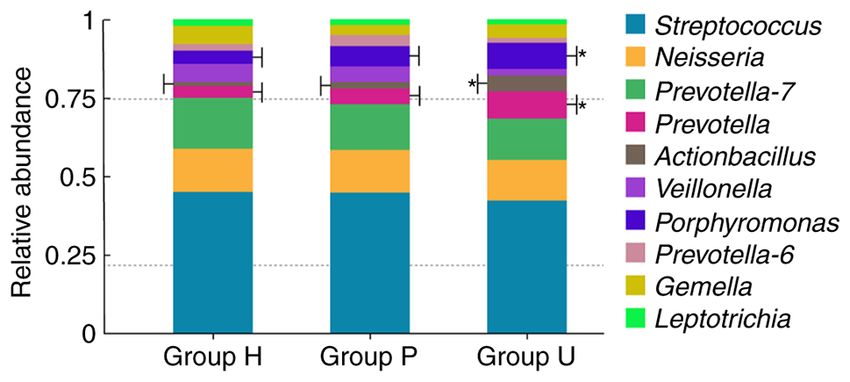
2                        ZHANG et al: EFFECTS OF PORCELAIN VENEERS ON GINGIVAL CREVICULAR FLORA

status of periodontal tissue. However, whether tooth prepara‑          treatment (including porcelain veneers, resin veneers and
tion affects periodontal health through changes in gingival            porcelain full crowns); iii) subjects unwilling to cooperate or
crevicular flora has not yet been reported. Compared with tradi‑       who ground their teeth at night; iv) female pregnant or lactating
tional bacterial culture methods, high‑throughput sequencing           subjects; v) subjects combined with known systemic diseases
technology has outstanding advantages such as high accuracy,           (AIDS, tuberculosis, hepatitis, known history of any other infec‑
high throughput, high sensitivity and low operating cost. It can       tious diseases, diabetes, ischemic heart disease, hypertension,
quickly and accurately reflect the composition and diversity           thyroid or other hormone disorders, autoimmune diseases and
of microorganisms and has been widely used in the field of             cancer); vi) subjects with abnormal coagulation function and
microbial research (13,14). The aim of the present study was to        vii) subjects who failed to cooperate with the follow‑up.
analyze the influence of tooth preparation on the composition              The study was approved by the Ethics Committee of
and diversity of gingival crevicular flora. High‑throughput            Nangang Branch of Heilongjiang Provincial Hospital and
sequencing technique was used to determine the influence of            written informed consent was obtained from all patients.
prepared porcelain veneers and unprepared porcelain veneers
on periodontal health.                                                 Therapeutic method. The therapeutic methods were as
                                                                       follows: Group P, after color comparison and photo recording,
Patients and methods                                                   tooth preparation, temporary restoration and veneering were
                                                                       carried out according to conventional procedures (14). The
Research subjects. A total of 20 patients were selected as             parameters were as follows: i) The thickness of tooth abrasion
research subjects, with a total of 40 teeth. They received             was 0.4‑0.6 mm; ii) dental prosthesis treatment was designed
treatment with anterior dental veneers at Nangang Branch,              with butt‑type cutting ends, with gingival margins; the neck
Heilongjiang Provincial Hospital (Harbin, China) from                  margin was designed as an angular shoulder, and the width was
January 2016 to December 2017. The group using unprepared              controlled at 0.3‑0.5 mm; iii) the veneer material was IPSe and
porcelain veneers was considered as the observation group              the Emax porcelain veneer was completed by the same producer
(group U). There were 9 patients (21 affected teeth in total)          (after 7 days, the patients were re‑examined and if necessary,
in this group, including 4 males (8 teeth), 5 females (13 teeth),      porcelain veneers could be adjusted or even re‑made); iv) the
14 anterior teeth and 7 mandibular anterior teeth. The group           tooth surface was treated with 37% phosphoric acid for 30 sec
using prepared porcelain veneers was considered as the control         and the porcelain veneer was treated with hydrofluoric acid
group (group P). There were 11 patients (19 affected teeth in          for 15 sec; and v) excess adhesive was carefully removed and
total) in this group, including 5 males (10 teeth), 6 females          polished when necessary. Group U, tooth preparation and tempo‑
(9 teeth), 12 anterior teeth and 7 mandibular anterior teeth.          rary restoration were not required. Veneer design requirements
Twenty healthy natural teeth of healthy people were selected           were the same as those of group P, except that the phosphoric
as healthy controls (group H). The patients were 18‑44 years           acid treatment time of the tooth surface was extended to 60 sec.
old, with an average age of (28.36±5.39) years.                            Both groups of patients were provided with oral health care
    Inclusion criteria for the porcelain veneer patients were as       guidance after treatment.
follows: i) All the patients met the clinical criteria of porcelain
veneer repair; ii) patients were in good mental state; iii) patients   Patient follow‑up. Follow‑up visits were conducted in the
who had not used antibiotics, hormone drugs or received                1st, 3rd, 6th, 12th and 24th month after surgery. The gingival
radiotherapy and chemotherapy in the past three months; and            health and the success rate of restorations were evaluated based
iv) patients who had no abnormal secretions in the mouth.              on the clinical evaluation criteria of the California Dental
Exclusion criteria were as follows: i) Patients complicated with       Association (15) and the modified Ryge evaluation criteria (16).
dental pulp and periodontal inflammation, odontatrophy, or             The success rate of restoration was calculated 2 years later.
loosened teeth before surgery; ii) patients with contraindica‑             Gingival health evaluation criteria (12,13) were as follows:
tions to surgery; iii) patients unwilling to cooperate or who          i) Healthy gingiva; ii) slight gingival inflammation and
ground their teeth at night; iv) female pregnant or lactating          a small amount of bleeding could be detected, and slight
patients; v) patients combined with known systemic diseases            gingival atrophy that did not affect the appearance could be
(AIDS, tuberculosis, hepatitis, known history of any other             observed; and iii) gingival swelling was obvious, bleeding and
infectious diseases, diabetes, ischemic heart disease, hyper‑          periodontal pockets deepened, accompanied by moderate to
tension, thyroid or other hormone disorders, autoimmune                severe gingival atrophy, affecting the appearance.
diseases and cancer); vi) patients with abnormal coagulation               Evaluation criteria for successful restoration (14) were as
function; and vii) patients who failed to cooperate with the           follows: The size and shape of the restored tooth were coor‑
follow‑up. There was no statistical difference in the general          dinated with the adjacent teeth and antagonist teeth; the color
data between the two groups (P>0.05) (Table I).                        was consistent, no discoloration occurred; the teeth were in
    Inclusion criteria for group H were as follows: i) All the         good occlusion, and there was no gap between the teeth under
subjects were aged 18‑65 years; ii) subjects were in good mental       naked eye observation; the occlusal performance of the teeth
state; iii) subjects who did not use antibiotics, hormone drugs or     was good, and there was no visible gap between the teeth;
receive radiotherapy and chemotherapy in the past three months;        there were no symptoms of swollen gums and sore teeth.
and iv) subjects who had no abnormal secretions in the mouth.
Exclusion criteria were as follows: i) Subjects complicated with       Sample collection. Samples were obtained 2 years after resto‑
dental pulp and periodontal inflammation, odontatrophy, or             ration from the affected tooth and healthy natural gingival
loosened teeth; ii) subjects who had received dental prosthesis        sulcus near the buccal profile. Subjects were advised to avoid
EXPERIMENTAL AND THERAPEUTIC MEDICINE 22: 777, 2021 3
Table I. Basic characteristics of patients.
Characteristics U group (n=21) P group (n=19) P‑value
Sex (male:female) 8 (38.1%):13 (61.9%) 10 (52.6%):9 (47.4%) 0.163
Age (years) 30.27±5.33 27.64±3.19 0.132
Blood results
WBC 6.02±2.03 5.91±1.56 0.093
CRP 1.19±0.40 1.96±1.33 0.151
ALT 10.40±3.90 12.92±8.43 0.560
AST 15.30±11.98,19.60) 16.91±12.83 0.118
BUN 3.64±1.92 3.21±1.97 0.106
Scr 94.15±16.35 96.55±14.51 0.474
P, prepared porcelain veneers; U, unprepared porcelain veneers.
oral cleaning such as tooth brushing and toothwash on the and GreenGene database (threshold: 0.8‑1.0) (7). QIIME soft‑
sampling day. Subjects were also requested to gargle with ware (v1.9.1) was applied to calculate the Alpha diversity value
sterile water 20 min before sampling to remove the stained (Shannon index) of a single sample (8).
supragingival plaque around the sampling area. Sterile cotton
balls were used for isolation. The sterile test paper of the same Statistical analysis. All data were expressed as the
specification was placed in the gingival crevicular for 30 sec. mean ± standard deviation. Independent sample t‑test
After the gingival crevicular fluid was acquired, it was quickly was used to compare the Alpha diversity among groups.
placed into a sterile transport tube, marked, and stored in a Wilcoxon rank sum test was used to compare the level of flora
‑80˚C freezer. Samples contaminated by blood stains and among groups. Veneer retention rate and periodontal health
saliva were excluded during sampling. were detected by chi‑square test using SPSS 22.0 software
(IBM Corp.). P4 ZHANG et al: EFFECTS OF PORCELAIN VENEERS ON GINGIVAL CREVICULAR FLORA
Table II. Evaluation of gingival health at different time‑points of reexamination.
Groups Gum health levela 1st month 3rd month 6th month 12th month 24th month Total adverse reactions P‑value
U (n/%) A 20 (100) 20 (100) 19 (95) 18 (90) 17 (85) 4 (20) 0.342
B 0 0 1 (5) 2 (5) 1 (5)
C 0 0 0 0 0
P (n/%) A 20 (100) 20 (100) 20 (100) 19 (95) 19 (95) 1 (5)
B 0 0 0 1 (95) 0
C 0 0 0 0 0
a
A, healthy gums; B, mild gum inflammation, a small amount of bleeding could be detected, mild atrophy of the gums that did not affect the
appearance could be observed; C, gums had obvious redness and swelling, bleeding, as well as deepened periodontal pockets, accompanied by
moderate to severe gum shrinkage which affected appearance. U, unprepared porcelain veneers; P, prepared porcelain veneers.
Table III. Comparison of the success rates of groups P and U after 2 years.
Groups Successful (n) Failed (n) Total (n) Success rate (%) P‑value
P 17 3 20 85 0.605
U 19 1 30 95
P, prepared porcelain veneers; U, unprepared porcelain veneers.
Figure 1. Length of distribution of raw reads. In total, 30 groups of original
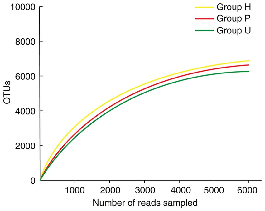
data were sequenced; the sequence length was concentrated at 450‑600 bp, Figure 2. Rarefaction curves of three groups. With the increase of sequencing
and the average length was >500 bp. number of the three groups of samples, the dilution curve gradually
increased. When the number of sequencing exceeded 3000, the dilution curve
reached the plateau stage, indicating that the sequencing depth had basically
covered all species in the sample, and the sampling and sequencing results
sampling and sequencing results could basically reflect the could reflect the real microbial community status of the obtained samples.
OTUs, operational taxonomic units; H, healthy control; P, prepared porcelain
real microbial community status of the obtained samples. veneers; U, unprepared porcelain veneers.
Analysis of flora diversity in samples from dental restoration
two years later. There was a significant difference in the
Alpha diversity index among the 3 groups of samples phylum with higher abundance at the phylum level were
(PEXPERIMENTAL AND THERAPEUTIC MEDICINE 22: 777, 2021 5
Figure 5. Relative abundance of species in oral microbial community of each
group at genus level. The top 10 genera with the largest relative abundance were
Streptococcus, Neisseria, Prevotella‑7, Prevotella, Actinobacillus, Veillonella,
Porphyromonas, Prevotella‑6, Gemella and Leptotrichia. Porphyromonas,
Prevotella and Actinobacillus in gingival crevicular fluid of group U were
Figure 3. Shannon index difference between groups. The Shannon indexes
significantly different from those in group P and group H (*P6 ZHANG et al: EFFECTS OF PORCELAIN VENEERS ON GINGIVAL CREVICULAR FLORA
pathogenic bacteria of periodontal inflammation in dental veneers and unprepared porcelain veneers on gingival crevic‑
implants (30,31). There was a significant difference in the ular microflora can only be analyzed to a certain extent in the
phylum level of Proteobacteria in gingival crevicular flora short term. A larger sample size and a longer study duration
among the three groups, and the relative abundance in group U are required to guide the proper clinical application of tooth
was higher than that in group H and group P, with a significant preparation.
difference. Porphyromonas, Prevotella and Actinobacillus
are closely related to the onset of the disease (26). in the Acknowledgements
present, the abundance of these three types of bacteria in
gingival crevicular fluid of group U was higher than that of Not applicable.
group P and group H, and the difference was statistically
significant. These results suggest that unprepared porcelain Funding
veneers may increase the number of pathogenic bacteria in
gingival crevicular fluid, which has certain adverse effects on This work was supported by Heilongjiang Provincial Health
periodontal health. The position and protrusion of the gingival Commission (2017‑492).
margin of porcelain veneers are important factors for the influ‑
ence of porcelain veneer treatment of periodontal tissue. The Availability of data and materials
subgingival margin is considered to have a negative impact
on periodontal health (29). A previous study suggests that The datasets used and/or analyzed during the present study are
the subgingival margin can change the normal protrusion of available from the corresponding author on reasonable request.
the root surface due to the marginal shape protrusion of the
prosthesis, affecting the periodontal tissue (32). Therefore, in Authors' contributions
order to prevent the subgingival edge from overstimulating
periodontal tissue, the gingival‑aligned edge was used in the RZ and XL conceived and designed this study. LS offered
present study. Due to the lack of tooth preparation, a thin administrative support. All the authors prepared the materials
veneer edge may form certain overhang, stimulate gums and for study. LS, DX and XL helped with data collection and
affect periodontal tissue. It can be observed from Table II summary. RZ and XL were responsible for data analysis and
that in the long‑term follow‑up, individual patients had minor interpretation. RZ wrote the manuscript. All the authors read
adverse reactions such as minor periodontal bleeding, but no and approved the final manuscript.
more serious periodontal complications such as grade C and
above, indicating that the long‑term prognosis of the patients Ethics approval and consent to participate
was favorable. One reason may be that unprepared porcelain
veneer treatment is minimally invasive, which minimizes the The study was approved by the Ethics Committee of Nangang
damage to the original periodontal microenvironment due to Branch of Heilongjiang Province Hospital. Signed written
the non‑grinding of tooth tissue. Although the periodontal flora informed consents were obtained from the patients.
may be in an unbalanced state due to the existence of over‑
hang, its fluctuation range was small. There was no obvious Patient consent for publication
pathological state from the macro perspective. However, the
follow‑up time of this study was short, and whether the current Not applicable.
flora distribution led to long‑term lesions remains unclear.
In previous studies, patients with unprepared porce‑ Competing interests
lain veneers had a higher success rate of restoration and
quality of life compared to those with prepared porcelain The authors declare that they have no competing interests.
veneers (33,34), however, some clinicians also maintain that
unprepared porcelain veneers would lead to oversize of teeth References
and impair oral esthetics. In addition, they can easily breed
bacteria, adversely affecting periodontal tissue (35‑38). The 1. Park DJ, Yang JH, Lee JB, Kim SH and Han JS: Esthetic improve‑
ment in the patient with one missing maxillary central incisor
present study also revealed that the unprepared porcelain restored with porcelain laminate veneers. J Adv Prosthodont 2:
veneers had a greater adverse impact on periodontal tissue in 77‑80, 2010.
terms of micro‑biology, and had potential risks to a certain 2. Horvath S and Dent DM: Minimally invasive restoration of
a maxillary central incisor with a partial veneer. Eur J Esthet
extent. At present, tooth preparation often exceeds enamel, Dent 7: 6‑16, 2012.
causing postoperative sensitivity, thus affecting the long‑term 3. Zhang H, Sun Y, Guo J, Meng M, He L, Tay FR and Zhang S:
curative effect. However, with the popularity of the concept The effect of food medium on the wear behaviour of veneering
porcelain: An in vitro study using the three‑body abrasion mode.
of precision medicine, personalized treatment programs could J Dent 83: 87‑94, 2019.
meet the needs of patients to a greater extent. The development 4. Mombelli A, Müller N and Cionca N: The epidemiology of
of micro‑minimally invasive technology and the progress of peri‑implantitis. Clin Oral Implants Res 23 (Suppl 6): S67‑S76, 2012.
5. Khan AS, Ng SHS, Vandeputte O, Aljanahi A, Deyati A,
related material technology have brought porcelain veneer Cassart JP, Charlebois RL and Taliaferro LP: A multicenter study
treatment a broader prospect and more selections. to evaluate the performance of high‑throughput sequencing for
The duration of the present study was 2 years, and the virus detection. mSphere 2: e00307‑e00317, 2017.
6. Coelho PG, Bonfante EA, Silva NR, Rekow ED and
number of cases involved was relatively small, which were the Thompson VP: Laboratory simulation of Y‑TZP all‑ceramic
limitations of the this study. The effect of prepared porcelain crown clinical failures. J Dent Res 88: 382‑386, 2009.EXPERIMENTAL AND THERAPEUTIC MEDICINE 22: 777, 2021 7
7. Eraslan O, Aykent F, Yücel MT and Akman S: The finite element 23. Kemp PF and Aller JY: Bacterial diversity in aquatic and other
analysis of the effect of ferrule height on stress distribution at environments: What 16S rDNA libraries can tell us. FEMS
post‑and‑core‑restored all‑ceramic anterior crowns. Clin Oral Microbiol Ecol 47: 161‑177, 2004.
Investig 13: 223‑227, 2009. 24. Peumans M, Van Meerbeek B, Lambrechts P and Vanherle G:
8. Baek K, Ji S and Choi Y: Complex intratissue microbiota forms Porcelain veneers: A review of the literature. J Dent 28: 163‑177,
biofilms in periodontal lesions. J Dent Res 97: 192‑200, 2018. 2000.
9. Carda‑Diéguez M, Bravo‑González LA, Morata IM, Vicente A 25. Aristidis GA and Dimitra B: Five‑year clinical performance of
and Mira A: High‑throughput DNA sequencing of microbiota at porcelain laminate veneers. Quintessence Int 33: 185‑189, 2002.
interproximal sites. J Oral Microbiol 12: 1687397, 2019. 26. D'Arcangelo C, De Angelis F, Vadini M and D'Amario M: Clinical
10. Cao Y, Qiao M, Tian Z, Yu Y, Xu B, Lao W, Ma X and Li W: evaluation on porcelain laminate veneers bonded with light‑cured
Comparative analyses of subgingival microbiome in chronic composite: Results up to 7 years. Clin Oral Investig 16: 1071‑1079,
periodontitis patients with and without IgA nephropathy by high 2012.
throughput 16S rRNA sequencing. Cell Physiol Biochem 47: 27. Dumfahrt H and Schaffer H: Porcelain laminate veneers. A
774‑783, 2018. retrospective evaluation after 1 to 10 years of service: Part II
11. Jensen A, Ladegaard Grønkjær L, Holmstrup P, Vilstrup H and clinical results. Int J Prosthodont 13: 9‑18, 2000.
Kilian M: Unique subgingival microbiota associated with peri‑ 28. Peumans M, De Munck J, Fieuws S, Lambrechts P, Vanherle G
odontitis in cirrhosis patients. Sci Rep 8: 10718, 2018. and Van Meerbeek B: A prospective ten‑year clinical trial of
12. Corrêa JD, Calderaro DC, Ferreira GA, Mendonça SM, porcelain veneers. J Adhes Dent 6: 65‑76, 2004.
Fernandes GR, Xiao E, Teixeira AL, Leys EJ, Graves DT 29. Beier US, Kapferer I, Burtscher D and Dumfahrt H: Clinical
and Silva TA: Subgingival microbiota dysbiosis in systemic performance of porcelain laminate veneers for up to 20 years. Int
lupus erythematosus: Association with periodontal status. J Prosthodont 25: 79‑85, 2012.
Microbiome 5: 34, 2017. 30. Chen JH, Shi CX, Wang M, Zhao SJ and Wang H: Clinical
13. Shin YH, Lee YN, Lee HH, Dong JK and Oh SC: Effect of evaluation of 546 tetracycline‑stained teeth treated with Cerinate
application of ZirLiner ® and blasting treatments on shear bond laminate veneers. Zhonghua Kou Qiang Yi Xue Za Zhi 38:
strength of zirconia‑veneered porcelain interface. J Dent Rehabil 119‑202, 2003 (In Chinese).
Appl Sci 24: 113‑127, 2008. 31. Costa FO, Ferreira SD, Cortelli JR, Lima RPE, Cortelli SC and
14. Deng B, Liu HC, Yi YF, Wang C, Wen N and Tian JM: Effects Cota LOM: Microbiological profile associated with peri‑implant
of veneering porcelain type on bending strength of dental diseases in individuals with and without preventive maintenance
Y‑TZP/porcelain bilayered structure. Adv Mater Res 105‑106: therapy: A 5‑year follow‑up. Clin Oral Investig 23: 3161‑3171,
524‑527, 2010. 2019.
15. Erdemir EO, Duran I and Haliloglu S: Effects of smoking on 32. Koyanagi T, Sakamoto M, Takeuchi Y, Maruyama N, Ohkuma M
clinical parameters and the gingival crevicular fluid levels of and Izumi Y: Comprehensive microbiological findings in
IL‑6 and TNF‑alpha in patients with chronic periodontitis. J Clin peri‑implantitis and periodontitis. J Clin Periodontol 40: 218‑226,
Periodontol 31: 99‑104, 2010. 2013.
16. Kim DM, Koszeghy KL, Badovinac RL, Kawai T, Hosokawa I, 33. Allen B, Kon M and Bar‑Yam Y: A new phylogenetic diversity
Howell TH and Karimbux NY: The effect of aspirin on gingival measure generalizing the Shannon index and its application to
crevicular fluid levels of inflammatory and anti‑inflammatory phyllostomid bats. Am Nat 174: 236‑243, 2009.
mediators in patients with gingivitis. J Periodontol 78: 1620‑1626, 34. Galloway‑Peña JR, Smith DP, Sahasrabhojane P, Ajami NJ,
2007. Wadsworth WD, Daver NG, Chemaly RF, Marsh L, Ghantoji SS,
17. Son J, Tze WTY and Gardner DJ: Thermal behavior of hydroxy‑ Pemmaraju N, et al: The role of the gastrointestinal microbiome
methylated resorcinol (HMR)‑treated maple veneer. Wood Fiber in infectious complications during induction chemotherapy for
Sci 37: 220‑231, 2005. acute myeloid leukemia. Cancer 122: 2186‑2196, 2016.
18. De Vasconcellos DK, Özcan M, Maziero Volpato CÂ, Bottino MA 35. Tsubota K: Ten‑year clinical observation of a porcelain laminate
and Yener ES: Strain gauge analysis of the effect of porcelain veneer seated with biological tissue adaptation (BTA) technique.
firing simulation on the prosthetic misfit of implant‑supported J Oral Sci 59: 311‑314, 2017.
frameworks. Implant Dent 21: 225‑229, 2012. 36. Marsh PD: Microbial ecology of dental plaque and its signifi‑
19. Singh S, Sharma P and Kumar M: Evaluation of the effects of cance in health and disease. Adv Dent Res 8: 263‑271, 1994.
0.05% sodium hypochlorite and 0.12% chlorhexidine gluco‑ 37. Reeves WG: Restorative margin placement and periodontium
nate twice daily rinse on periodontal parameters and gingival health. J Prosthet Dent 66: 733‑736, 1991.
crevicular fluid HSV1 and CMV levels in patients with chronic 38. Kawai K, Urano M and Ebisu S: Effect of surface roughness of
periodontitis: A multicentric study. Med J Armed Forces India 2: porcelain on adhesion of baeteria and their synthesizing glueans.
102‑109, 2020. J Prosthet Dent 83: 664‑667, 2000.
20. Chai J, McGivney GP, Munoz CA and Rubenstein JE: A multi‑
center longitudinal clinical trial of a new system for restorations. This work is licensed under a Creative Commons
J Prosthet Dent 77: 1‑11, 1997. Attribution-NonCommercial-NoDerivatives 4.0
21. Gemlamaz D and Ergin S: Clinical evalution of all‑ceramic International (CC BY-NC-ND 4.0) License.
crowns. J Prosthet Dent 89: 189‑196, 2002.
22. Ryge G: Clinical criteria. Int Dent J 30: 347‑358, 1980.You can also read