Anatomic Patterns of the Facial Nerve in Parotidectomized Patients
←
→
Page content transcription
If your browser does not render page correctly, please read the page content below
American Journal of Otolaryngology and Head and Neck Surgery                                                                                     Research Article
                                                                                                                                           Published: 15 Jul, 2021

Anatomic Patterns of the Facial Nerve in Parotidectomized
                         Patients
                                             Pacheco-Ojeda Luis1*, Moncayo-Young Victoria2, Merlo-Cifuentes Felipe3, Del Salto-
                                             Aguagallo María4 and Zabala-Parreño Andres2,3,4,5
                                             1Department     of Head and Neck Surgery, Hospital Metropolitano, Ecuador
                                             2Pontificia   Universidad Católica, Ecuador
                                             3Hospital   de Especialidades Carlos Andrade Marín, Ecuador
                                             4Hospital   Eugenio Espejo, Ecuador
                                             5Clinical   Microbiology and Molecular Epidemiology MCEP

                                             Abstract
                                             Introduction: The main challenge of parotid surgery is dissection and preservation of the facial
                                             nerve. Many studies about its surgical anatomy, based on cadaver dissections, have been reported,
                                             but less frequently on patients undergoing parotid surgery.
                                             Objective: The current study aimed to study the diagrammed facial nerves and three morphological
                                             parameters.
                                             Methods: The clinical records of patients who underwent parotid surgery at a third level hospital in
                                             Quito, Ecuador were reviewed. The facial nerve was diagrammed after dissection, a picture taken,
                                             and anatomic measurements registered.
                                             Results: The most common anatomic branching types were I and III, and IA and IB, according to
                                             Davis’ and Katz’s classifications, respectively. Mean value of the angle between the anterior border
                                             of the mastoid process and the trunk of the facial nerve was 64o. Mean value of the length of the
                                             trunk of the facial nerve was 17 mm, and mean value of the distance between the stylomastoid
                                             foramen and the tip of the mastoid process, was 18 mm.

                    OPEN ACCESS              Conclusion: Two surgical landmarks to identify the facial nerve during parotidectomy, not
                                             previously described, were analyzed. Firs table, the angle between the anterior border of the mastoid
                    *Correspondence:         process and the trunk of the facial nerve which may be an important guide for dissection but
      Pacheco-Ojeda Luis, Department         may vary anatomically or according to the position of an adjacent tumor. Secondly, the distance
   of Head and Neck Surgery, Hospital        from the tip of the mastoid process to the stylomastoid foramen which is extremely important for
  Metropolitano, Sarmiento de Gamboa         identification on the trunk at the beginning of the surgical procedure.
      Oe 538, 170510, Quito, Ecuador,
   E-mail: luispacheco.o@hotmail.com
                                             Keywords: Parotid gland; Facial nerve; Anatomy
          Received Date: 22 Jun 2021
                                             Introduction
          Accepted Date: 12 Jul 2021
         Published Date: 15 Jul 2021             The main challenge of parotid surgery is dissection and preservation of the facial nerve.
                                Citation:    Therefore, the knowledge of the anatomy of this nerve is of great importance. A great number of
    Luis P-O, Victoria M-Y, Felipe M-C,      studies on the surgical anatomy of the intra and extraparotid facial nerve dissected on cadavers have
     Salto-Aguagallo María D, Parreño        been reported [1-4]. However, these types of study on patients who have undergone parotid surgery
    Andres Z. Anatomic Patterns of the       have been somewhat less frequent [5].
      Facial Nerve in Parotidectomized           Anatomic branching of the facial was described in the classical studies of Davis and Katz
      Patients. Am J Otolaryngol Head        [1,5]. Branching patterns according to Davis description were as follows: Type I, no anastomosis
          Neck Surg. 2021; 4(6): 1143.       between branches of the facial nerve; type II, anastomotic connection between branches of the
     Copyright © 2021 Pacheco-Ojeda          temporofacial division; type III, a single anastomosis between the temporofacial and cervicofacial
     Luis. This is an open access article    divisions; type IV, a combination of type II and type III; type V, two anastomotic rami passed from
           distributed under the Creative    the cervicofacial division to interwine with the branches of the temporofacial division; and type
   Commons Attribution License, which        VI, plexiform arrangement (Figure 1). In a study of parotid dissection patients, Katz divided facial
   permits unrestricted use, distribution,   nerve configurations into five main types: straight branching pattern (type I); a loop involving the
       and reproduction in any medium,       zygomatic division (type II); a loop involving the buccal division (type III); a complex pattern with
   provided the original work is properly    multiple interconnections (type IV); and two main trunks, one major and one minor (type V)
                                    cited.   (Figure 2).

Remedy Publications LLC.                                                       1                                         2021 | Volume 4 | Issue 6 | Article 1143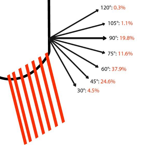
Pacheco-Ojeda Luis, et al., American Journal of Otolaryngology and Head and Neck Surgery
Figure 1: Davis’ classification.
T: Temporal branch; Z: Zygomatic branch; B: Buccal branch; M: Mandibular
branch; C: Cervical branch
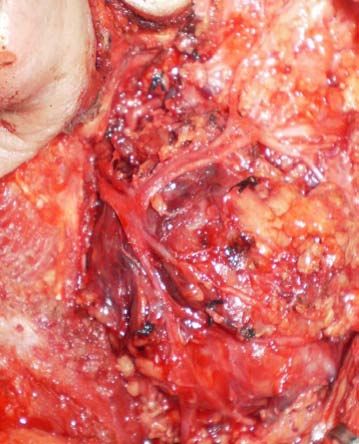
Figure 3: Type III Davis and II Katz anatomic distribution of the facial nerve
after superficial parotidectomy.
surgery by a single surgeon at the third level public hospital, Social
Security Hospital, and some private hospitals in Quito, Ecuador,
South America, from 1980 to 2020, were reviewed. A Hospital Board
permission was obtained. All these patients underwent a superficial,
total, or extended parotidectomy (Figure 3) performed as previously
described [6]. All of them signed an informed consent before
surgery. Mean age of patients of 49 years ± 19.4; 236 were males and
204 females; and 234 were operated for benign lesions and 136 for
malignancies.
The facial nerve was diagrammed after dissection, a picture taken,
and additional anatomic elements were measured with a mm-scale
ruler and a protractor, at the end of the surgical procedure. Then,
Figure 2: Katz’s classification. data was registered in the clinical records. This study was started
T: Temporal branch; Z: Zygomatic branch; B: Buccal branch; M: Mandibular prospectively in 1988.
branch; C: Cervical branch
The clinical information of the patients was organized in a database,
On the other hand, several surgical landmarks to identify the for subsequent tabulation and analysis using descriptive statistics to
facial nerve during parotidectomy have been described, including the determine the percentage frequencies to achieve a distribution of the
tympanomastoid suture line, the tragal pointer, the posterior belly of study variables. They were represented in frequency tables, with the
the digastric muscle, the styloid process, and the retrograde dissection aim of doing a contrast of observed frequencies with those expected.
[6]. The current study aimed to review the facial nerve branching Results
pattern and some dissection useful parameters of the facial nerve
morphology in a selected group of patients submitted to parotid The extratemporal facial nerve branching pattern was
surgery. diagrammed in 348 parotidectomies. Anatomic branching of the
facial nerve was classified according to Davis’ and Katz’s descriptions.
Material and Methods The most common type found in Davis’ classification was type I
The clinical records of 440 patients who underwent parotid (57%), followed by type III (19%) and II (18%) (Table 1). In Katz’s
Table 1: Anatomic type distribution of facial nerve branching according to Davis’ classification [1].
Author(s) Number of cases Type (%)
I II III IV V VI
Davis et al. [1] 350 13 20 28 24 9 6
Bernstein et al. [2] 35 9 9 25 19 22 16
Myint et al. [3] 79 11 16 34 19 7 13
Weerapant et al. [4] 100 1 10 20 18 29 21
The present study 348 57 18 19 3.5 2 0.3
Remedy Publications LLC. 2 2021 | Volume 4 | Issue 6 | Article 1143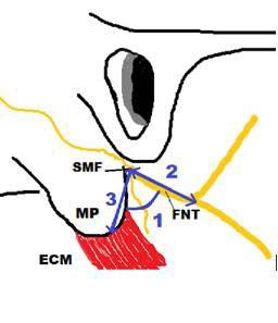
Pacheco-Ojeda Luis, et al.,                                                                        American Journal of Otolaryngology and Head and Neck Surgery

Table 2: Anatomic type distribution of facial nerve branching according to Katz’s classification [5].
             Author(s)                      Number of cases                                                          Type (%)

                                                                          IA        IB        II        IIIA         IIIB          IIIC         IVA          IVB         V

Katz et al. [5]                                    100                    18        6        14          25           13            6            10           4          3

The present study                                  348                    59        13        8          8             1            2            5            4          0

                                                                                          Figure 5: Anatomic parameters: 1. Angle between the anterior borders of the
                                                                                          mastoid process and the upper part of sternocleidomastoid muscle and the
                                                                                          trunk of the facial nerve, 2. Length of the trunk of the facial nerve, 3. Distance
  Figure 4: Values of the angle between the anterior borders of the mastoid               between the stylomastoid foramen and the tip of the mastoid process.
  process and the upper part of sternocleidomastoid muscle, and the trunk of              SMF: Stylomastoid Foramen; MP: Mastoid Process; FNT: Facial Nerve
  the facial nerve.                                                                       Trunk; SCM: Sternocleidomastoiid Muscle

classification, type IA as the most frequent (59%), followed by IB                       facial nerve. Malignant tumors are treated with total parotidectomy
(13%) and IIIA (8.1%) (Table 2).                                                         with preservation of the facial nerve if it is not involved with tumor.
                                                                                         Otherwise, it must be sacrificed, and a reconstruction attempted. Neck
    The following anatomic parameters were also registered:
                                                                                         dissection is indicated if clinical lymph nodes are present according
   1. The angle between the anterior borders of the mastoid                              to National Comprehensive Cancer Network (NCCN) guidelines.
process and the upper part of the Sternocleidomastoid Muscle (SCM)                       Tumors with adverse features are treated with adjuvant radiation
and the Facial Nerve Trunk (FNT) was measured in 354 patients. The                       therapy and occasionally with systemic therapy. Parotidectomy can
mean value was 64° ± 18.2, ranging from 30° to 120° (Figure 4). The                      also be performed in case of intraparotid or periparotid metastatic
most common value was 60° (Figure 4).                                                    lymph node from another tissue or organ primary, and for chronic
                                                                                         sialoadenitis or sialolithiasis.
    2. The length of the trunk of the facial nerve between
the stylomastoid foramen and its division in temporofacial and                               Regarding the classic surgical landmarks, the tympanomastoid
cervicofacial branches was measured in 301 patients. Mean value 17                       suture line lies between the mastoid and tympanic segments of the
mm ± 4.54, ranging from 8 mm to 40 mm (Figure 5).                                        temporal bone and is approximately 6 mm to 8 mm lateral to the
                                                                                         stylomastoid foramen. The main trunk of the nerve can be found
    3. The distance between the stylomastoid foramen and the tip                         midway between the cartilaginous tragal pointer of the external
of the mastoid process was also measured in 301 patients. The mean                       auditory canal and the posterior belly of the digastric muscle. The
value was 18 mm ± 4.08, ranging from 8 mm to 32 mm (Figure 5).                           nerve is usually located inferior and medial to the tragal pointer [6].
Discussion                                                                                    Borle proposed an anatomic triangle to reliably identify the Facial
     The approach to the facial nerve is vitally important during                        Nerve Trunk (FNT). Description of the lines of this triangle was as
surgical procedures of the parotid gland for benign or malignant                         following: The first starts at the tip of the mastoid process, running
lesions. This applies also to facial plastic surgery, surgery of vascular                along the superior border of the posterior belly of the digastric
lesions, and trauma [7-9]. The main concern of the surgeon is the                        muscle; a second line along the posterior border of the ramus of the
identification and preservation of the facial nerve as well as an                        mandible; and a third line, from the tip of the mastoid process, (angle
                                                                                         b) running anteriorly, till it joins the second line. The mean distance
adequate parotidectomy including the complete removal of the tumor
                                                                                         of FNT from angle b was found to be 12.18 mm [10].
and the surrounding salivary tissue. Benign tumors are managed with
superficial parotidectomy if located in the superficial lobe, or total                      Other landmarks and anatomic descriptions of the different
parotidectomy if located in the deep lobe taking care to preserve the                    branches of the facial nerve have been reported due to the particular

Remedy Publications LLC.                                                            3                                               2021 | Volume 4 | Issue 6 | Article 1143
Pacheco-Ojeda Luis, et al., American Journal of Otolaryngology and Head and Neck Surgery
attention given when operating in specific regions of the face and, of Salame who found a length of 16.44 ± 3.2 mm in a study in 46
consequently, to minimize damage to them [8]. cadavers. The trunk must be dissected as far as the pes anserinus,
the structure marking the separation of the cervicofacial (lower)
For the temporal branch, Pitanguy and Ramos described a line
and temporofacial (upper) divisions. These main branches and the
starting from a point 0.5 cm below the tragus that extended in the
smaller distal rami are then followed carefully to map their relation to
direction of the brow, passing 1.5 cm above the lateral extremity of
the tumor and of dissecting free of it.
the eyebrow [11].
The angle between the anterior border of the mastoid process
We era pant described three landmarks: The vertical distance
and the trunk of the facial nerve may be a guide to the direction of
from the mandibular angle to the marginal mandibular branch, the
the dissection of the trunk of the nerve. The mean value was 60°,
horizontal distance from the lateral palpebral line to the otobasion
ranging from 30° to 120°. The most common value was 60° (38%).
superius, and the distance from the lateral palpebral line to the apex
This angle may vary anatomically but, most importantly, according
of the parotid gland [4].
to the position of an adjacent tumor that can deviate the FNT upward
Sanderson described the average distance, 3.21 cm, from the apex or downward. We have not found another report of this anatomic
of the tragus to the point where the frontotemporal branch of the presentation in the literature.
facial nerve crosses the inferior border of the zygomatic arch [12].
It is well known for the variability of facial nerve branching
Furnas also described the vagino-mastoid to guide the exposition patterns. For anatomic branching pattern description, we used
of the FNT and the upper and outermost aspect of the eyebrow and the classifications of Davis and Katz. We have not found new
the point where a projection of the hairline crosses the zygoma, as a classifications in the literature. Davis classified the facial nerve
landmark for the temporal branch [13]. configuration in six types based on 350 cadaveric dissections. On
the other hand, Katz classified the patterns in 5 main types and 9
Tayebi Meybodi et al. [14] proposed the digastric branch of the
subtypes based on surgical dissections of 100 patients undergoing
facial nerve as a landmark to localize the extratemporal portion of the
parotidectomy. In cadavers, dissections could extend further to the
facial nerve [14].
boundaries of the parotid gland. On the other hand, a surgeon must
As Marginal Mandibular Nerve (MMN) injuries have been dissect very carefully the facial nerve to remove, adequately and
reported in many head and neck procedures (parotidectomy, excision completely, salivary tissue according to the type of parotidectomy,
of the submandibular gland, neck dissection, etc.) several reference superficial or total, and the type tumor, benign or malignant.
points have been described to avoid these lesions. Among them: The
In Davis’ classification, a type was assigned according to the
gonion, the posterior border of the antegonial notch, the superior arc
presence and number of anastomotic rami between branches of the
of the antegonial notch, the anterior border of the antegonial notch,
temporofacial division, or between the main temporofacial and the
and the facial artery. Only its relationship with the anterior facial vein
cervicofacial divisions. In Katz’s classification, types and subtypes
seems to be constant. This nerve almost always runs superficially [15].
were assigned according to the aforementioned anastomosis and,
A modern and remarkably interesting method for studying anatomy,
additionally, to the origin of the secondary divisions. In any case, the
Anatomage (Anatomage, Inc., San Jose, CA), was used to study the
surgeon must dissect and try to preserve even the anastomotic rami.
course of the MMN in the area where it crosses the facial vessels [16].
In our experience, the occasional sacrifice of very tiny anastomotic
The current study describes practical and handy landmarks, rami has not resulted in an apparent partial deficit of mobility of the
observed through a long period of surgical practice, to safely identify face.
and dissect the facial nerve. These are the angle between the anterior
Kehrer reappraised the zygomaticobuccal branch system
borders of the mastoid process and of the sternomastoid muscle and
concerning anastomoses and crossings that could be relevant for
the trunk of the facial nerve, the length of the trunk of the facial nerve
facial reanimation surgery [17].
between the stylomastoid foramen and its division in temporofacial
and cervicofacial branches, and the distance between the stylomastoid Roostaeian described the safe planes of dissection during surgical
foramen and the tip of the mastoid process. undermining to minimize the risk of facial nerve injury during
rhytidectomy [7]. An interesting point in this report is the attention
The identification of the facial nerve is the most important step at
to the cervical branch; one of the most commonly injured branches,
the beginning of the surgical procedure. Once the tail of the parotid
because of its relatively superficial nature and intimate attachment to
gland has been elevated off the SCM until the posterior belly of the
the platysma. It can pucker the lower lip. Fortunately, the complete
digastric muscle and then separated from the cartilage of the tragus
return of function has been reported in nearly all cases of injury of
and the external auditory canal, great care must be taken to identify
this branch.
the nerve. The main concern is to know how deep the surgeon should
dissect to find it. The distance from the tip of the mastoid process Distribution of FN branching pattern comparing to Davis types
to the stylomastoid foramen through which the nerve emerges may was very variable in several series (Table 1). In our study, type
vary a lot. So, we measured this distance in our patients. The mean I classification was the most common (57%) and type VI the least
value was 18 mm but it ranged from 8 mm to 32 mm. Sometimes, frequent. Type III was more common in Davis’, Bernstein's, and
identifying the nerve takes time and palpation of the styloid process Myint’s series but type IV was the most frequent in Weerapant’s
can help. report.
The length of the trunk of the facial nerve may also vary In Katz's original description, 44% of patients had a type III
anatomically. In our patients, the mean value was 17 ± 4.54 mm, pattern, 24% had an unbranched pattern (type I), and 3% of the
ranging from 8 mm to 40 mm. This finding is similar to the report patients had two main trunks (type V). We did not find any patient
Remedy Publications LLC. 4 2021 | Volume 4 | Issue 6 | Article 1143Pacheco-Ojeda Luis, et al., American Journal of Otolaryngology and Head and Neck Surgery
with the latter type and most of our patients had an unbranched 5. Katz AD, Catalano P. The clinical significance of the various anastomotic
pattern (Table 2). branches of the facial nerve. Arch Otolaryngol Head Neck Surg.
1987;113(9):959-62.
Adidharma used an in vivo Facial Nerve Mapping (FNM) during
6. Langerman A, Netterville J. Parotidectomy. Medscape Updated. 2019.
Vascular Anomaly (VAN) surgeries involving the Facial Nerve (FN)
in 67 patients and simplified the facial nerve patterns into two groups: 7. Roostaeian J, Rohrich RJ, Stuzin JM. Anatomical considerations to prevent
Type 1A included FN patterns with 2 anastomoses (anatomical types facial nerve injury. Plast Reconstr Surg. 2015;135(5):1318-27.
I to III) and type 2A contained FN patterns with ≥ 2 anastomoses 8. Myckatyn TM, Mackinnon SE. A review of facial nerve anatomy. Semin
(anatomical types IV to VI). Additionally, he classified the nerve Plast Surg. 2004;18(1):5-11.
relationship with mass as through, adherent or separate and the 9. Adidharma L, Bly RA, Theeuwen HA, Holdefer RN, Slimp J, Kinney
surgical approach as direct, anterograde, or retrograde [9]. GA, et al. Facial nerve branching patterns vary with vascular anomalies.
Laryngoscope. 2020;130(11):2708-13.
Magnetic resonance Diffusion Weighted Imaging (DWI) was
conducted on 2 parotid-healthy cadaveric patients, tracking the 10. Borle RM, Hadhav A, Bohla N, Hingnikar P, Gaitwad P. Borle's triangle: A
extracranial course of the facial nerve to provide a reliable facial nerve reliable anatomical landmark for ease of identification of facial nerve trunk
map [18]. Correlations between imaged tracts and the anatomic during parotidectomy. J Oral Biol Craniofac Res. 2019;9(1):33-6.
course of the extracranial facial nerve were identified to an accuracy 11. Pitanguy I, Silveira Ramos AM. The frontal branch of the facial nerve:
of 1 mm. According to the authors, this visualization could have The importance of its variations in face lifting. Plast Reconstr Surg.
diagnostic implications in differentiating benign from malignant 1966;38(4):352-6.
tumors and, crucially, neural involvement. 12. Sanderson KG, Conti A, Colussi M, Connolly C. A simple clinical
application for locating the frontotemporal branch of the facial nerve using
Conclusion the zygomatic arch and the tragus. Am Soc Aesthetic Plast Surg. Aesthet
Anatomical landmarks can be important to visualize and identify Surg J. 2020;40(5):NP223-7.
the facial nerve during head and neck surgery. We have included 13. Furnas DW. Landmarks for the trunk and the temporofacial division of
additional measurements that have offered a better understanding of the facial nerve. Br J Surg. 1965;52(9):694-6.
the facial nerve anatomy during parotidectomy. 14. Tayebi Meybodi A, Borba Moreira L, Lawton MT, Preul MC. Anatomical
The branching patterns of the facial nerve seem to be highly assessment of the digastric branch of the facial nerve as a landmark
to localize the extratemporal facial nerve trunk. Surg Radiol Anat.
variable as in previous studies and ours. The main and secondary
2019;41(6):657-62.
divisions must be properly identified to avoid injuries during parotid
surgery. 15. Marcuzzo AV, Šuran-Brunelli AN, Dal Cin E, Rigo S, Piccinato A, Boscolo
Nata F, et al. Surgical anatomy of the marginal mandibular nerve: A
References systematic review and meta-analysis. Clin Anat. 2019.
1. Davis RA, Anson BJ, Budinger JM, Kurth LR. Surgical anatomy of the 16. Stranzias P, Botou A, Manoli A, Skandalakis PN, Filipou D. Variation of
facial nerve and parotid gland based upon a study of 350 cervicofacial marginal mandibular nerve in a caucasian male cadaver: A study using the
halves. Surg Gynecol Obstet. 1956;102(4):385-412. anatomage table. Cureus. 2019;11(11):e6168.
2. Bernstein L, Nelson RH. Surgical anatomy of the extraparotid distribution 17. Kehrer A, Engelmann S, Ruewe M, Geis S, Taeger C, Kehrer M, et al.
of the facial nerve. Arch Otolaryngol. 1984;110(3):177-83. Anatomical study of the zygomatic and buccal branches of the facial nerve:
Application to facial reanimation procedures. Clin Anat. 2019;32(4):480-8.
3. Myint K, Azian AL, Khairul FA. The clinical significance of the branching
pattern of the facial nerve in Malaysian subjects. Med J Malaysia. 18. Kininy W El, Roddy D, Davy S, Roman E, O’Keefe V, O’Hanlon E, et
1992;47(2):114-21. al. Magnetic resonance diffusion weighted imaging using constrained
spherical deconvolution-based tractography of the extracranial course
4. Weerapant E, Bunaprasert T, Chokrungvaranont P, Chentanez V.
of the facial nerve. Oral Surg Oral Med Oral Pathol Oral Radiol.
Anatomy of the facial nerve branching patterns, the marginal mandibular
2020;130(2):e44-56.
branch and its extraparotid ramification in relation to the lateral palpebral
line. Asian Biomed. 2010;4(4):603-8.
Remedy Publications LLC. 5 2021 | Volume 4 | Issue 6 | Article 1143You can also read